. 2007 Oct;81(19):10437-50.
doi: 10.1128/JVI.00399-07. Epub 2007 Jul 18.

A tyrosine-based signal plays a critical role in the targeting and function of adenovirus RIDalpha protein

Affiliations

A tyrosine-based signal plays a critical role in the targeting and function of adenovirus RIDalpha protein

Nicholas L Cianciola et al. J Virol. 2007 Oct.

Abstract

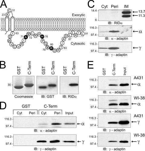
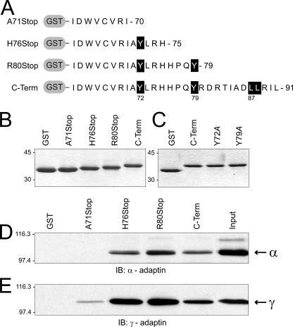
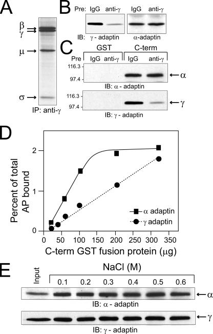
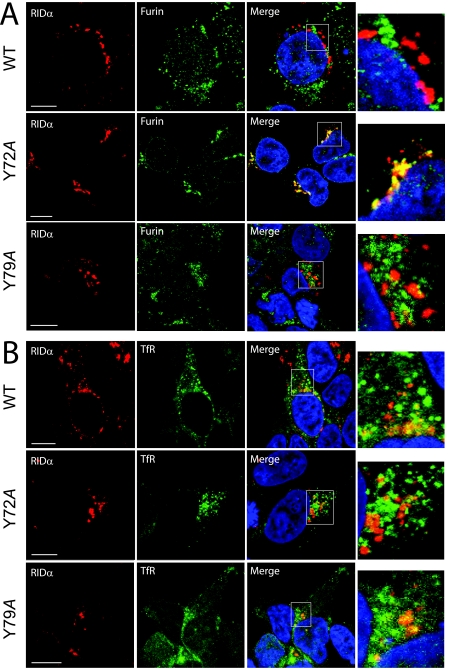
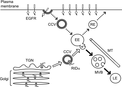
Early region 3 genes of human adenoviruses contribute to the virus life cycle by altering the trafficking of cellular proteins involved in adaptive immunity and inflammatory responses. The ability of early region 3 genes to target specific molecules suggests that they could be used to curtail pathological processes associated with these molecules and treat human disease. However, this approach requires genetic dissection of the multiple functions attributed to early region 3 genes. The purpose of this study was to determine the role of targeting on the ability of the early region 3-encoded protein RIDalpha to downregulate the EGF receptor. A fusion protein between the RIDalpha cytoplasmic tail and glutathione S-transferase was used to isolate clathrin-associated adaptor 1 and adaptor 2 protein complexes from mammalian cells. Deletion and site-directed mutagenesis studies showed that residues 71-AYLRH of RIDalpha are necessary for in vitro binding to both adaptor complexes and that Tyr72 has an important role in these interactions. In addition, RIDalpha containing a Y72A point mutation accumulates in the trans-Golgi network and fails to downregulate the EGF receptor when it is introduced into mammalian cells as a transgene. Altogether, our data suggest a model where RIDalpha is trafficked directly from the trans-Golgi network to an endosomal compartment, where it intercepts EGF receptors undergoing constitutive recycling to the plasma membrane and redirects them to lysosomes.


Figures

FIG. 1.
FIG. 1.
Binding of RIDα cytosolic tail to AP complexes. (A) Schematic showing membrane topology of RIDα and the complete amino acid sequence (in single-letter code) of the carboxyl terminus (Ile63-Leu91) that was linked to GST for expression in Escherichia coli. Tyr72 and Tyr79 or residues that were changed to stop codons (Fig. 2) are highlighted in gray or black, respectively. Nuclear magnetic resonance studies have shown that residues 87-LLRIL comprise an amphipathic helix stabilized by interactions with membrane mimetic micelles (45). Two additional residues are presented in the exocytic loop connecting the two membrane spanning helices: Ala23, comprising the amino terminus of the 11.3-kDa cleaved form of the protein produced cotranslationally by signal peptidase, and Cys31, which forms intermolecular disulfide bonds (27). (B) GST or the C-terminal (C-Term) fusion protein was purified from E. coli by glutathione affinity chromatography and resolved by SDS-PAGE for detection by Coomassie staining or by immunoblotting (IB) with GST or RIDα-specific antibodies. (C) Ad-infected cells were fractionated into crude cytosol (Cyt) and peripheral (Peri) and integral membrane (IM) proteins, as described in Materials and Methods. Equal aliquots of total cell equivalents were resolved by SDS-PAGE and immunoblotted with antibodies to RIDα, α-adaptin, or γ-adaptin. (D) GST or C-terminal fusion proteins were incubated with crude cytosol or peripheral membrane proteins isolated from N-PLC-PRF/5 cells, and bound proteins were immunoblotted with adaptin-specific antibodies. (E) Fusion proteins were incubated with peripheral membrane proteins from A431 or WI-38 cells and analyzed as described for panel D. Molecular weight standards: β-galactosidase, 116,300; phosphorylase B, 97,400; carbonic anhydrase, 30,000; lysozyme, 14,400; aprotinin, 6,000.
FIG. 2.
FIG. 2.
Mapping the sites of interaction of RIDα with AP complexes using truncation mutants. (A) Schematic showing sequences of cytoplasmically truncated GST fusion proteins used in the mapping studies. Critical residues in putative sorting signals are highlighted by a black background. (B and C) Proteins were purified from E. coli by glutathione affinity chromatography and resolved by SDS-PAGE for detection by Coomassie staining. Proteins in panel B were used in panels D to E of this figure, and those in panel C were used to obtain the data in Table 1. (D and E) Fusion proteins shown in panel B were incubated with peripheral membrane proteins isolated from N-PLC-PRF/5 cells, and bound proteins were immunoblotted (IB) with adaptin-specific antibodies. C-Term, C terminal.
FIG. 3.
FIG. 3.
Lack of competition between AP-1 and AP-2 for RIDα C-terminal binding. (A) Peripheral membrane proteins from cells that were metabolically labeled with l-[35S]cysteine for 16 h were incubated with the γ-adaptin monoclonal antibody, which immunoprecipitates (IP) intact AP-1 complexes comprised of β, μ, and σ subunits in addition to γ-adaptin. (B and C) Peripheral membrane fractions were preincubated with nonspecific mouse immunoglobulin G (IgG) or with the γ-adaptin monoclonal antibody, and the immunodepleted samples were then incubated with GST or C-terminal (C-term) fusion proteins. Aliquots of the immunodepleted samples (B) and proteins bound to glutathione beads (C) were analyzed by immunoblotting (IB) with adaptin-specific antibodies. (D) Equal aliquots of peripheral membrane proteins were added to increasing concentrations of the C-terminal fusion protein indicated on the x axis. Bound proteins were transferred to nitrocellulose filters, which were incubated with 125I-labeled secondary antibodies after an initial incubation with primary antibodies to α-adaptin or γ-adaptin for quantification by phosphor storage autoradiography. The amount of bound protein was calculated as a percentage of total adaptin proteins in the peripheral membrane fractions, based on immunoblots of total protein quantified using the same method. The experiment was carried out twice with similar results. (E) C-terminal/adaptin complexes were washed five times with wash buffers supplemented with various concentrations of NaCl, as indicated in the figure.
FIG. 4.
FIG. 4.
Colocalization of wild-type (WT), Y72A, and Y79A RIDα with membrane-compartment markers. HEK-293 cells transiently expressing wild-type, Y72A, or Y79A mutant proteins were costained with antibodies to the FLAG-tagged viral protein (red channel) and to the TGN marker furin (A) or the endocytic marker TfR (B) (green channel) and DAPI (blue channel) for analysis by confocal microscopy. Red and green channels were merged, and “yellow” indicates the overlap of red and green fluorescence. Higher-magnification images of outlined areas are shown to the right of each set of panels. Size markers, 10 μm.
FIG. 5.
FIG. 5.
Subcellular distribution of wild-type and Y72A RIDα proteins. (A) CHO cells were fractionated on Percoll gradients, and individual fractions were assayed for β-hexosaminidase activity (open circles). Cells were also fractionated after they had been incubated with 125I-transferrin for 30 min on ice (open squares) or after they were transfected with a human EGFR expression plasmid and incubated with 125I-EGF for 30 min on ice 48 h later (open triangles). Enzyme activity or radioactivity was plotted as a percentage of total activity or radioactivity detected across the entire gradient. (B) Equal aliquots of total cellular protein from individual cell fractions were immunoblotted (IB) with antibodies to well-characterized markers of the Golgi body (gp125) and of the plasma membrane and early endosomes (TfR). (C) CHO cells were transfected with wild-type (WT) or Y72A RIDα plasmids, and immune complexes from individual Percoll gradient fractions were analyzed by immunoblotting with a RIDα-specific antibody. (D and E) CHO cells transfected as described for panel C and nontransfected CHO cells were pulse-labeled and then switched to chase medium to allow proteins to reach their steady-state localization. Proteins were immunoprecipitated and analyzed by SDS-PAGE under standard reducing conditions (+DTT) or were immunoprecipitated from cells subjected to surface reducing (+) or sham (−) conditions followed by SDS-PAGE under nonreducing conditions (−DTT).
FIG. 6.
FIG. 6.
Effect of nocodazole on RIDα protein subcellular localization. CHO cells transiently expressing wild-type or Y72A mutant proteins were pretreated with dimethyl sulfoxide vehicle (Veh) or nocodazole (Noc; 100 μM) for 30 min prior to staining with antibody to the FLAG-tagged viral protein (red channel) and with DAPI (blue channel). Cell outlines (in green) were made with the MetaMorph program using phase contrast images as a guide. Size markers, 10 μm.
FIG. 7.
FIG. 7.
Effect of Y72A and Y79A point mutations on RIDα expression and function in mammalian cells. CHO cells were cotransfected with plasmid expressing the human EGFR along with a plasmid expressing wild-type RIDα (WT), plasmids expressing RIDα with a Y72A or Y79A substitution, or an empty vector (Sham). (A) Equal aliquots of total cell protein were resolved by SDS-PAGE, transferred to nitrocellulose, and immunoblotted with a RIDα-specific antibody. (B) The cells were radiolabeled with [35S]cysteine for 30 min, starting at 48 h posttransfection, and harvested either immediately or following a 5-h chase in medium with an excess of nonradioactive cysteine. EGFR immune complexes were resolved by SDS-PAGE and detected by autoradiography. IP, immunoprecipitation.
FIG. 8.
FIG. 8.
Summary model. Data presented in this study suggest that newly synthesized RIDα is delivered directly from the TGN to endosomes (open arrows) where it encounters EGFRs undergoing constitutive recycling to the plasma membrane (dashed arrows). Since RIDα proteins with a defective AP binding site accumulate in the Golgi body and TGN, we conclude that newly synthesized RIDα proteins encounter AP-1 necessary for incorporation into clathrin-coated vesicles (CCV), which are necessary for exiting the biosynthetic compartment. Data also show that RIDα is delivered directly to endosomes and not the plasma membrane, suggesting that the ability of the viral protein to bind AP-2 serves a quality control function in which proteins that “leak” to the plasma membrane are quickly endocytosed and retargeted to their appropriate intracellular locations. Although the EGFR and RIDα are both transported to late endosomes (LE) via multivesicular body (MVB) transport intermediates (solid arrows), only EGFR is degraded. EE and RE, early and recycling endosomes, respectively; MT, microtubule.

Similar articles

Cited by

References

    1. Avila, M. M., G. Carballal, H. Rovaletti, B. Ebekian, M. Cusminsky, and M. Weissenbacher. 1989. Viral etiology in acute lower respiratory infections in children from a closed community. Am. Rev. Respir. Dis. 140:634-637. - PubMed
    1. Bonifacino, J., and L. Traub. 2003. Signals for sorting of transmembrane proteins to endosomes and lysosomes. Annu. Rev. Biochem. 72:395-447. - PubMed
    1. Brandt, C. D., H. W. Kim, A. J. Vargosko, B. C. Jeffries, J. O. Arrobio, B. Rindge, R. H. Parrott, and R. M. Chanock. 1969. Infections in 18,000 infants and children in a controlled study of respiratory tract disease. I. Adenovirus pathogenicity in relation to serologic type and illness syndrome. Am. J. Epidemiol. 90:484-500. - PubMed
    1. Bretscher, A. 1989. Rapid phosphorylation and reorganization of ezrin and spectrin accompany morphological changes in A431 cells induced by EGF. J. Cell Biol. 108:921-930. - PMC - PubMed
    1. Carlin, C. R., and B. B. Knowles. 1986. Biosynthesis and glycosylation of the epidermal growth factor receptor in human tumor-derived cell lines A431 and Hep 3B. Mol. Cell. Biol. 6:257-264. - PMC - PubMed

Publication types

MeSH terms

LinkOut - more resources