. 2007 Oct;81(19):10506-14.
doi: 10.1128/JVI.02853-06. Epub 2007 Jul 18.

Host cell cathepsins potentiate Moloney murine leukemia virus infection

Affiliations

Host cell cathepsins potentiate Moloney murine leukemia virus infection

Pankaj Kumar et al. J Virol. 2007 Oct.

Abstract

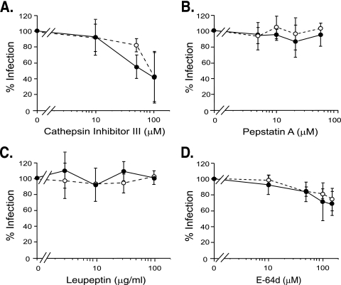
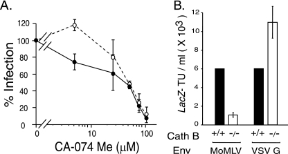
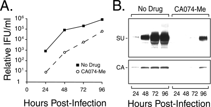
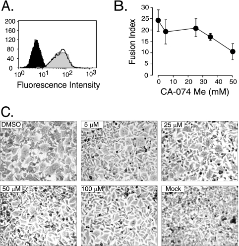
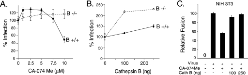
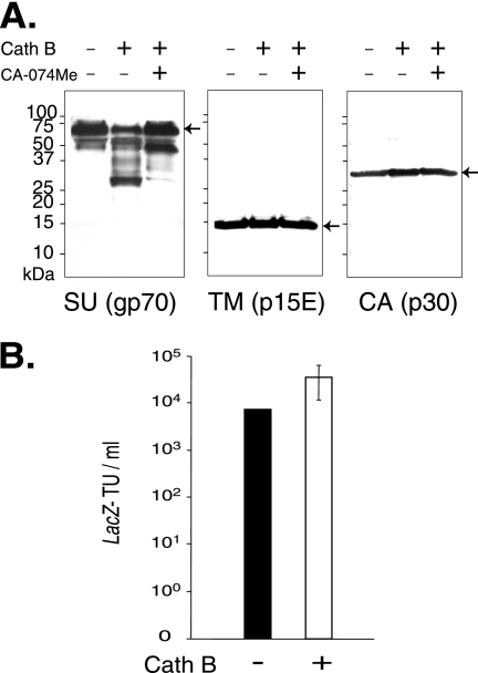
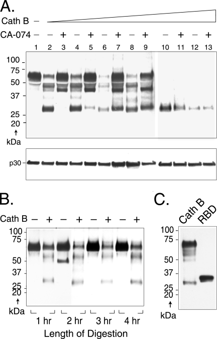
The roles of cellular proteases in Moloney murine leukemia virus (MLV) infection were investigated using MLV particles pseudotyped with vesicular stomatitis virus (VSV) G glycoprotein as a control for effects on core MLV particles versus effects specific to Moloney MLV envelope protein (Env). The broad-spectrum inhibitors cathepsin inhibitor III and E-64d gave comparable dose-dependent inhibition of Moloney MLV Env and VSV G pseudotypes, suggesting that the decrease did not involve the envelope protein. Whereas, CA-074 Me gave a biphasic response that differentiated between Moloney MLV Env and VSV G at low concentrations, at which the drug is highly selective for cathepsin B, but was similar for both glycoproteins at higher concentrations, at which CA-074 Me inhibits other cathepsins. Moloney MLV infection was lower on cathepsin B knockout fibroblasts than wild-type cells, whereas VSV G infection was not reduced on the B-/- cells. Taken together, these results support the notion that cathepsin B acts at an envelope-dependent step while another cathepsin acts at an envelope-independent step, such as uncoating or viral-DNA synthesis. Virus binding was not affected by CA-074 Me, whereas syncytium induction was inhibited in a dose-dependent manner, consistent with cathepsin B involvement in membrane fusion. Western blot analysis revealed specific cathepsin B cleavage of SU in vitro, while TM and CA remained intact. Infection could be enhanced by preincubation of Moloney MLV with cathepsin B, consistent with SU cleavage potentiating infection. These data suggested that during infection of NIH 3T3 cells, endocytosis brings Moloney MLV to early lysosomes, where the virus encounters cellular proteases, including cathepsin B, that cleave SU.


Figures

FIG. 1.
FIG. 1.
Effects of protease inhibitors on infection of NIH 3T3 cells. Naïve NIH 3T3 cells were exposed to lacZ-transducing, replication-defective pseudovirions for 2 h in the presence of the indicated amounts of inhibitor. Closed circles, Moloney MLV Env-pseudotyped MLV. Open circles, VSV G-pseudotyped MLV. (A) Cathepsin inhibitor III, a broad-spectrum inhibitor of cathepsins, was used at 10, 50, 100, and 150 μM. (B) Pepstatin A, an inhibitor of aspartyl proteases, was used at 5, 10, 20, and 50 μM. (C) Leupeptin, an inhibitor of serinyl and cysteinyl proteases, was used at 3, 10, 30, and 100 μg/ml. (D) E-64d, an inhibitor of cysteinyl cathepsins (all known cathepsins except D, E, and G), was used at 10, 50, 100, and 150 μM. The values shown are the mean percent infection ± standard deviation of three independent experiments for cathepsin inhibitor III, pepstatin A, and E-64d in which cells (quadruplicate samples) were preincubated with inhibitor for 30 min prior to virus exposure. For leupeptin, the values shown are the mean percent infection ± standard deviation of three independent experiments performed without preincubation of host cells with inhibitor; a fourth experiment was performed with leupeptin preincubation for 1 h. Similar results were observed in all experiments.
FIG. 2.
FIG. 2.
Host cell cathepsin B is important to ecotropic Moloney MLV infection. (A) Quadruplicate wells of naïve NIH 3T3 cells were preincubated with the indicated amounts of CA-074Me (5, 25, 50, 75, and 100 μM) for 30 min prior to the addition of Moloney MLV Env-pseudotyped MLV (closed circles) or VSV G-pseudotyped MLV (open circles) for 2 h. The inhibitor was maintained during virus exposure. The values shown are the mean percent infection ± standard deviation of three independent experiments. (B) Cathepsin B knockout cells are less susceptible to Moloney MLV (MoMLV) but not to control VSV G pseudovirions. Quadruplicate wells of naïve cathepsin B−/− MEFs were exposed to serially diluted Moloney MLV pseudovirions or to serially diluted VSV G-pseudotyped MLV, and the lacZ-transducing titer was calculated from the end point dilution 48 h later. For comparison, aliquots of the serial virus dilutions were applied to parental wild-type cathepsin MEFs (B+/+). The values shown are the mean LacZ-transducing units (TU)/ml ± standard deviations of three independent experiments.
FIG. 3.
FIG. 3.
Replication-competent Moloney MLV is sensitive to inhibition. Naïve NIH 3T3 cells were preincubated in the presence or absence of 75 μM CA-074 Me for 4 h prior to the addition of replication-competent Moloney MLV. After a 1-h incubation, the unbound virus and inhibitor were washed from the cells. The culture medium was harvested every 24 h for 4 days and analyzed by end point dilution titration for infectivity. (B) Immunoblot analysis of SU and CA expression in culture medium samples from panel A. The results are representative of four independent experiments. IFU, infectious units.
FIG. 4.
FIG. 4.
Inhibition of cathepsin B influences membrane fusion. (A) CA-074Me does not down-regulate virus binding sites. The mean fluorescence intensity was 63.1 for 99% of NIH 3T3 cells preincubated in the vehicle DMSO (white peak and solid black line) compared to 63.7 for 99% of NIH 3T3 cells treated with the inhibitor (gray peak), 5.6 for 99% of NIH 3T3 cells with mock virus plus primary and secondary antibodies (solid black peak) and receptor-negative human 293 cells, and 3.6 for 100% of NIH 3T3 cells without antibodies. Similar results were obtained in a second independent experiment. (B) Quadruplicate samples of XC cells were preincubated for 30 min with CA-074 Me (0, 5, 10, 25, 35, and 50 μM) prior to the addition of replication-defective Moloney MLV pseudovirions. Cell-cell fusion was scored by light microscopy. The values shown are the mean fusion index ± standard deviation (n = 4) calculated as follows: (number of nuclei in syncytium/total number of nuclei) × 100. Similar results were obtained in three additional experiments. (C) Inhibition of cathepsin B markedly reduced the number of nuclei per syncytium. Light micrographs of XC cells exposed to the indicated concentrations of CA-074 Me and then to Moloney MLV (DMSO, 5 μM, 25 μM, 50 μM, and 100 μM) or mock virus (no virus). Micrographs taken at a magnification of ×80.
FIG. 5.
FIG. 5.
Further evidence supporting a role for cathepsin B in the membrane fusion step of infection. (A) Cathepsin B−/− MEFs are not sensitive to inhibition by CA-074 Me in the range that is selective for inhibition of cellular cathepsin B. Quadruplicate samples of B−/− and control B+/+ MEFs were preincubated with 0, 1, 2.5, 5, 7.5, and 10 μM CA-074 Me for 1 h prior to the addition of replication-defective virus plus the inhibitor; 6 h later, the cells were fixed, stained, and then scored by light microscopy. Similar results were obtained in two additional experiments. One hundred percent infection (no CA-074 Me) was 1 × 10³ ± 0.1 × 10³ for B+/+ cells and 4.8 × 10² ± 0.1 × 10² LacZ TU/ml for the poorly susceptible B−/− cells. The error bars indicate standard deviations. (B) Addition of purified exogenous cathepsin B increased infection of B−/− MEFs. Triplicate samples of B−/− and B+/+ cells were exposed to virus mixed with the indicated amounts of purified cathepsin B. Similar results were observed in two additional independent experiments. One hundred percent infection (no cathepsin B added) was 3.8% ± 0.8% of B+/+ cells infected and 0.8% ± 0.2% of B−/− cells. The standard deviations are not visible for the 100-ng B+/+ and 250-ng B−/− samples because their error bars were smaller than the symbol for the data point. (C) Addition of exogenous cathepsin B overcame the inhibition of fusion resulting from CA-074 Me. Quadruplicate wells of NIH 3T3 cells were preincubated with 50 μM CA-074 Me for 2 h to irreversibly inhibit endogenous cathepsin, and then the cells were rinsed once in DMEM, and virus plus the indicated amounts of purified cathepsin B were applied to the cells for 6 h. A similar restoration of syncytium induction was seen in an independent experiment.
FIG. 6.
FIG. 6.
Cathepsin B (Cath B) cleaves the envelope SU on virions. Aliquots of Moloney MLV pseudovirions purified by high-speed ultracentrifugation for 45 min were incubated with or without purified cathepsin B (68 μg/ml) for 1 h in acetate buffer, pH 5.5, in the presence or absence of 100 μM CA-074Me. (A) Viral proteins in 10 microliters of each reaction mixture analyzed by separation on 10 to 20% Tris-glycine gradient gels (Invitrogen) were immunoblotted to anti-SU antiserum, and the immunoblot was stripped and reprobed with anti-TM antibody (a gift of Alan Rein) and then stripped a second time and reprobed with anti-CA antiserum. Similar results were obtained in a second independent experiment. (B) Effect of preincubation with cathepsin B on virus infection. The values shown are the mean lacZ-transducing units (TU)/ml ± standard deviations from six independent experiments. Virus was prepared and incubated with cathepsin B as for panel A, except that after incubation with the protease, or with acetate reaction buffer alone, the reaction mixtures were diluted to their original volumes (1× with respect to the virus concentration in the original virus stock) in regular growth medium and then serially diluted and applied to quadruplicate wells of naïve NIH 3T3 cells.
FIG. 7.
FIG. 7.
Cathepsin B (Cath B) cleaves virion SU at specific sites in a dose-dependent manner. (A) Aliquots of replication-competent Moloney MLV purified by high-speed ultracentrifugation for 90 min were incubated with increasing amounts of cathepsin B for 1 h in acetate buffer, pH 5.5, in the presence or absence of 100 μM CA-074Me and then immunoblotted to anti-SU antiserum (top), and the immunoblot was subsequently stripped and reprobed with anti-CA antiserum (bottom). Lane 1, no cathepsin B; lanes 2 and 3, 1.25 μg/ml; lanes 4 and 5, 12.5 μg/ml; lanes 6 and 7, 68 μg/ml; lanes 8 and 9, 125 μg/ml; lanes 10 and 11, 250 μg/ml; lanes 12 and 13, 375 μg/ml cathepsin B. Similar results were obtained in a second independent experiment. (B) Virus was incubated in the presence or absence of cathepsin B in buffer, pH 5.5. Aliquots were removed at hourly intervals through 4 h, and digestion was terminated as described in Materials and Methods. Samples were analyzed by immunoblot analysis to anti-SU antiserum. (C) The RBD of SU is similar in antiserum reactivity and molecular mass to the 28-kDa fragment. Purified recombinant RBD (residues 1 to 236 plus 6 additional residues remaining from an affinity purification tag) from the closely related Friend MLV was immunoblotted to the anti-SU antiserum. For comparison, a sample of Moloney MLV incubated with cathepsin B (68 μg/ml for 1 h) as described above was included.

Similar articles

Cited by

References

    1. Andersen, K. B. 1987. Cleavage fragments of the retrovirus surface protein gp70 during virus entry. J. Gen. Virol. 68:2193-2202. - PubMed
    1. Andersen, K. B. 1983. Leupeptin inhibits retrovirus infection in mouse fibroblasts. J. Virol. 48:765-769. - PMC - PubMed
    1. Andersen, K. B., and B. A. Nexo. 1983. Entry of murine retrovirus into mouse fibroblasts. Virology 125:85-98. - PubMed
    1. Andersen, K. B., and H. Skov. 1989. Retrovirus-induced cell fusion is enhanced by protease treatment. J. Gen. Virol. 70:1921-1927. - PubMed
    1. Bacheler, L., and H. Fan. 1981. Isolation of recombinant DNA clones carrying complete integrated proviruses of Moloney murine leukemia virus. J. Virol. 37:181-190. - PMC - PubMed

Publication types

LinkOut - more resources