. 2007 Oct;18(10):3776-87.
doi: 10.1091/mbc.e07-01-0034. Epub 2007 Jul 18.

ADD66, a gene involved in the endoplasmic reticulum-associated degradation of alpha-1-antitrypsin-Z in yeast, facilitates proteasome activity and assembly

Affiliations

ADD66, a gene involved in the endoplasmic reticulum-associated degradation of alpha-1-antitrypsin-Z in yeast, facilitates proteasome activity and assembly

Craig M Scott et al. Mol Biol Cell. 2007 Oct.

Abstract

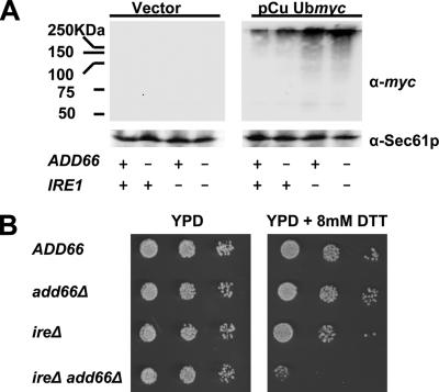
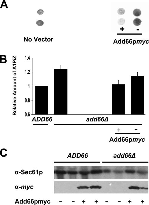
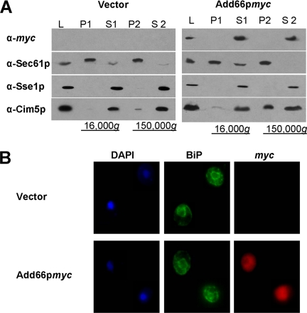
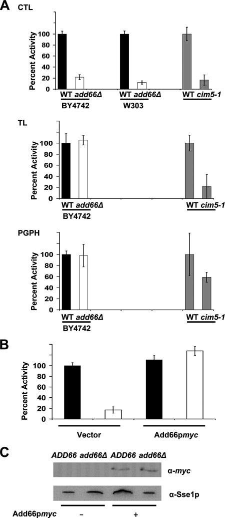
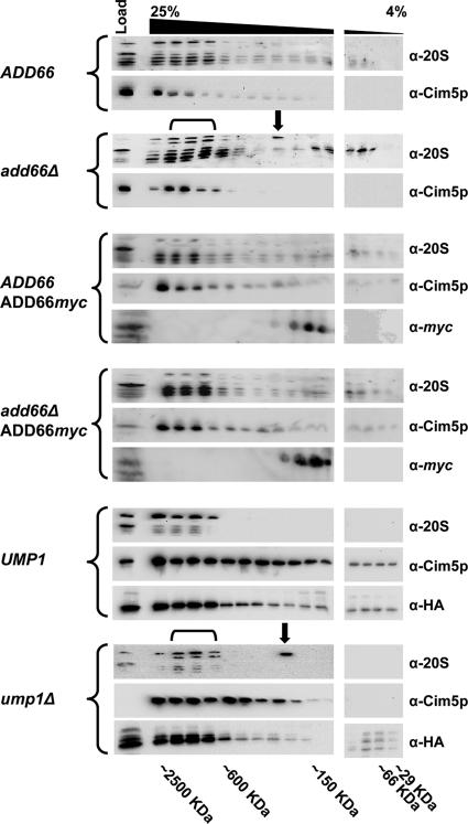
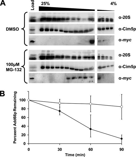
Antitrypsin deficiency is a primary cause of juvenile liver disease, and it arises from expression of the "Z" variant of the alpha-1 protease inhibitor (A1Pi). Whereas A1Pi is secreted from the liver, A1PiZ is retrotranslocated from the endoplasmic reticulum (ER) and degraded by the proteasome, an event that may offset liver damage. To better define the mechanism of A1PiZ degradation, a yeast expression system was developed previously, and a gene, ADD66, was identified that facilitates A1PiZ turnover. We report here that ADD66 encodes an approximately 30-kDa soluble, cytosolic protein and that the chymotrypsin-like activity of the proteasome is reduced in add66Delta mutants. This reduction in activity may arise from the accumulation of 20S proteasome assembly intermediates or from qualitative differences in assembled proteasomes. Add66p also seems to be a proteasome substrate. Consistent with its role in ER-associated degradation (ERAD), synthetic interactions are observed between the genes encoding Add66p and Ire1p, a transducer of the unfolded protein response, and yeast deleted for both ADD66 and/or IRE1 accumulate polyubiquitinated proteins. These data identify Add66p as a proteasome assembly chaperone (PAC), and they provide the first link between PAC activity and ERAD.


Figures

Figure 1.
Figure 1.
ADD6 and IRE1 synthetically interact. ADD66, add66Δ, ire1Δ, and ire1Δ add66Δ strains were transformed with a control plasmid or a plasmid expressing a ubiquitin-myc fusion protein under the transcriptional control of a copper inducible promoter (pCu Ubmyc). (A) Representative Western blots of extracts from cells containing a vector control or the ubiquitin expression vector are shown. Extracts were prepared after cells had been treated with 100 μM CuSO4 for 1 h at 30°C. Blots were probed with anti-myc and anti-Sec61p (as a loading control). (B) Serial dilutions of the indicated strains (Table 1) were grown on YPD in the presence or absence of 8 mM DTT, as indicated, for 48 h at 30°C. Of note, we chose to examine DTT sensitivity on YPD medium at pH 6.5 to reduce alternate stresses, although previously published work demonstrated a greater sensitivity to DTT in the ire1Δ strain when grown using other conditions (Frand and Kaiser, 1998; Pollard et al., 1998).
Figure 2.
Figure 2.
The A1PiZ degradation defect is rescued in add66Δ strains expressing Add66p-myc. (A) A colony-blot immunoassay was performed with anti-antitrypsin antiserum on add66Δ strains expressing A1PiZ and that lacked a vector or that were transformed with an empty vector (−) or with an ADD66-myc expression plasmid (+). (B) The results from three independent colony-blot immunoassays were quantified for wild-type yeast (ADD66) and add66Δ yeast that lacked a vector or that were transformed with the ADD66-myc expression vector (+) or a vector control (−). Data were quantified from signals detected in the linear range of the analysis. (C) Proteins extracts were prepared from ADD66 and add66Δ yeast transformed with a vector control (−) or with the ADD66myc expression plasmid (+) and total proteins were resolved by SDS-PAGE. The blots were probed with anti-myc and anti-Sec61 anti-sera. Duplicate colonies were analyzed and are shown here.
Figure 3.
Figure 3.
Add66p is cytosolic. (A) add66Δ strains were transformed with a control plasmid or with a plasmid engineered for the constitutive expression of Add66p-myc. Cell lysates (L) were subjected to 16,000 × g and 150,000 × g centrifugations. Total proteins in the pellets (P1 and P2) and supernatants (S1 and S2) were resolved by SDS-PAGE, and then they were analyzed by Western blot analysis with anti-myc, anti-Sec61p (ER membrane protein), anti-Sse1p (a primarily cytosolic protein; Goeckeler et al., 2002), and anti-Cim5p (a regulatory subunit of the 26S proteasome with cytosolic and ER membrane subcellular localizations) antisera. (B) Indirect immunofluorescence of add66Δ strains transformed with the plasmids described in A were stained with 4,6-diamidino-2-phenylindole (nuclear staining), and then they were probed with anti-BiP (ER perinuclear and peripheral staining) and with anti-myc antisera, and signals were detected as described in Materials and Methods.
Figure 4.
Figure 4.
The chymotrypsin-like activity of the 26S proteasome is reduced in extracts prepared from the add66Δ strain. The CTL, TL, and PGPH activities in clarified extracts from ADD66 (black bar) or add66Δ (white bar) yeast in two different strain backgrounds (BY4742 and W303) were determined. Proteasome activities in CIM5 and cim5-1 (gray bar) strains were used as a positive control. The relative activity was determined by normalizing the fluorescent signals to the levels corresponding to the wild-type strains, as described in Materials and Methods. (B) Constitutive expression of Add66p-myc restores the CTL activity in the add66Δ strain. Wild-type and add66Δ strains were transformed with an empty vector (−) or a vector engineered for the expression of Add66p-myc (+), and the CTL activity was analyzed as described in A. (C) Cytosolic proteins from the strains in B were resolved by SDS-PAGE, and then they were probed for Add66p-myc and Sse1p expression by Western blot analysis. Sse1p served as a loading control.
Figure 5.
Figure 5.
Yeast deleted for ADD66 accumulate a 20S intermediate and unprocessed 20S subunits. Cell extracts were prepared from an ADD66 and add66Δ strain containing either a control plasmid or a plasmid engineered for the endogenous expression of Add66p-myc, and from a UMP1 (JD133) and ump1Δ (JD134) strain. In total, 5 mg of protein was then resolved on a linear glycerol gradient (4–25%), and fractions were collected. Proteins in every other fraction were examined for the presence of 20S subunits, a component of the 19S subunit (Cim5p), and Add66p-myc by Western blot analysis. The migrations of molecular mass markers, which were analyzed in parallel, are indicated below the gel, the black downward bracket indicates fractions containing immature 20S subunits (a slower migrating doublet), and the black downward arrow indicates the migration of a 20S assembly intermediate. Note that the later two were observed only in extracts prepared from add66Δ and ump1Δ cells. The immunoreactive HA species in the UMP1 and ump1Δ gradients represents Pre2p (Table 1).
Figure 6.
Figure 6.
Add66p is degraded by the 26S proteasome. A pdr5Δ strain transformed with a plasmid designed for the constitutive expression of ADD66-myc was incubated either with 100 μM MG-132 or the equivalent volume of DMSO for 1 h at 30°C. (A) Cell extracts from each strain were prepared, and 5 mg of protein was resolved on a linear glycerol gradient (4–25%), and fractions were collected. Proteins in every other fraction were immunoblotted for 20S subunits, a component of the 19S subunit (Cim5p), and Add66p-myc. Molecular mass markers, which were analyzed in parallel, are indicated below the gel. Note that these blots were purposely overexposed (compared with those in Figure 5). (B) The strains described in A were harvested at the indicated time points after the addition of cycloheximide, and cell extracts were prepared and subjected to SDS-PAGE and immunoblotted for Add66p-myc and Sec61p (as a loading control). The amount of Add66p-myc at the start of the chase in each strain, after standardization to the amount of Sec61p at each time point, was set to 100%. ○, MG-132; •, DMSO control.

Similar articles

Cited by

References

    1. Adams A., Gottschling D. E., Kaiser C. A., Stearns T. Plainview, NY: Cold Spring Harbor Laboratory Press; 1997. Methods in Yeast Genetics.
    1. Arendt C. S., Hochstrasser M. Eukaryotic 20S proteasome catalytic subunit propeptides prevent active site inactivation by N-terminal acetylation and promote particle assembly. EMBO J. 1999;18:3575–3585. - PMC - PubMed
    1. Aridor M., Hannan L. A. Traffic jam: a compendium of human diseases that affect intracellular transport processes. Traffic. 2000;1:836–851. - PubMed
    1. Aridor M., Hannan L. A. Traffic jams II: an update of diseases of intracellular transport. Traffic. 2002;3:781–790. - PubMed
    1. Babbitt S. E., et al. ATP hydrolysis-dependent disassembly of the 26S proteasome is part of the catalytic cycle. Cell. 2005;121:553–565. - PubMed

Publication types

MeSH terms