. 2007 Oct;18(10):3788-99.
doi: 10.1091/mbc.e06-10-0930. Epub 2007 Jul 18.

A unique element in the cytoplasmic tail of the type II transforming growth factor-beta receptor controls basolateral delivery

Affiliations

A unique element in the cytoplasmic tail of the type II transforming growth factor-beta receptor controls basolateral delivery

Stephen J Murphy et al. Mol Biol Cell. 2007 Oct.

Abstract

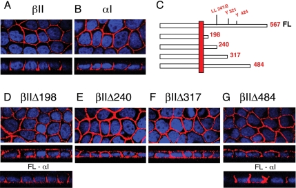
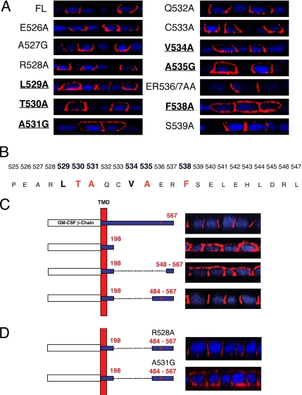
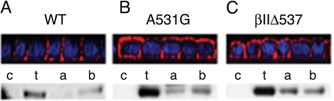
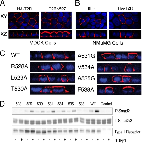
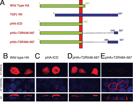
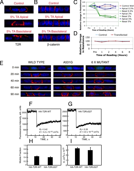
Transforming growth factor (TGF)-beta receptors stimulate diverse signaling processes that control a wide range of biological responses. In polarized epithelia, the TGFbeta type II receptor (T2R) is localized at the basolateral membranes. Sequential cytoplasmic truncations resulted in receptor missorting to apical surfaces, and they indicated an essential targeting element(s) near the receptor's C terminus. Point mutations in the full-length receptor confirmed this prediction, and a unique basolateral-targeting region was elucidated between residues 529 and 538 (LTAxxVAxxR) that was distinct, but colocalized within a clinically significant signaling domain essential for TGFbeta-dependent activation of the Smad2/3 cascade. Transfer of a terminal 84 amino-acid fragment, containing the LTAxxVAxxR element, to the apically sorted influenza hemagglutinin (HA) protein was dominant and directed basolateral HA expression. Although delivery to the basolateral surfaces was direct and independent of any detectable transient apical localization, fluorescence recovery after photobleaching demonstrated similar mobility for the wild-type receptor and a missorted mutant lacking the targeting motif. This latter finding excludes the possibility that the domain acts as a cell membrane retention signal, and it supports the hypothesis that T2R sorting occurs from an intracellular compartment.


Figures

Figure 1.
Figure 1.
The TGFβ type II receptor is trafficked to the basolateral membranes by a mechanism independently of conventional tyrosine or dileucine motifs. MDCK cell clones were plated at 5 × 10⁴ cells/12-mm transwell and allowed to fully polarize over 72 h, as described in Materials and Methods. MD-1 cells were stained for βII (A) or αI chimeric receptors (B) by using primary antibodies to the external GM-CSF β or α chains, respectively, and secondarily tagged with Cy3 (red). (C) Truncated chimeric βII receptors depicting the location of potential trafficking motifs stably expressed in MDCK cells (D–G). MDCK clones βIIΔ198 (D) and βIIΔ484 (G) additionally express the full-length αI chimeric receptor (FL-αI) (D and G, bottom). Images are represented as the horizontal XY flat sections above lower perpendicular XZ cross-sectional images. Nuclei (blue) were stained with DAPI.
Figure 2.
Figure 2.
The basolateral localizing signal of the TGFβ type II receptor is located at the C-terminal in the region of amino acid 537. (A) The basolateral targeting signal is located between amino acids 484 and 567. (B–E) Serial 10 amino acid C-terminal truncation mutants (βIIΔ527, βIIΔ537, βIIΔ547, and βIIΔ557) were stably expressed in MDCK cells and imaged using the GM-CSF β antibody and the Cy3-tagged secondary (red). Nuclei were additionally stained with DAPI. Images are represented as the horizontal XY flat sections above lower perpendicular XZ cross-sectional images of polarized cultures.
Figure 3.
Figure 3.
Basolateral retention of the TGFβ type II receptor is dependent on six residues located between amino acids 529-538. (A) Alanine or glycine point mutations were engineered into the βII receptor between amino acid residues 525-547. Mutant receptors were transiently transfected into fully polarized MDCK monolayers for 6 h before staining with the GM-CSF β antibody. Confocal images are presented for the full-length (FL) T2R or mutants spanning the region 526-539. (B) The predicted basolateral targeting signal motif LTAxxVAxxF is illustrated with red, indicating the four most critical residues. (C) Stable MDCK clones expressing full-length (βII; first construct), membrane-truncated (βIIΔ198; second construct), or internal sequence deletion mutant βII receptors (βIIΔ198-547 or βIIΔ198-483; third and fourth constructs, respectively) or βIIΔ198-483 constructs (D) containing R528A or A531G point mutations were stained as described in Figures 1 and 2. The location of the targeting motif defined in A and B is indicated by the red * in the 484–567 fragment. Nuclei were stained with DAPI and images are represented as perpendicular XZ cross sections.
Figure 4.
Figure 4.
Biotin T2R labeling at apical or basolateral membrane domains. Stable MDCK cell lines expressing wild type (A), A531G point-mutated (B) or βIIΔ537-truncated (C) βII chimeric receptor constructs were biotin labeled apically (a) or basolaterally (b) as polarized monolayers or as a nonpolarized monolayer for total labeling (t). Biotinylated protein was extracted on streptavidin-agarose beads and Western blotted using a receptor specific antibody. Bottom, control nonlabeled cells are designated c. Top, XZ cross sections of parallel immunofluorescently stained polarized monolayers.
Figure 5.
Figure 5.
Chimeric and endogenous type II TGFβ receptors similarly localize and induce Smad phosphorylation. (A) Stable MDCK clones expressing either the HA-tagged wild-type (HA-T2R) or amino acid 527-truncated (T2RΔ527) native T2R were generated and stained using the primary HA antibody (12CA5) and the Cy3-tagged secondary (red). (B) Expression of the chimeric βII (βIIR) and wild-type HA-T2R in fully polarized NMuMG epithelial cells. Six hours after transient transfection cells were stained for chimeric (βIIR) or native (HA-T2R) receptor expression by using the GM-CSF β or HA (12CA5) primary antibodies, respectively, and secondary Cy3 (red). (C) Native WT or containing the indicated point mutation HA-tagged native type II receptors were transiently transfected into fully polarized MDCK cells. Receptor expression was determined 5 h later using 12CA5 as described, and expression is represented as perpendicular XZ cross-sectional images. Nuclei in panels A-C were additionally stained with DAPI. (D) DR26 epithelial cells (do not express T2Rs) were used directly (control) or transfected with the indicated point mutant or wild-type (WT) T2Rs depicted in C. Cultures were left untreated (−) or stimulated (+) with 10 ng/ml TGFβ1 for 45 min before being processed for Western analysis (150 μg). After protein transfer, the membranes were sequentially probed for phospho-Smad2 (top) and type II receptor expression (bottom) before stripping and reprobing with a total-Smad2/3 antibody (middle) to control for protein loading. Identical results are obtained with the analogous chimeric T2R mutants (data not shown).
Figure 6.
Figure 6.
The TGFβ type II receptor basolateral localizing signal is dominant over the apical targeting signal in the influenza virus HA protein. (A) Influenza virus HA protein chimeras were constructed with terminal fragments of the T2R fused to the HA transmembrane domain. The full-length type II receptor (TGFβ RII) is provided for orientation and the location of the identified targeting sequence is indicated by a red *. (B–E) Fully polarized MDCK cell monolayers were transfected for 5 h with plasmids expressing either wild-type HA, an ICD-deleted HA protein (pHA-ICD), or chimeras consisting of pHA-ICD expressing type II receptor terminal fragments 548-567 or 484-567 (pHA+T2R548-567 and pHA+T2R484-567, respectively). Receptors and nuclei were visualized as described. Images are represented as horizontal XY flat sections focused at either the apical surface (top) or the central nuclei plains (middle), above lower perpendicular XZ cross sections through the plain of the cell.
Figure 7.
Figure 7.
The type II TGFβ receptor traffics directly to the basolateral membrane and loss of the targeting signal does not affect lateral membrane diffusion. Fully polarized MDCK cell monolayers on 12-mm transwells were transfected with chimeric βII receptors and incubated at 37°C for 3 h. The cells were then transferred to 18°C for 3 h before incubation with 5% tannic acid (TA) for 10 min in the apical or basolateral reservoirs as labeled. Cells were then washed three times with PBS before returning to 37°C for 1 h in full medium before cell-surface receptor (A) or β-catenin (B) staining as described. Monolayers were additionally monitored for any change in transepithelial resistance after tannic acid (C) or transfection (D) treatments for the times specified. (E) Polarized MDCK cell monolayers transfected with the wild-type, A531G point mutant, or 6 x Mutant βII receptors were stained for cell-surface receptor expression at the indicated times after 37°C release from Golgi block. (F–I) FRAP studies on the lateral diffusion of HA-T2R-WT and HA-T2RΔ527 in MDCK cells. Cells were transfected with expression vectors for the above-mentioned proteins, and the cell-surface receptors were labeled in the cold by fluorescent monovalent Fab′ (see Materials and Methods). FRAP studies were conducted at 16°C in HBSS/HEPES/BSA. (F and G) Typical FRAP curves depicting the lateral diffusion of HA-T2R-WT (F) and HA-T2RΔ527 (G). Solid lines are the best fit to the lateral diffusion equation (Petersen and Elson, 1986). (H and I) Average Rf and D values, respectively, derived from multiple FRAP measurements (mean ± SEM of 30–40 measurements in each case). No significant differences were detected between HA-T2R-WT and HA-T2RΔ527 in either Rf (p > 0.1, Student's t test) or D (p > 0.2).

Similar articles

Cited by

References

    1. Anders R. A., Arline S. L., Dore J. J., Leof E. B. Distinct endocytic responses of heteromeric and homomeric transforming growth factor β receptors. Mol. Biol. Cell. 1997;8:2133–2143. - PMC - PubMed
    1. Anders R. A., Dore J. J., Jr, Arline S. L., Garamszegi N., Leof E. B. Differential requirement for type I and type II transforming growth factor β receptor kinase activity in ligand-mediated receptor endocytosis. J. Biol. Chem. 1998;273:23118–23125. - PubMed
    1. Anders R. A., Leof E. B. Chimeric granulocyte/macrophage colony-stimulating factor/transforming growth factor-beta (TGF-β) receptors define a model system for investigating the role of homomeric and heteromeric receptors in TGF-β signaling. J. Biol. Chem. 1996;271:21758–21766. - PubMed
    1. Aroeti B., Kosen P. A., Kuntz I. D., Cohen F. E., Mostov K. E. Mutational and secondary structural analysis of the basolateral sorting signal of the polymeric immunoglobulin receptor. J. Cell Biol. 1993;123:1149–1160. - PMC - PubMed
    1. Aroeti B., Okhrimenko H., Reich V., Orzech E. Polarized trafficking of plasma membrane proteins: emerging roles for coats, SNAREs, GTPases and their link to the cytoskeleton. Biochim. Biophys. Acta. 1998;1376:57–90. - PubMed

Publication types

MeSH terms

Substances

LinkOut - more resources