. 2007 Sep;18(9):3620-34.
doi: 10.1091/mbc.e06-10-0917. Epub 2007 Jul 18.

Chlamydomonas outer arm dynein alters conformation in response to Ca2+

Affiliations

Chlamydomonas outer arm dynein alters conformation in response to Ca2+

Miho Sakato et al. Mol Biol Cell. 2007 Sep.

Abstract

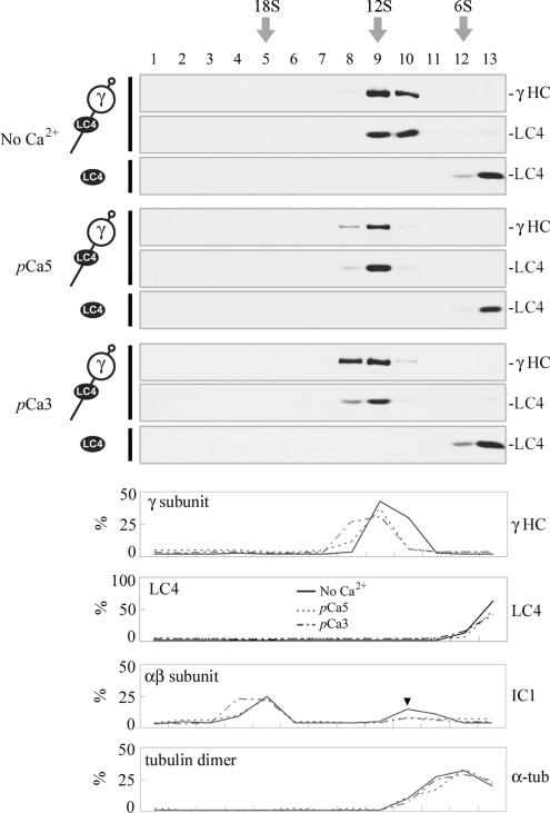
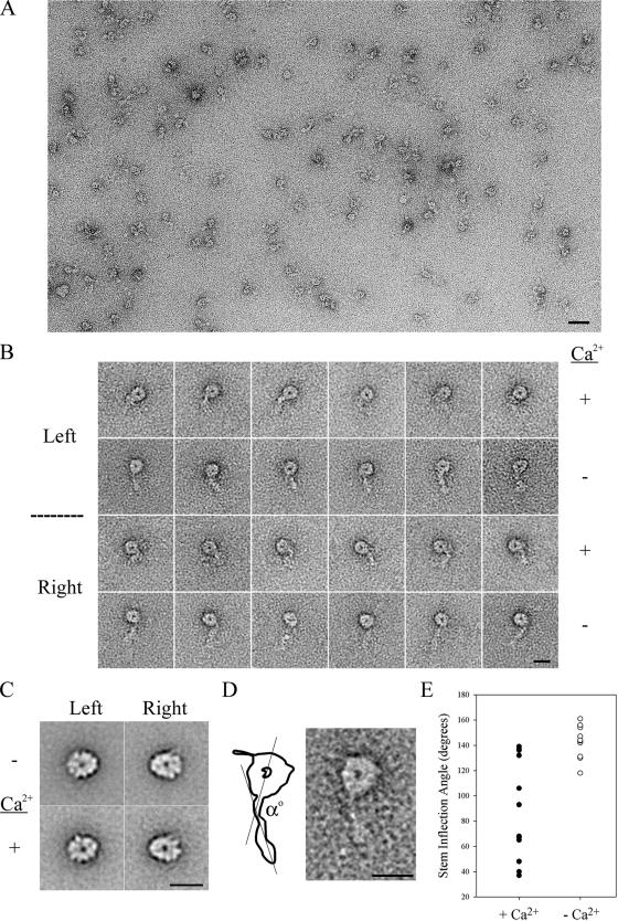
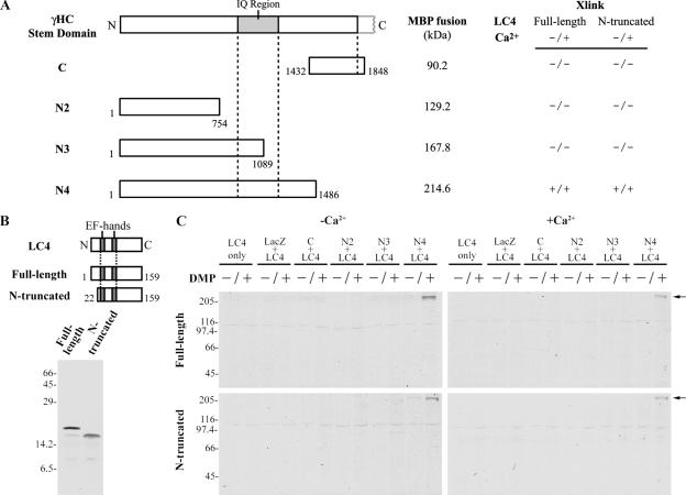
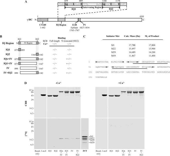
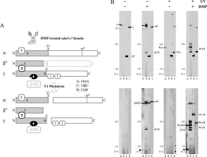
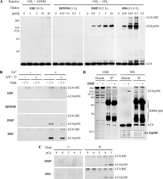
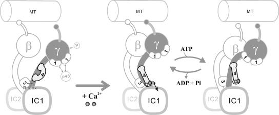
We have previously shown that Ca(2+) directly activates ATP-sensitive microtubule binding by a Chlamydomonas outer arm dynein subparticle containing the beta and gamma heavy chains (HCs). The gamma HC-associated LC4 light chain is a member of the calmodulin family and binds 1-2 Ca(2+) with K(Ca) = 3 x 10(-5) M in vitro, suggesting it may act as a Ca(2+) sensor for outer arm dynein. Here we investigate interactions between the LC4 light chain and gamma HC. Two IQ consensus motifs for binding calmodulin-like proteins are located within the stem domain of the gamma heavy chain. In vitro experiments indicate that LC4 undergoes a Ca(2+)-dependent interaction with the IQ motif domain while remaining tethered to the HC. LC4 also moves into close proximity of the intermediate chain IC1 in the presence of Ca(2+). The sedimentation profile of the gamma HC subunit changed subtly upon Ca(2+) addition, suggesting that the entire complex had become more compact, and electron microscopy of the isolated gamma subunit revealed a distinct alteration in conformation of the N-terminal stem in response to Ca(2+) addition. We propose that Ca(2+)-dependent conformational change of LC4 has a direct effect on the stem domain of the gamma HC, which eventually leads to alterations in mechanochemical interactions between microtubules and the motor domain(s) of the outer dynein arm.


Figures

Figure 1.
Figure 1.
Characterization of the CT61 antibody against LC4. (A) Axonemes prepared from wild-type and mutant strains (see Table 1) were electrophoresed in a 5–15% acrylamide gradient gel and stained with Coomassie blue (CBB, top). Similar samples were transferred to nitrocellulose and probed with blot-purified CT61 antibody (bottom). A single major band corresponding to LC4 was observed. This protein was missing only in mutants that lack the outer dynein arm (oda2 and oda3). Several minor bands were also detected in whole axonemal samples. Mr standards (×103) are indicated at left. (B) Left, electrophoretic analysis of purified intact outer arm dynein (including the docking complex) in a 5–15% acrylamide gradient gel; Coomassie blue stain. Right, an immunoblot of an identical sample probed with CT61. Only a single band that comigrates with native LC4 was detected. (C) Top, intact outer arm dynein and the αβ and γ HC subunits electrophoresed in a 3–5% acrylamide 3–8 M urea gel to separate the individual HCs; silver stain. The band marked α* is a proteolytic fragment previously referred to as “band 11” (Pfister et al., 1982) that is derived by cleavage at a site located ∼90 kDa from the C-terminus of the intact HC (King and Witman, 1988a). Similar samples were also electrophoresed in a 5–15% acrylamide gradient gel and probed with CT61 (bottom). The LC4 protein is present only in the intact dynein particle and the isolated γ HC subunit.
Figure 2.
Figure 2.
Cytoplasmic preassembly of the LC4/γ HC complex. (A) Axonemes from wild type and the oda2 mutant strain, purified intact outer arm dynein, and the αβ and γ HC subparticles were probed with the CT240 antibody raised against the γ HC N-terminal domain. This antibody specifically reacts with the γ HC and did not recognize other HCs within either oda2 axonemes or the αβ HC subparticle from the outer dynein arm. (B) The CT240 antibody was used to immunoprecipitate the γ HC from a wild-type cytoplasmic extract. Immunoblot analysis (WB) revealed that the pellet contained the γ HC (detected using the 12γB mAb) and LC4; neither protein was present in the preimmune control sample. (C) To further address the preassembly state of these components, cytoplasmic extracts (CE) prepared from wild-type, oda2, and oda3 cells were incubated with CT240 antibody, and the resulting immunoprecipitates (IP) were analyzed for the presence of the γ HC, LC4, IC1, LC5, and DC1 using antibodies 12γB, CT61, 1878A, R4929, and anti-DC1, respectively. Note that LC4 was not detected in the original extracts at a loading of 20 μg, but could be observed when protein amount was increased and exposure times were lengthened (not shown).
Figure 3.
Figure 3.
Association of LC4 with the γ HC N-terminal domain. (A) Map of the γ HC indicating the origin of the products generated by photocleavage at the V1 site and the location at which LC4 binds; this interaction may be stabilized by crosslinking. (B) The purified γ HC subunit was treated with 0–10 mM DMP in the presence of 1 mM ATP and 100 μM vanadate, electrophoresed in 4% acrylamide 4 M urea gels, and either stained with silver (SS) or blotted and probed with 12γB and CT61 to reveal the γ HC and LC4, respectively (top). Similar samples were photocleaved at the V1 site within the HC before crosslinking (bottom). The N- and C-terminal γ HC photocleavage products are indicated at left. LC4 is crosslinked to the smaller, N-terminal fragment.
Figure 4.
Figure 4.
The native LC4/γ HC complex is stable in both high and low Ca2+. The purified γ HC complex was sedimented in a 5–20% sucrose density gradient in the absence of Ca2+ (top) or at pCa 5 and pCa 3 (middle and bottom); the bottom of the gradient is at left. Equal volumes of each fraction were electrophoresed and the γ HC and LC4 detected by immunoblotting with 12γB and CT61 antibodies, respectively. Recombinant LC4 was processed in parallel to ensure that it did not sediment at ∼12S in the absence of the γ HC. LC4 does not dissociate from the γ HC in a Ca2+-dependent manner. However, there is a small shift in the sedimentation coefficient of the complex toward a more rapidly migrating species at high Ca2+ levels. To further quantify this shift, the % of the total γ HC, recombinant LC4, the outer arm αβ subunit, and tubulin dimer present in each fraction was determined in the absence of Ca2+ and at pCa 5 and 3; the latter two samples were sedimented in additional gradients (blots not shown). Unlike the γ subunit, neither LC4 nor tubulin shift upon Ca2+ addition; the αβ subunit peak did not shift at pCa 5, but spread out by one additional fraction only at pCa 3. The arrowhead in the αβ subunit plot indicates a small peak of dissociated IC/LC complex detected by the IC1 antibody (1878A).
Figure 5.
Figure 5.
Electron microscopic analysis of the γ heavy chain subunit. (A) Survey micrograph of γ HC subunit particles prepared in the presence of Ca2+ and negatively stained with uranyl acetate. Scale bar, 40 nm. (B) Montage of class-averaged images of isolated γ HC subunit particles prepared for negative stain electron microscopy in either the presence or absence of Ca2+; both left and right views are shown. Scale bar, 15 nm. (C) Image averages of left and right views of the γ HC motor domain in the presence and absence of Ca2+ are shown. Scale bar, 15 nm. (D) Enlargement of the left view of one γ HC subunit particle class average imaged in the absence of Ca2+ and interpretative diagram illustrating the method used to measure the stem inflection angle α. Scale bar, 15 nm. (E) The stem inflection angle α was measured for particles prepared in the presence and absence of Ca2+. In the absence of ligand, a low angular dispersion (between ∼120 and 160°) about the inflection point was observed, whereas considerably greater variability in α (between ∼35 and 140°) was found for particles prepared in the presence of Ca2+.
Figure 6.
Figure 6.
A Ca2+-independent LC4-binding region on the γ HC. (A) Map of the γ HC N-terminal region indicating the location and mass of the various segments used to assess LC4 binding; the interaction properties of each fragment in the presence and absence of Ca2+ are indicated (see also C). (B) Map of the LC4 constructs used and autoradiograph of the 35S-labeled in vitro–translated full-length and N-terminal–truncated proteins. (C) After incubation of LC4 with the γ HC stem domain fusion proteins, interactions were stabilized by DMP crosslinking, and the samples were electrophoresed. Only the γ HC N4 segment was competent to bind LC4 (both full-length and truncated forms) in a Ca2+-independent manner (arrows). No interaction was observed between LC4 and other parts of the γ HC N-terminal domain.
Figure 7.
Figure 7.
Ca2+-dependent interaction of LC4(22-159) with the γ HC IQ region. (A) Map of the γ HC, indicating the location and sequence of the two IQ motifs. (B) Map indicating the various γ HC MBP fusion proteins used and whether they bound either full-length or the N-terminal–truncated form of LC4 in the presence or absence of Ca2+. Constructs containing the intervening (IV) region plus one or both IQ motifs bound LC4(22-159) well only in the absence of Ca2+ (++); the intervening region between the IQ motifs exhibited detectable binding (+). (C) Sequence of LC4 indicating the location of Met residues (bold) at which translation initiation occurred and the calculated mass and measured Mr of the products. The two EF hands are underlined. (D) MBP fusion proteins containing various segments of the IQ motif region and MBP-LacZ and bead-only controls were incubated with in vitro–translated LC4 (IVT) in the presence and absence of 1 mM Ca2+. Full-length LC4 did not bind to any of the constructs. In contrast, N-terminal truncated LC4 obtained by translation initiation at M22 bound to this γ HC region in a Ca2+-dependent manner.
Figure 8.
Figure 8.
Chemical crosslinking defines intradynein interactions. (A) Diagram illustrating the crosslinked products generated by DMP treatment and subsequent V1 photocleavage of oda4-s7 dynein, which contains a truncated form of the β HC (β*). The relative size of the HC fragments corresponds to their apparent size after electrophoresis; it does not directly relate to their actual mass based on sequence. (B) Outer arm dynein containing a ∼160-kDa truncated form of the β HC motor domain from oda4-s7 was incubated with 1 mM ATP plus 100 μM vanadate, and half the samples were irradiated with UV light to cleave the α and γ HCs at their V1 sites. After the photolysis reaction, proteins were then subject to crosslinking with 10 mM DMP or were treated with solvent alone. Samples were electrophoresed and probed with 18αA, 18βC, and 12γB to detect the N-terminal regions of the three HCs, and with R5932, R4930, CT61, and R4924, which recognize the HC-associated LC1, LC3, LC4, and LC5 proteins, respectively. The top series of blots indicate the location of HC bands and LC3, whereas the other LCs were analyzed with respect to LC3 in the bottom series. In the presence of DMP, LC3 is crosslinked to both the β and γ HCs (labeled β*/3 and γ/3); after photolysis the γ HC/LC3 product (N-γ/3) lacks the C-terminal motor unit and consequently migrates more rapidly. Further analysis identified a crosslinked band containing the γ HC and LC1, LC3, and LC4 (γ/4/3/1). After photolysis, this complex yields two products: the γ HC N-terminal region crosslinked to LC3 and LC4 (N-γ/4/3) and a γ HC C-terminal domain linked to LC1 (C-γ/1). The arrowhead marks an additional product containing LC4 that is obtained in enhanced yield after UV irradiation to cleave the HCs at the V1 site; this product is further analyzed in Figure 9.
Figure 9.
Figure 9.
Ca2+-dependent crosslinking of LC4 to IC1. (A) Purified outer arm dynein was treated with the carbodiimide EDC or with the amine-selective reagents DFDNB, DMP, and DSS in the presence of 1 mM Ca2+. After electrophoresis in 8% acrylamide SDS gels, samples were probed for the presence of LC4. Crosslinked products containing LC4 and either the γ HC or p100 are evident in the EDC, DMP, and DSS samples; upon prolonged exposure a very small amount of product was detectable in the DFDNB-treated sample (not shown). (B) Purified outer arm dynein in the presence or absence of 1 mM ATP/100 μM vanadate, and 1 mM Ca2+ was treated with the indicated crosslinking reagents (20 mM EDC, 1 mM DFDNB, 10 mM DMP, and 0.01 mM DSS). Formation of the LC4-γ HC product was not dependent on either nucleotide or Ca2+. In contrast, LC4-p100 crosslinking was dramatically increased in the presence of Ca2+, and the product yield was also nucleotide-dependent. (C) Purified dynein was incubated at pCa 9–pCa 3 in the presence or absence of 10 mM DMP or 0.01 mM DSS. Significant levels of the LC4-p100 product were only obtained at and above pCa 4. (D) Immunoprecipitation of crosslinked LC4-p100 using the CT61 antibody. Purified outer arm dynein was crosslinked with 10 mM DMP in the presence of Ca2+ (Dynein), denatured, refolded, and immunoprecipitated (IP). Samples were electrophoresed in 10% tricine SDS gels and either stained with Coomassie blue (CBB) or transferred and probed with the CT61 antibody (WB). A LC4-p100 crosslinked product was immunoprecipitated in addition to LC4 as indicated at right. An inset at the bottom right shows an enlarged image of the boxed region of the Coomassie blue–stained gel. Mass spectrometry identified p100 as IC1. The asterisk indicates non-crosslinked IC1 that migrates with Mr78,000.
Figure 10.
Figure 10.
A model for LC4 interactions within outer arm dynein. In the absence of Ca2+, LC4 is tightly bound to the γ HC (left); this complex cannot efficiently form a rigor bond with MTs (Sakato and King, 2003). After Ca2+ addition, LC4 changes conformation and its Ca2+-bound EF hand motifs detach from the IQ region and come into proximity of or attach to IC1 (center). This is predicted to lead to an alteration in the N-terminal stem domain of the γ HC and the activation of motor activity. After ATP addition, a further change in γ HC conformation occurs, causing LC4 to move somewhat away from IC1 (right). Other outer arm dynein components including the α HC are not incorporated into this diagram because their functional interactions with either the γ HC, LC1, or LC4 have not been defined.

Similar articles

Cited by

References

    1. Adams G., Huang B., Piperno G., Luck D. Central pair microtubule complex of Chlamydomonas flagella: polypeptide composition as revealed by analysis of mutants. J. Cell Biol. 1981;91:69–76. - PMC - PubMed
    1. Bahler M., Rhoads A. Calmodulin signaling via the IQ motif. FEBS Lett. 2002;513:107–113. - PubMed
    1. Baron D. M., Kabututu Z. P., Hill K. L. Stuck in reverse: loss of LC1 in Trypanosoma brucei disrupts outer dynein arms and leads to reverse flagellar beat and backward movement. J. Cell Sci. 2007;120:1513–1520. - PubMed
    1. Benashski S. E., King S. M. Investigation of protein-protein interactions within flagellar dynein using homobifunctional and zero-length crosslinking reagents. Methods. 2000;22:365–371. - PubMed
    1. Benashski S. E., Patel-King R. S., King S. M. Light chain 1 from the Chlamydomonas outer dynein arm is a leucine-rich repeat protein associated with the motor domain of the γ heavy chain. Biochemistry. 1999;38:7253–7264. - PubMed

Publication types

LinkOut - more resources