. 2007 Oct;18(10):3835-44.
doi: 10.1091/mbc.e06-07-0625. Epub 2007 Jul 18.

The amyloid-beta precursor protein is phosphorylated via distinct pathways during differentiation, mitosis, stress, and degeneration

Affiliations

The amyloid-beta precursor protein is phosphorylated via distinct pathways during differentiation, mitosis, stress, and degeneration

Zoia Muresan et al. Mol Biol Cell. 2007 Oct.

Abstract

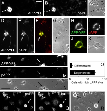
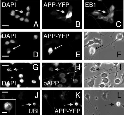
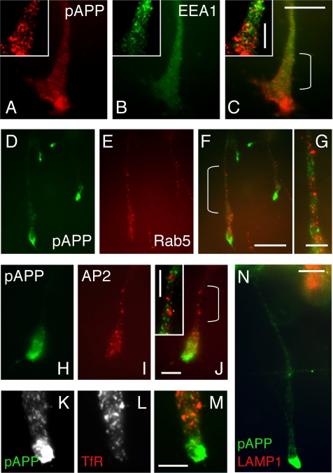
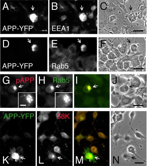
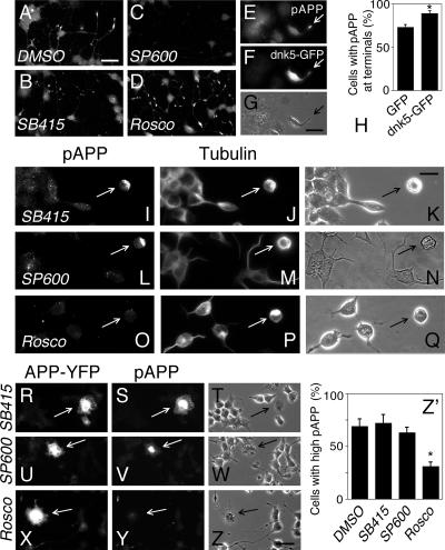
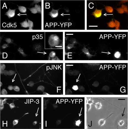
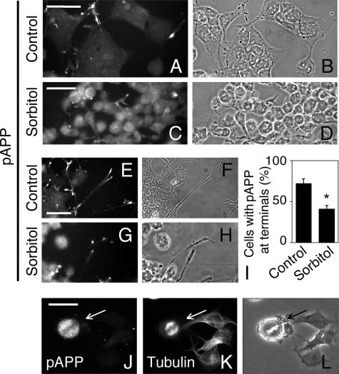
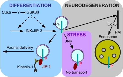
Phosphorylation of amyloid-beta precursor protein (APP) at Thr(668) is a normal process linked to neurite extension and anterograde transport of vesicular cargo. By contrast, increased phosphorylation of APP is a pathological trait of Alzheimer's disease. APP is overexpressed in Down's syndrome, a condition that occasionally leads to increased APP phosphorylation, in cultured cells. Whether phosphorylation of APP in normal versus high APP conditions occurs by similar or distinct signaling pathways is not known. Here, we addressed this problem using brainstem-derived neurons (CAD cells). CAD cells that ectopically overexpress APP frequently show features of degenerating neurons. We found that, in degenerating cells, APP is hyperphosphorylated and colocalizes with early endosomes. By contrast, in normal CAD cells, phosphorylated APP (pAPP) is excluded from endosomes, and localizes to the Golgi apparatus and to transport vesicles within the neurites. Whereas the neuritic APP is phosphorylated by c-Jun NH(2)-terminal kinase through a pathway that is modulated by glycogen synthase kinase 3beta, the endosomal pAPP in degenerated CAD cells results from activation of cyclin-dependent kinase 5. Additional signaling pathways, leading to APP phosphorylation, become active during stress and mitosis. We conclude that distinct pathways of APP phosphorylation operate in proliferating, differentiating, stressed, and degenerating neurons.


Figures

Figure 1.
Figure 1.
APP phosphorylation in degenerating neurons. (A–K) Degenerating CAD cells that overexpress APP also show high levels of pAPP (arrows). CAD cells were transfected with APP-YFP, and the transfected fusion protein was detected via the YFP tag. Note that pAPP largely colocalizes with APP-YFP (D–K), with over 70% of vesicles that contain APP-YFP (green; H–K) also staining for pAPP (red; H–K). (L–N) Transfected CAD cells that show differentiated phenotype do not contain increased pAPP levels compared with nontransfected cells. Arrow points to a cell (extending two processes) that overexpresses APP-YFP. (O) Quantitative measurement of pAPP accumulation in the cell body of CAD cells that overexpress APP-YFP. Cells with differentiated or degenerated phenotype were analyzed separately. The graph shows the percentage of APP-YFP–overexpressing cells in each group that showed increased pAPP levels. Error bars, SEM. (P–R) Nontransfected, degenerating CAD cells, identified here by their rounded shape and the abnormal microtubule cytoskeleton (arrows), show increased levels of pAPP. Microtubules are detected with an anti-tubulin antibody. (C, G, K, N, and R) are phase-contrast micrographs. Scale bars, 40 μm (A–G and L–N); 20 μm (H–K and P–R).
Figure 2.
Figure 2.
CAD cells that overexpress APP do not show nuclear fragmentation (A–F), but contain ubiquitinated inclusions (J–L). Nontransfected (G–I) or APP-YFP transfected cells (A–F and J–L) were stained for DNA (DAPI; A, D, and G), exogenously expressed APP-YFP (with anti-GFP antibody; B, E, and K), pAPP (H), EB1 (C), and ubiquitin (UBI; J and inset). Note that an apoptotic cell, with fragmented nucleus, does not show increased pAPP levels (G–I). The inset in J shows that ubiquitin is detected in large inclusions, typical for neurodegenerating neurons that contain mis-folded proteins. Also note that the level of EB1 (a cytoplasmic microtubule-binding protein) is not increased in APP-overexpressing cells (A–C). Arrows point to the relevant cells. (F, I, and L) Phase-contrast micrographs. Scale bars, 20 μm (A–L); 5 μm (inset).
Figure 3.
Figure 3.
Most neuritic pAPP is not localized to early endosomes or lysosomes, in differentiated CAD cells. Differentiated CAD cells were double labeled for pAPP and one of the following endosomal or lysosomal marker proteins: EEA1 (A–C), Rab5 (D–G), AP2 (H–J), transferrin receptor (TfR; K–M), or LAMP1 (N). Each image shows a distal neurite with the terminal, at high magnification. Note that, although endocytic markers are present at neurite terminals, as reported for primary neurons, they do not significantly colocalize with pAPP. Quantitatively, in each case, <15% of the pAPP present at the terminal or along the neurites colocalized with the endocytic marker (see also Supplementary Figure 3). Insets have been adjusted for contrast and brightness to allow clear visualization of particles containing pAPP and endocytic markers. Panel G corresponds to the region marked by the bracket in F. Insets correspond to the marked regions, indicated by brackets, in (C and J). Scale bars, 5 μm (G–M, and insets in A–C and J); 10 μm (A–C and N); 20 μm (D–F).
Figure 4.
Figure 4.
pAPP localizes to endosomes in degenerating CAD cells. CAD cells were transfected with APP-YFP (A–F and K–N) or nontagged APP (G–J), followed by immunolabeling. APP-YFP was detected via its tag (A, D, K, and M). (C, F, J, and N) Phase-contrast micrographs. Insets in G and H show quasi-identical particulate localization of pAPP and Rab5. Also note the increased level of endocytic markers in cells that overexpress APP, suggesting increased endocytosis in these cells. The Golgi apparatus maintains normal morphology and is only slightly up-regulated in degenerating cells that overexpress APP-YFP (L and M). Note that APP-YFP is distributed throughout the cell, whereas the Golgi apparatus is a compact structure confined to a small area adjacent to the nucleus. Also note that the APP-YFP–expressing cell is vesiculated (N). Arrows point to cells overexpressing APP-YFP (A–F and K–N) or APP (G–J). Scale bars, 20 μm (A–N); 5 μm (insets in G and H).
Figure 5.
Figure 5.
Inhibition of Cdk5 decreases APP phosphorylation in degenerating CAD cells (I–Z′) but not in differentiated cells (A–H). (A–D) Detection of pAPP in CAD cell cultures treated with inhibitors of GSK3β (SB415286), JNK (SP600125), or Cdk5 (roscovitine). A DMSO control is also shown (A). We estimated the number of neurites that contained pAPP above a determined threshold level (see Materials and Methods and Supplementary Figure 4). Quantitative data, derived from thresholded images (generated to eliminate low-intensity labeling), showed pAPP accumulation in only 36% of neurites, in cultures treated with SB415286. This compares with 66 and 80% of neurites containing pAPP at terminals, in cultures treated with DMSO and roscovitine, respectively. These data are derived from one set of experiments, but similar results were obtained in two other sets of experiments. Thresholded images are not shown. (E–G) Inhibition of Cdk5 by transfection of CAD cells with the dominant negative construct, dnk5-GFP, does not prevent APP phosphorylation and accumulation at neurite terminals (arrows). (H) Quantitative measurement of the effect of dnk5-GFP expression on pAPP localization at neurite terminals. Control cells were transfected with GFP. Percentages of cells with neurites that showed pAPP at terminals are indicated. Error bars, SEM; *p < 0.01. (I–Z) Inhibition of Cdk5 (O–Q and X–Z), but not GSK3β (I–K and R–T) or JNK (L–N and U–W) inhibits APP phosphorylation in degenerating CAD cells, in nontransfected cultures (I–Q) or cells transfected with APP-YFP (R–Z). In nontransfected cultures, degenerating cells were identified by their spherical shape and abnormal microtubule cytoskeleton. Arrows point to degenerating cells. APP-YFP and dnk5-GFP were detected with an anti-GFP antibody. (G, K, N, Q, T, W, and Z) are phase-contrast micrographs. The different appearance of cells in N is due to the accidental use of an incorrect phase ring. Note that, to avoid saturation of fluorescence images containing brightly labeled cells (I–Z), micrographs have been acquired at exposure times that allow only poor visualization of neuritic pAPP. Scale bars, 40 μm (A–D and R–Z); 20 μm (E–G and I–Q). (Z′) Quantitative measurement of the effect of kinase inhibitors on pAPP accumulation in the cell body of cells transfected with APP-YFP. The graph shows the percentage of transfected cells that showed increased pAPP levels. Error bars, SEM; *p < 0.005 (roscovitine vs. DMSO).
Figure 6.
Figure 6.
Abnormal distribution of Cdk5 and p35/p25, but not JNK and JIP-3, in APP-YFP–overexpressing CAD cells with degenerated phenotype. Detection of Cdk5 (A–C), p35/p25 (D and E), phosphorylated, active JNK (pJNK; F and G), and JIP-3 (H–J) in CAD cells overexpressing APP-YFP (arrows). The relevant cell in D (arrow) is shown in the inset, at higher magnification. APP-YFP was detected with an anti-GFP antibody. (J) A phase-contrast micrograph. Scale bars, 20 μm (A–J); 10 μm (inset in D).
Figure 7.
Figure 7.
APP is phosphorylated in cells subjected to stress, or during mitosis. (A–H) CAD cells were cultured with (C, D, G, and H) or without (A, B, E, and F) sorbitol, and immunolabeled for pAPP. Note the increased amount of pAPP in the cell body, and the diminished accumulation of pAPP at neurite terminals in sorbitol-treated cells (compare G with E). (I) Quantitative measurement of the effect of sorbitol treatment on pAPP localization at neurite terminals. Percentages of cells with neurites that showed pAPP at terminals are indicated. Error bars, SEM; *p < 0.005. The graph does not take into account that the pAPP levels at neurite terminals of sorbitol-treated cells, when present, are significantly lower than in nontreated cells. Moreover, a significantly smaller area of the growth cone is occupied by pAPP in sorbitol-treated, compared with control cells (compare G with E). (J–L) A mitotic cell (arrow) shows intense, punctate distribution of pAPP (J). The mitotic spindle was detected with an anti-tubulin antibody (K). (B, D, F, H, and L) Phase-contrast micrographs. Scale bars, 40 μm (A–D); 20 μm (E–H and J–L).
Figure 8.
Figure 8.
Distinct pathways of APP phosphorylation in differentiated and degenerating neurons. The left side of the diagram illustrates the JNK-dependent APP phosphorylation that is facilitated by JIP-3, up-regulated by GSK3β (which may act either upstream or downstream of JNK), and down-regulated by Cdk5. As depicted, the inhibitory effect of Cdk5 occurs through inactivation—likely indirect—of GSK3β (for a possible mechanism, see Morfini et al., 2004). This pathway leads to recruitment of kinesin-1, and transport of pAPP into neurites (Muresan and Muresan, 2005b). The precise role of GSK3β in this pathway remains to be established. The right side of the diagram illustrates targeting of APP to endosomes, and its phosphorylation by Cdk5, in degenerating neurons. The precise site of APP phosphorylation by Cdk5 remains to be established. Also shown is the stress-activated pathway that leads to APP phosphorylation by JNK. Activation of this pathway likely blocks axonal transport of pAPP, possibly through phosphorylation of kinesin-1 (Morfini et al., 2006). APP phosphorylation during mitosis is not shown.

Similar articles

Cited by

References

    1. Ando K., Oishi M., Takeda S., Iijima K., Isohara T., Nairn A. C., Kirino Y., Greengard P., Suzuki T. Role of phosphorylation of Alzheimer's amyloid precursor protein during neuronal differentiation. J. Neurosci. 1999;19:4421–4427. - PMC - PubMed
    1. Aplin A. E., Gibb G. M., Jacobsen J. S., Gallo J. M., Anderton B. H. In vitro phosphorylation of the cytoplasmic domain of the amyloid precursor protein by glycogen synthase kinase-3beta. J. Neurochem. 1996;67:699–707. - PubMed
    1. Ball C. L., Hunt S. P., Robinson M. S. Expression and localization of alpha-adaptin isoforms. J. Cell Sci. 1995;108:2865–2875. - PubMed
    1. Bennett B. L., et al. SP600125, an anthrapyrazolone inhibitor of Jun N-terminal kinase. Proc. Natl. Acad. Sci. USA. 2001;98:13681–13686. - PMC - PubMed
    1. Bu B., Li J., Davies P., Vincent I. Deregulation of cdk5, hyperphosphorylation, and cytoskeletal pathology in the Niemann-Pick type C murine model. J. Neurosci. 2002;22:6515–6525. - PMC - PubMed

Publication types

MeSH terms

Substances

LinkOut - more resources