. 2007 Jul 18;27(29):7654-62.
doi: 10.1523/JNEUROSCI.0203-07.2007.

The microtubule-associated protein tumor overexpressed gene/cytoskeleton-associated protein 5 is necessary for myelin basic protein expression in oligodendrocytes

Affiliations

The microtubule-associated protein tumor overexpressed gene/cytoskeleton-associated protein 5 is necessary for myelin basic protein expression in oligodendrocytes

Victor P Francone et al. J Neurosci. .

Abstract

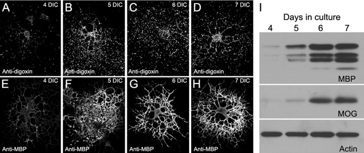
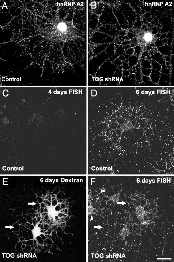
Tumor overexpressed gene (TOG) protein, encoded by cytoskeleton-associated protein CKAP5, is a microtubule-associated protein that binds to heterogeneous nuclear ribonucleoprotein (hnRNP) A2. hnRNP A2 is an RNA trafficking factor that associates with myelin basic protein (MBP) mRNA. In oligodendrocytes, TOG, hnRNP A2, and MBP mRNA colocalize in granules that assemble in the perikaryon and are transported to the peripheral network of processes that extends from it. MBP accumulates preferentially in the membrane of the medial and distal portions of these cellular processes. MBP expression was reduced when TOG level was lowered by short-hairpin (sh) RNA. The reduction in TOG did not affect overall cell morphology or the assembly, transport, localization, or number of MBP mRNA-containing granules. Reduced levels of TOG did not affect another oligodendrocyte-specific component, myelin oligodendrocyte glycoprotein, which is expressed at the same time as MBP but translated from mRNA localized in the cell body. Expression in a neural cell line of a green fluorescent protein (GFP)-MBP fusion protein derived from a construct containing GFP and the full-length cDNA for the rat 14 kDa MBP was reduced when TOG level was lowered by shRNA treatment. Expression of GFP, derived from GFP mRNA containing the hnRNP A2 binding element of MBP mRNA, was similarly reduced in cells with low TOG levels. These data indicate that TOG is necessary for efficient translation of MBP mRNA and suggest that this role is mediated by its interaction with hnRNP A2.


Figures

Figure 1.
Figure 1.
TOG expression is reduced by shRNA treatment. A, Sequence, target localization, and putative folding of the short hairpin RNAs (Open Biosystems, top; produced in our laboratory, bottom) used to target TOG RNA. B–E, Four-day-old oligodendrocytes were microinjected with a mixture of TOG shRNA and Alexa Fluor 546-conjugated dextran (B, C) or control shRNA and Alexa Fluor 546-conjugated dextran (D, E) (microinjected and Alexa Fluor 546-conjugated dextran-positive cells are indicated by arrows). Cells were fixed after 24 h and immunostained for TOG (B, D). Arrowheads indicate non-microinjected cells. B and C are the same field. D and E are the same field. Scale bar, 20 μm.
Figure 2.
Figure 2.
Cell morphology and microtubule organization are unchanged in TOG shRNA-treated oligodendrocytes. Oligodendrocytes were microinjected with a mixture of control shRNA and Alexa Fluor 546-conjugated dextran (A) or with TOG shRNA and Alexa Fluor 546-conjugated dextran (B), grown, fixed 48 h later, and immunostained with anti-TOG (C, D) and anti-tubulin (E, F). A, C, and E are the same cell; B, D, and F are the same cell.
Figure 3.
Figure 3.
Developmental appearance of MBP and MOG in cultured oligodendrocytes. Oligodendrocytes isolated from mixed cultures of rat brain cells were grown and analyzed for the presence of MBP transcripts by FISH (anti-digoxin) (A–D) and immunostained for MBP (anti-MBP) (E–H) at 4, 5, 6, and 7 d in culture (DIC). Homogenates of the same cultures were processed for Western blot to detect the presence of MBP, MOG, and actin (I).
Figure 4.
Figure 4.
MBP expression is reduced in TOG shRNA-treated oligodendrocytes. Oligodendrocytes were microinjected (cells marked by arrows) with a mixture of control shRNA and Alexa Fluor 546-conjugated dextran (red) (A, C, E) or with TOG shRNA and Alexa Fluor 546-conjugated dextran (red) (B, D, F), grown, and fixed according to the schedule depicted on the left. Open arrows mark the initiation of treatment; filled arrows mark the end of treatment (formaldehyde fixation). A, B, Immunostaining with anti-MBP (green); C, D, immunostaining with anti-MOG (green); E, F, immunostaining with anti-PLP (green). Scale bar, 30 μm.
Figure 5.
Figure 5.
Distribution of cytoplasmic hnRNP A2 and MBP transcripts is unaffected in TOG shRNA-treated oligodendrocytes. A, B, Immunodetection of hnRNP A2 in 6-d-old control (A) and 6-d-old TOG shRNA-treated (B) oligodendrocytes. C–F, Detection of MBP transcripts by FISH in 4- and 6-d-old control oligodendrocytes (C, D, respectively) and in 6-d-old oligodendrocytes that were TOG shRNA treated at 4 d in culture (F, arrows), and indicated by the presence of Alexa Fluor 546-conjugated dextran (E, arrows). Noninjected cells are marked by arrowheads in F. Scale bar, 30 μm.
Figure 6.
Figure 6.
Translation of exogenous GFP–MBP is reduced in TOG shRNA-treated B104 neuroblastoma cells. B104 neuroblastoma cells were injected first with control shRNA (A, C, E) or TOG shRNA (B, D, F) and, 24 h later, with GFP–MBP cDNA mixed with Alexa Fluor 546-conjugated dextran (A, B). Twenty-four hours after the second injection, cells were fixed and immunostained for TOG (C, D). GFP–MBP protein (E, F). A, C, and E are the same field; B, D, and F are the same field. Cells injected with control shRNA and GFP–MBP cDNA mixed with dextran or TOG shRNA and GFP–MBP cDNA mixed with dextran are marked by an arrow in A, B, C and in B, D, F, respectively. The ratio of the fluorescence intensity of GFP–MBP to dextran (translational efficiency) for individual cell is plotted as a function of TOG fluorescence intensity in the same cell (G). Each symbol represents one cell. Cells treated with TOG shRNA, TOG shRNAmir, control shRNA, and no shRNA are represented by filled squares, filled circles, open squares, and open circles, respectively.
Figure 7.
Figure 7.
Translation of a mutated A2RE-containing mRNA is not affected by TOG shRNA. A, B104 cells were injected first with control shRNA or with TOG shRNA to knock down TOG expression and, 24 h later, with MBP mRNA mixed with Alexa Fluor 546-conjugated dextran. Twenty-four hours after the second injection, cells were fixed and immunostained for TOG. Images were collected simultaneously in three channels and the fluorescence intensity of dextran, GFP–MBP, and TOG measured in each cell. Translation efficiency, Ratio of GFP–MBP fluorescence intensity over that of dextran fluorescence intensity. Each filled circle represents the values of one cell. B, B104 cells were injected first with TOG shRNA to knockdown TOG expression and, 24 h later, with Alexa Fluor 546-conjugated dextran mixed with A2RE–GFP mRNA or A8G–MBP mRNA. Cells were then processed and analyzed as in A. Filled circles, A2RE–GFP mRNA-injected cells; open squares, A8G–GFP mRNA-injected cells.

Similar articles

Cited by

References

    1. Ainger K, Avossa D, Diana A, Barry C, Barbarese E, Carson J. Transport and localization elements in myelin basic protein mRNA. J Cell Biol. 1997;138:1077–1087. - PMC - PubMed
    1. Akhmanova A, Hoogenraad C. Microtubule plus-end-tracking proteins: mechanisms and functions. Curr Opin Cell Biol. 2005;17:47–54. - PubMed
    1. Amur-Umarjee S, Schonmann V, Campagnoni A. Neuronal regulation of myelin basic protein mRNA translocation in oligodendrocytes is mediated by platelet-derived growth factor. Dev Neurosci. 1997;19:143–151. - PubMed
    1. Barbarese E, Brumwell C, Kwon S, Cui H, Carson J. RNA on the road to myelin. J Neurocytol. 1999;28:263–270. - PubMed
    1. Browne G, Proud C. Regulation of peptide-chain elongation in mammalian cells. Eur J Biochem. 2002;269:5360–5368. - PubMed

Publication types

MeSH terms

LinkOut - more resources