. 2007 Jul 18;27(29):7740-50.
doi: 10.1523/JNEUROSCI.1604-07.2007.

Presynaptic contributions of chordin to hippocampal plasticity and spatial learning

Affiliations

Presynaptic contributions of chordin to hippocampal plasticity and spatial learning

Mu Sun et al. J Neurosci. .

Abstract

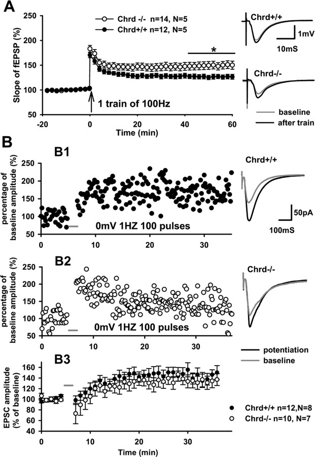
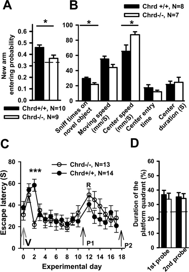
Recently, several evolutionary conserved signaling pathways that play prominent roles in regulating early neurodevelopment have been found to regulate synaptic remodeling in the adult. To test whether adult neuronal expression of bone morphogenic protein (BMP) signaling components also plays a postnatal role in regulating neuronal plasticity, we modulated BMP signaling in mice both in vivo and in vitro by genetic removal of the BMP inhibitor chordin or by perfusing recombinant BMP signaling pathway components onto acute hippocampal slices. Chordin null mice exhibited a significant increase in presynaptic transmitter release from hippocampal neurons, resulting in enhanced paired-pulse facilitation and long-term potentiation. These mice also showed a decreased acquisition time in a water maze test along with less exploratory activity during Y-maze and open-field tests. Perfusion of BMP ligands onto hippocampal slices replicated the presynaptic phenotype of chordin null slices, but bath application of Noggin, another antagonist of BMP signaling pathway, significantly decrease the frequency of miniature EPSCs. These results demonstrate that the BMP signaling pathway contributes to synaptic plasticity and learning likely through a presynaptic mechanism.


Figures

Figure 1.
Figure 1.
A, Expression pattern of BMP signaling pathway components in the mouse hippocampus. Anti-pSmad staining in adult wild-type mouse hippocampus; cell body layers showed strong staining, including CA1, CA3, and dentate gyrus (DG) regions. B, Anti-pSmad staining under higher magnification; signals are seen in pyramidal neurons of CA1, inhibitory neurons (parvalbumin positive), and astrocytes (typical morphology). Blue channel shows 4′,6′-diamidino-2-phenylindole (DAPI) nuclear counterstaining. Note that not all cell bodies show pSmad accumulation. Striatum pyramidal (s.p.) and striatum radiatum (s.r.) are labeled. C, D, Anti-Chrd staining in CA1 of Chrd +/+ and Chrd −/− slices. Note the strong Chrd signal in the wild-type pyramidal cells (C) but not in the mutant (D). E1–E4, BMPRII can be detected on 1-week-old hippocampal pyramidal neuronal cultured cells. E1, Map2 staining of dendrites. E2, E3, BMPRII staining in dendrites (E2) and the axon synapsin I-stained synaptic structure (E3). E4, Overlay of E1–E3. F, Higher magnification showed colocalization of synapsin I and BMPRII at synaptic structures.
Figure 2.
Figure 2.
Normal dendritic architecture and spine density is seen in the hippocampus of Chrd −/− mice. A, B, Map2 and GFAP double staining of the CA1 region from Chrd +/+ (A) and Chrd −/− (B) mice. Map2 staining (green) showed a similar dendrite branching pattern from both genotypes; GFAP staining (red) showed a similar distribution and density of astrocytes both genotypes. C, D, Golgi staining showing apical tertiary dendrites of CA1 pyramidal neurons from Chrd +/+ and Chrd −/−, respectively.
Figure 3.
Figure 3.
Increased PPF and mEPSC frequency in Chrd −/− slices. A, Relationship between the slope of fEPSP and amplitude of presynaptic fiber volley for Chrd −/− and Chrd +/+ mice. Data are expressed as mean ± SEM. Representative traces of fEPSPs evoked with different stimulus strengths are shown in the insets. B, PPF was measured as the ratio between the slopes of fEPSPs evoked by the second and first pulses and plotted for several ISIs. Field EPSPs were evoked with a stimulus that evoked 30% of the maximal fEPSP. Values represent mean ± SEM; *p < 0.05, **p < 0.01 (unpaired t test). Representative traces of fEPSPs evoked with 25 mS ISI are shown in the insets. C, Increase in the frequency of mEPSCs in Chrd −/− mice. Examples of mEPSCs recorded in Chrd −/− and Chrd +/+ mice are shown in the left. Note the significant difference in the mean frequency of mEPSCs recorded in slices from Chrd −/− and Chrd +/+ mice (***p < 0.005, unpaired test). Data are presented as mean ± SEM. There is no significant difference for mEPSC amplitude between genotypes.
Figure 4.
Figure 4.
Ultrastructural change of hippocampal synapses in Chrd −/− mice. A, Transmission electron microscopy of asymmetric synapse of Chrd +/+ in CA1 region; symbol ↕ points to the synaptic cleft, and symbol { indicates the length of active zone. Arrows point to docked vesicles. At, Axon terminal; Sp, spine; PSD, postsynaptic density. B, Asymmetric synapse of Chrd −/− in CA1 region. (Values are listed in Table 1.)
Figure 5.
Figure 5.
Effect of perfusion of BMPs and BMP antagonists on mEPSCs and PPF. A, Perfusion of BMP6 on Chrd +/+ slices increased the frequency but not the amplitude of mEPSCs. Left shows representative sweeps from control and 15 min after treatment. Right shows significant increase of frequency but not amplitude after perfusion of BMP6. B, BMP2 at a similar concentration had no effect on mEPSCs after 15 min perfusion. Representative traces of mEPSCs are shown on the left. Right shows time-lapse change of amplitude and frequency of mEPSCs at 3 min analysis interval. There is no significant change of either mEPSCs amplitude or frequency. C, Perfusion of Noggin, an antagonist of BMP signaling, inhibited release possibilities but not amplitude of mEPSC after 15 min (paired t test, p < 0.05). D, PPF of wild-type slices treated with BMP6. Slices were incubated in a mini-interface chamber with 200 ng/ml BMP6 or vehicle for 30 min. They were then transferred to an interface recording chamber to test paired-pulse facilitation. At 50 mS ISI, BMP6-treated slices showed higher PPF. Normalized representative traces were shown as inset.
Figure 6.
Figure 6.
Increase in high-frequency tetanus-induced LTP in Chrd −/− slices. A, Chrd −/− mice show higher LTP with 100Hz, 1 s protocol. Mean slope of fEPSPs recorded 0–20 min before induction of LTP was set as baseline. Number of analyzed slices (n) and animals (N) are indicated. Representative sweeps are shown on the right. B, Pairing protocol-induced postsynaptic-specific LTP is normal in Chrd −/− slices. B1, Sample recording from Chrd +/+ slice. B2, Sample recording from Chrd −/− slice. B3, Accumulated data from two genotypes for pairing protocol-induced LTP. As shown in the right, there is no difference between genotypes after 30 min.
Figure 7.
Figure 7.
Transition from early-phase LTP to late-phase LTP. A, TBS protocol induced a similar level of LTP in Chrd −/− and Chrd +/+ slices. Mean amplitudes of fEPSPs recorded 0–10 min before induction of LTP were set as a baseline. Above the summary plot are shown an average baseline sweep and a sweep 1 h after induction. B, A train of 100 Hz in 1 s given four times with a 5 min interval induces late-phase LTP at the same level from both Chrd −/− and Chrd +/+ slices. Averaged sweeps of baseline and 3 h after induction are shown above the accumulated plot. C, In a submerged recording chamber, early-phase LTP induced by one train of 100 Hz lasts longer than 3 h. The average slope of fEPSP of the last 20 min showed a significant difference between Chrd −/− and Chrd +/+ slices. (158.8 ± 12.8 vs 120.6 ± 5.7; p < 0.05, t test)
Figure 8.
Figure 8.
Cognitive behavior in Chrd −/− mice. A, Chrd −/− mice showed less entry time to the formerly closed arm during a recall test of Y-maze. Dashed line is the random possibility for the new arm 0.33. B, Chrd −/− mice showed less interaction with a novel object and faster running speed inside the center of an open field. Chrd −/− mice show no difference in locomotive speed in areas outside the center compared with Chrd +/+ mice. Chrd −/− mice showed no significant change of center entry time and center staying time. C, In a water maze test, Chrd −/− mice showed shorter escape latency on the second day of learning. C shows the escape latency plotted against training day. On day 0, (V) refers to cued platform; on days 1–17, a hidden platform was used. P1 and P2 indicate day 11 probe test and day 18 probe test. R means start of reversal test, in which the hidden platform was moved to an adjacent quadrant. D, On day 11, the platform was removed from the pool and mice were given 60 s to explore the pool. Both Chrd −/− and Chrd +/+ mice showed a similar higher preference for exploring the quadrant in which the platform used to be located. After the reversal on day 13, a second probe test was given on day 18, and again no difference between genotypes is seen in their preference for exploring the quadrant in which the platform used to be located. Dashed line indicates the 25% possibility of random selection for the target quadrant.

Similar articles

Cited by

References

    1. Abbott LF, Regehr WG. Synaptic computation. Nature. 2004;431:796–803. - PubMed
    1. Aberle H, Haghighi AP, Fetter RD, McCabe BD, Magalhaes TR, Goodman CS. wishful thinking encodes a BMP type II receptor that regulates synaptic growth in Drosophila . Neuron. 2002;33:545–558. - PubMed
    1. Bachiller D, Klingensmith J, Kemp C, Belo JA, Anderson RM, May SR, McMahon JA, McMahon AP, Harland RM, Rossant J, De Robertis EM. The organizer factors Chordin and Noggin are required for mouse forebrain development. Nature. 2000;403:658–661. - PubMed
    1. Bachiller D, Klingensmith J, Shneyder N, Tran U, Anderson R, Rossant J, De Robertis EM. The role of chordin/Bmp signals in mammalian pharyngeal development and DiGeorge syndrome. Development. 2003;130:3567–3578. - PubMed
    1. Baines RA. Synaptic strengthening mediated by bone morphogenetic protein-dependent retrograde signaling in the Drosophila CNS. J Neurosci. 2004;24:6904–6911. - PMC - PubMed

Publication types

MeSH terms

LinkOut - more resources