. 2007 Jul 18;27(29):7751-61.
doi: 10.1523/JNEUROSCI.1661-07.2007.

Minocycline alleviates death of oligodendrocytes by inhibiting pro-nerve growth factor production in microglia after spinal cord injury

Affiliations

Minocycline alleviates death of oligodendrocytes by inhibiting pro-nerve growth factor production in microglia after spinal cord injury

Tae Y Yune et al. J Neurosci. .

Abstract

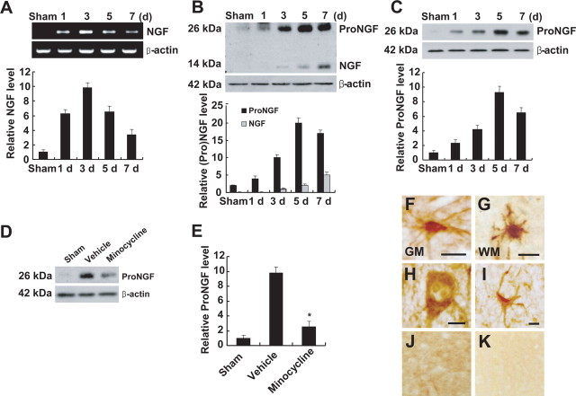
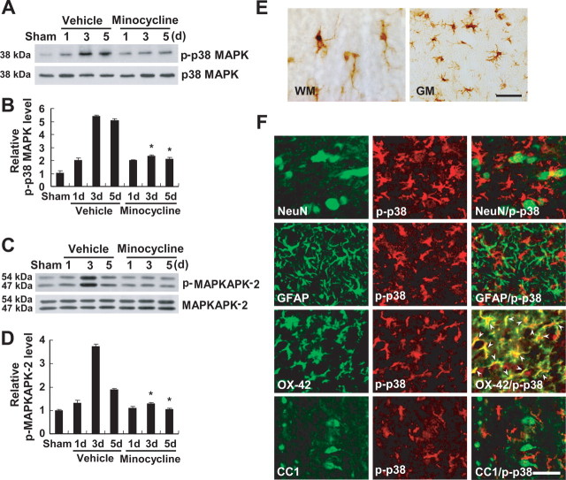
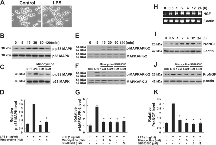
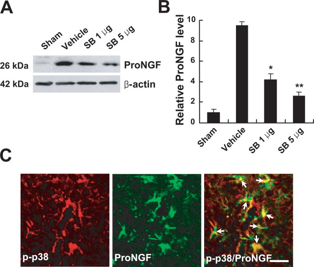
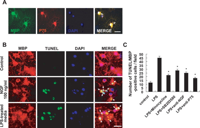
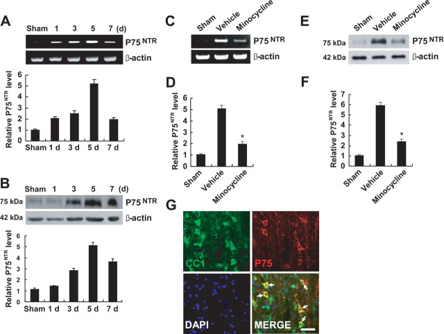
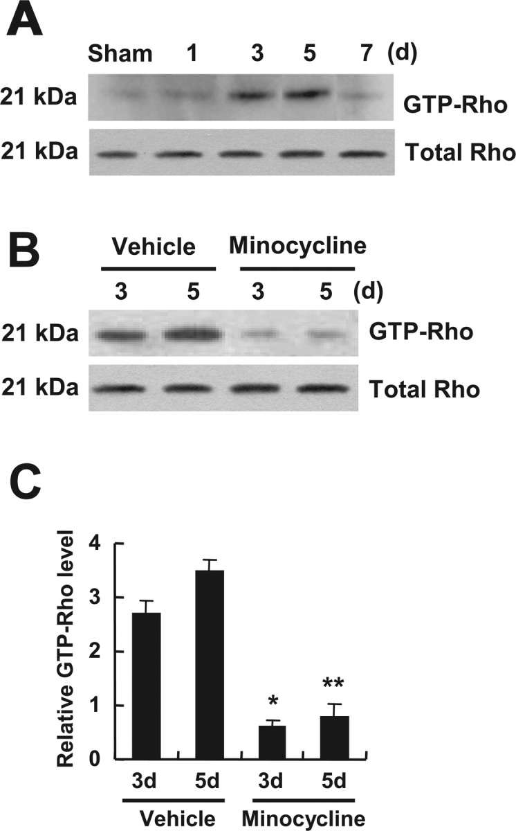
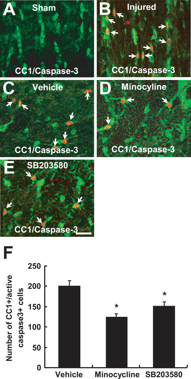
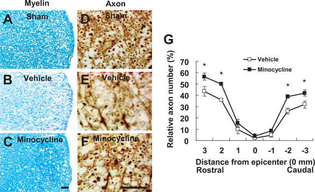
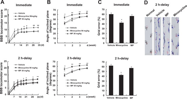
Spinal cord injury (SCI) causes a permanent neurological disability, and no satisfactory treatment is currently available. After SCI, pro-nerve growth factor (proNGF) is known to play a pivotal role in apoptosis of oligodendrocytes, but the cell types producing proNGF and the signaling pathways involved in proNGF production are primarily unknown. Here, we show that minocycline improves functional recovery after SCI in part by reducing apoptosis of oligodendrocytes via inhibition of proNGF production in microglia. After SCI, the stress-responsive p38 mitogen-activated protein kinase (p38MAPK) was activated only in microglia, and proNGF was produced by microglia via the p38MAPK-mediated pathway. Minocycline treatment significantly reduced proNGF production in microglia in vitro and in vivo by inhibition of the phosphorylation of p38MAPK. Furthermore, minocycline treatment inhibited p75 neurotrophin receptor expression and RhoA activation after injury. Finally, minocycline treatment inhibited oligodendrocyte death and improved functional recovery after SCI. These results suggest that minocycline may represent a potential therapeutic agent for acute SCI in humans.


Figures

Figure 1.
Figure 1.
Minocycline inhibits proNGF expression after SCI. A, B, NGF mRNA (A) and protein expression (B) were increased after SCI. B, C, Anti-mature-NGF antibody detected both mature (14 kDa) and proNGF (26 kDa) (B), and anti-proNGF antibody detected the high-molecular-weight band of 26 kDa (C). Note that the extent of induction of proNGF was greater than that observed with mature NGF (B). D, Minocycline treatment decreased the level of proNGF expression compared with that observed in vehicle-treated control at 5 d after injury. E, Quantitative analysis of Western blots shows that minocycline significantly inhibited proNGF expression when compared with that in vehicle control at 5 d after injury. Values are mean ± SD of three separate experiments. *p < 0.001. F–I, Immunocytochemical analysis shows that microglia in the white (WM) (F) and gray (GM) (G) matters, and neuron (H) and astrocyte (I) located near the lesion site expressed proNGF at 5 d after injury. J, K, Sham-operated (J) and negative control (K) were negative for proNGF. Double labeling using specific cell markers revealed that proNGF-positive cells shown in F–I were indeed microglia (F, G), neuron (H), and astrocyte (I), respectively (data not shown). Scale bars, 10 μm.
Figure 2.
Figure 2.
Minocycline inhibits p38MAPK and MAPKAPK-2 activation after SCI. A, C, SCI increased the levels of p-p38MAPK (A) and p-MAPKAPK-2 (C). B, D, Quantitative analysis of Western blots shows that minocycline treatment significantly inhibited both p-p38MAPK and p-MAPKAPK-2 when compared with those observed in vehicle control at 3 and 5 d after injury. Values are mean ± SD of three separate experiments. *p < 0.001. E, Immunocytochemical analysis shows that cells resembling microglia in the white (WM) and gray (GM) matters expressed p-p38MAPK at 5 d after injury. Double-labeled immunocytochemical analysis shows that only OX-42-positive microglia expressed p-p38MAPK (arrowheads), whereas neurons (NeuN), astrocytes (GFAP), and oligodendrocytes (CC1) were negative at 5 d after injury (F). Scale bars, 20 μm.
Figure 3.
Figure 3.
Minocycline inhibits p38MAPK-dependent proNGF expression in microglia, BV2 cells. A, Cells were plated onto six-well plates and treated with LPS. Note that BV2 cell treated with LPS for 4 h exhibited cellular processes, a characteristic of activated microglial cells. B, The level of p-p38MAPK was increased and peaked at 30 min after LPS treatment. C, Minocycline (1 and 5 nm) treatment decreased the level of p-p38MAPK at 30 min after LPS treatment when compared with that in cultures treated with LPS only. D, Quantitative analysis of Western blots shows that minocycline significantly inhibited p-p38MAPK expression at 30 min after LPS treatment when compared with that in cultures treated with LPS only. Values are mean ± SD of three separate experiments. *p < 0.001. E, The level of p-MAPKAPK-2 was increased and peaked at 30 min after LPS treatment. F, Minocycline (1 and 5 nm) or SB203580 treatment (1 and 5 μm), an inhibitor of p38MAPK, decreased the level of p-MAPKAPK-2 at 30 min after LPS treatment when compared with that in cultures treated with LPS only. G, Quantitative analysis of Western blots shows that minocycline and SB203580 significantly inhibited p-MAPKAPK-2 expression at 30 min after LPS treatment when compared with that in cultures treated with LPS only. Values are mean ± SD of three separate experiments. *p < 0.001. H, I, NGF mRNA (H) and proNGF protein (26 kDa) (I) expression were increased and peaked at 2 and 4 h, respectively, after LPS treatment. J, Minocycline (1 and 5 nm) and SB203580 (1 and 5 μm) treatment decreased the level of proNGF expression at 4 h after LPS treatment compared with that in cultures treated with LPS only. K, Quantitative analysis of Western blots shows that minocycline and SB203580 significantly inhibited proNGF expression at 4 h after LPS treatment when compared with that in cultures treated with LPS only. Values are mean ± SD of three separate experiments. *p < 0.001.
Figure 4.
Figure 4.
SB203580 inhibits proNGF expression after SCI. SB203580 (1 and 5 μg) was injected directly into the spinal cord at the lesion epicenter after injury. Spinal cord tissues were harvested at 5 d after injury. A, SB203580 treatment decreased the level of proNGF expression when compared with that in vehicle-treated control after injury. B, Quantitative analysis of Western blots shows that SB203580 significantly inhibited proNGF expression compared with that in vehicle-treated control after injury. Values are mean ± SD of three separate experiments. *p < 0.05; **p < 0.001. C, Double-labeled immunocytochemical analysis showed that p-p38MAPK-positive microglia expressed proNGF (arrows) at 5 d after injury. Scale bar, 20 μm.
Figure 5.
Figure 5.
A, Microglia-derived proNGF induces apoptosis of oligodendrocytes in culture. Immunocytochemical analysis shows that MBP-positive oligodendrocytes expressed p75NTR (arrows). Scale bar, 20 μm. Differentiated oligodendrocytes were treated with LPS-treated BV2 cell culture medium. After 24 h, cells were processed for TUNEL and MBP staining. B, Representative photographs show that LPS-treated BV2 cell culture medium or recombinant NGF (as a positive control) induced the apoptotic cell death of oligodendrocytes as revealed by the presence of both TUNEL- and MBP-positive cells (arrows). Note that control shows no TUNEL/MBP-positive cells. Scale bar, 20 μm. C, Quantitative analyses of TUNEL-positive oligodendrocytes show that LPS-induced oligodendrocyte cell death was significantly inhibited by minocycline or SB203580 treatment. Also, oligodendrocyte cell death was significantly attenuated when LPS-treated BV2 cell culture medium subjected to immunoprecipitation using a neutralizing anti-NGF polyclonal antibody or when oligodendrocytes were treated with anti- p75NTR antibody before treatment with LPS-treated BV2 cell culture medium (C). Values are mean ± SD of three separate experiments. *p < 0.001 compared with LPS.
Figure 6.
Figure 6.
Minocycline inhibits p75NTR expression after SCI. A, B, The levels of p75NTR mRNA (A) and protein (B) were increased and peaked at 5 d after SCI. C, E, Minocycline treatment decreased the levels of p75NTR mRNA (C) and protein (E) expression compared with those observed in vehicle control at 5 d after injury. D, F, Quantitative analyses of Western blots show that minocycline significantly inhibited p75NTR mRNA (D) and protein (F) expression when compared with those observed in vehicle control at 5 d after injury. Values are mean ± SD of three separate experiments. *p < 0.001. G, Immunocytochemical analysis showed that CC1-positive oligodendrocytes expressed p75NTR at 5 d after injury (arrows). Scale bar, 20 μm.
Figure 7.
Figure 7.
Minocycline inhibits RhoA activation after SCI. GTP-bound RhoA was isolated by pull-down assay and detected by Western blot using anti-Rho antibody as described in Materials and Methods. A, RhoA was activated and peaked at 5 d after injury. B, Minocycline decreased the level of RhoA activation compared with that in the vehicle control at 3 and 5 d after injury. Total Rho level determined from total tissue lysates was not changed after injury (A, B). C, Quantitative analysis of Western blots shows that minocycline significantly inhibited RhoA activation when compared with that in the vehicle control at 3 and 5 d after injury. Values are mean ± SD of three separate experiments. *p < 0.05; **p < 0.001.
Figure 8.
Figure 8.
Minocycline and SB203580 inhibits apoptosis of oligodendrocytes after SCI. Rats receiving the 25 mm insult were treated twice per day with minocycline, beginning 2 h after injury. SB203580 (5 μg) was injected directly into the spinal cord at the lesion epicenter at 2 h after injury. After 5 d injury, spinal cord sections were processed for double labeling using CC1 antibody, a marker for oligodendrocytes, and anti-cleaved capase-3 antibody. A, B, Immunocytochemical analysis shows that a number of cleaved (activated) caspase-3-positive oligodendrocytes (red, arrows) was observed in the injured spinal cord (B), whereas no cleaved caspase-3-positive cell was observed in the sham-operated cord (A) (longitudinal sections). C–E, Minocycline (D) or SB203580 (E) treatment decreased the number of cleaved caspase-3-positive oligodendrocytes (arrows) compared with that observed in the vehicle control (C) (transverse sections). Scale bar, 20 μm. F, Quantitative analysis of caspase-3-positive oligodendrocytes shows that minocycline or SB203580 significantly inhibited the death of oligodendrocytes when compared with that in the vehicle control after injury. Cleaved caspase-3-positive oligodendrocytes were counted as described in Materials and Methods. *p < 0.001.
Figure 9.
Figure 9.
Minocycline reduces myelin and axonal loss after SCI. Spinal cords at 38 d after injury were processed for Luxol fast blue and neurofilament staining. Transverse cryosections were selected 2000 μm rostral to the lesion site. A, B, Luxol fast blue staining shows that myelin loss in lateral funiculus was extensive in the vehicle control (B) when compared with that in sham control (A) after injury. C, Minocycline treatment decreased the extent of myelin loss after injury. Scale bar, 30 μm. D, E, Neurofilament staining shows that fewer axons were observed in the vehicle control (E) when compared with those in sham control (D) after injury. F, Minocycline treatment decreased the extent of axonal loss after injury. Scale bar, 30 μm. G, Quantitative analysis of neurofilament-stained axons within the vestibulospinal tract showed that the number of axons in minocycline-treated spinal cord was significantly higher than that in the vehicle control. Neurofilament-positive axons were counted as described in Materials and Methods. *p < 0.05.
Figure 10.
Figure 10.
Minocycline improves functional recovery after SCI. After SCI, minocycline or MP was administered either immediately or 2 h after injury, and recovery was assessed by BBB, inclined plane test, footprint analysis, and grid walk test (n = 20). A, B, Minocycline treatment (given immediately or after a 2 h delay) significantly improved locomotor function as assessed by BBB score (A) and inclined plane test (B) when compared with that of vehicle control. Note that MP treatment had no significant effect on recovery. *p < 0.05; **p < 0.01. C, The minocycline-treated group (given immediately or after a 2 h delay) shows a significantly lower error percentage when compared with that of vehicle-treated group, whereas MP-treated group shows no significant improvement in the grid walk test. The grid walk test was performed at 5 weeks after injury. *p < 0.05. D, Representative footprints obtained from each group at 5 weeks after SCI show that minocycline treatment (given after a 2 h delay) improved foot coordination, whereas vehicle- and MP-treated animals showed inconsistent coordination and toe drags.

Similar articles

Cited by

References

    1. Ackery A, Robins S, Fehlings MG. Inhibition of Fas-mediated apoptosis through administration of soluble Fas receptor improves functional outcome and reduces posttraumatic axonal degeneration after acute spinal cord injury. J Neurotrauma. 2006;23:603–616. - PubMed
    1. Balentine JD. Pathology of experimental spinal cord trauma. I. The necrotic lesion as a function of vascular injury. Lab Invest. 1978;39:236–253. - PubMed
    1. Barrett GL. The p75 neurotrophin receptor and neuronal apoptosis. Prog Neurobiol. 2000;61:205–292. - PubMed
    1. Basso DM, Beattie MS, Bresnahan JC. A sensitive and reliable locomotor rating scale for open field testing in rats. J Neurotrauma. 1995;12:1–21. - PubMed
    1. Beattie MS, Farooqui AA, Bresnahan JC. Review of current evidence for apoptosis after spinal cord injury. J Neurotrauma. 2000;17:915–925. - PubMed

Publication types

MeSH terms