. 2007 Sep;14(9):1165-72.
doi: 10.1128/CVI.00157-07. Epub 2007 Jul 18.

Feasibility of a multiplex flow cytometric bead immunoassay for detection of anti-epoetin alfa antibodies

Affiliations

Feasibility of a multiplex flow cytometric bead immunoassay for detection of anti-epoetin alfa antibodies

John Ferbas et al. Clin Vaccine Immunol. 2007 Sep.

Abstract

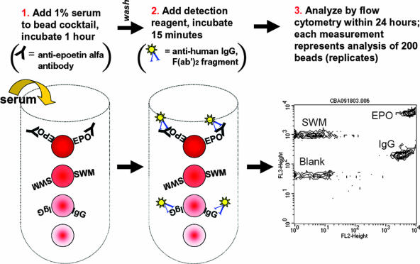
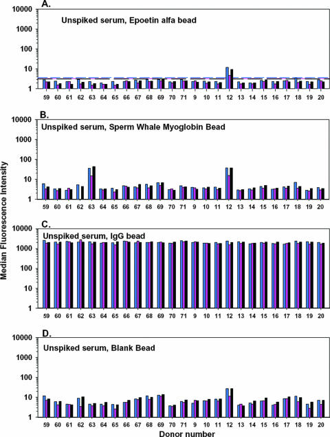
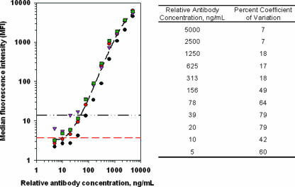
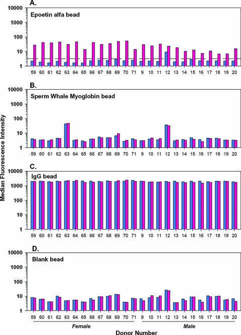
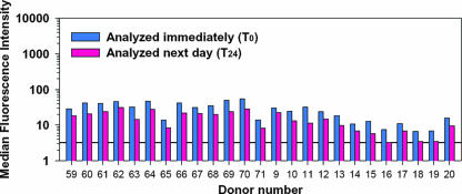
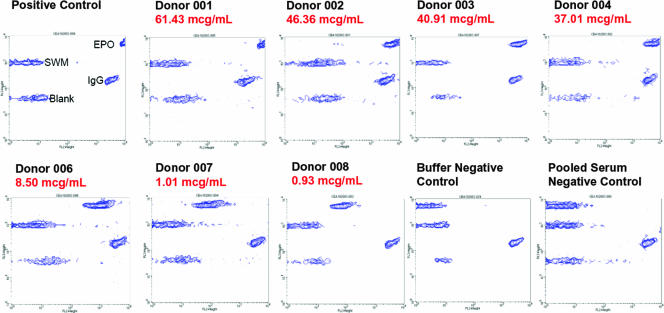
Immunogenicity profiles of recombinant therapeutic proteins are important to understand because antibodies raised against these molecules may have important clinical sequelae. The purpose of the present study was to demonstrate that a flow cytometric bead array could be used to detect clinically relevant antibodies with specificity to such therapeutics. We chose to evaluate well-characterized specimens from persons treated with epoetin alfa that developed antibody-mediated pure red blood cell aplasia as a means to demonstrate the utility of this platform. Our data show that this assay is capable of detecting anti-epoetin alfa antibodies with a relative antibody concentration of 50 ng/ml, where 25 of 25 sera spiked with antibodies at this concentration scored positive. Moreover, the assay was designed to include positive and negative control beads for each specimen that is processed to ensure the specificity of the signal when detected. Measurement of interassay precision supports quantitative estimates of relative antibody concentrations in the range of 313 to 5,000 ng/ml, where the percent coefficient of variation did not exceed 20%. With respect to clinical specimens, antibodies with specificity for epoetin alfa could be easily detected in a set of specimens from persons with pure red blood cell aplasia that had prior exposure to the EPREX brand of recombinant epoetin alfa. Further development and validation of this approach may facilitate successful widespread application of the method for detection of anti-epoetin alfa antibodies, as well as antibodies directed against other recombinant therapeutic proteins.


Figures

FIG. 1.
FIG. 1.
Flow cytometric bead immunoassay for anti-epoetin alfa antibodies. This multiplexed assay was designed with 4 beads of distinct fluorescence intensity that had epoetin alfa (EPO), sperm whale myoglobin (SWM; a negative control), or human IgG (IgG; a positive control) covalently attached to their surfaces by standard amine chemistry. A fourth bead was left unconjugated and served as a reference bead control. The assay involved incubation with 1% serum, followed by addition of a fluorescent detection reagent [phycoerythrin-conjugated anti-human IgG, F(ab′)2] and analysis on a standard flow cytometer. The intensity of fluorescence is therefore proportional to the amount of serum antibody captured by each of the respective beads. The address for each bead corresponds to the position in the diagram and is labeled as such in the rightmost dot plot from the flow cytometer.
FIG. 2.
FIG. 2.
Characterization of background binding of human serum IgG in the flow cytometric bead immunoassay. In this experiment, sera from 25 persons without prior exposure to an erythropoietic agent were tested to determine the background binding to each bead. The blue, pink, and black bars represent data from three independent experiments. The assay cutoffs, described as the mean plus the standard deviations, are displayed for each assay run as corresponding, color-matched dashed lines. See the text for a detailed discussion of the data.
FIG. 3.
FIG. 3.
Serial dilution of a human serum specimen that was positive for anti-epoetin alfa antibodies. Serial dilution of this specimen on four independent experiments (coded by unique symbols) demonstrated a dose-response relationship between anti-epoetin antibody concentration and signal generated from the flow cytometer. The average regression line (black dashed line calculated in Sigmaplot version 8.0) from these four experiments is plotted for reference. The horizontal red dashed line indicates the mean plus three standard deviations from the negative control specimens; the horizontal black dashed/dotted line indicates where the 50-ng/ml concentration falls. This relative antibody concentration was subsequently used to document the sensitivity of the assay (Fig. 4).
FIG. 4.
FIG. 4.
Demonstration that the flow cytometric bead immunoassay is of adequate sensitivity for clinical application. In this experiment, the positive control specimen from Fig. 3 was spiked into the 25 serum specimens studied for background binding in Fig. 2 at a final relative antibody concentration of 50 ng/ml. A signal in excess of the mean plus three standard deviations of the unspiked specimens was demonstrated for all specimens. The signal was specific to the bead coupled to epoetin alfa; an increase in signal was not apparent in any of the other three beads in the multiplexed array.
FIG. 5.
FIG. 5.
Degradation of signal in specimens held for an extended time period. To demonstrate stability of positive signals in this assay, the specimens from Fig. 4 were held overnight at 5°C and analyzed the following day. The drop-off in signal (3 to 26 MFI units; 12 MFI units on average) was consistent between samples and would not impact identification of positive specimens, provided that their relative antibody concentration was >50 ng/ml.
FIG. 6.
FIG. 6.
Validation of assay performance with seven sera known to be positive for anti-epoetin alfa antibodies. Seven sera from patients treated with EPREX and previously characterized (5, 22) to contain anti-epoetin alfa antibodies were tested in the flow cytometric bead immunoassay to demonstrate that the assay can detect clinically relevant antibody species. For each specimen, the relative antibody concentration, as determined in a Biacore immunoassay (22), is indicated in red. The specimens are rank ordered, from the highest antibody concentration to the lowest. Visual inspection of the fluorescence intensity of signal from the epoetin alfa (EPO) bead shows that the flow cytometric bead immunoassay is in concordance with the relative antibody concentration estimated by the Biacore method.

Similar articles

Cited by

References

    1. Bennett, C. L., D. Cournoyer, K. R. Carson, J. Rossert, S. Luminari, A. M. Evens, F. Locatelli, S. M. Belknap, J. M. McKoy, E. A. Lyons, B. Kim, R. Sharma, S. Costello, E. B. Toffelmire, G. A. Wells, H. A. Messner, P. R. Yarnold, S. M. Trifilio, D. W. Raisch, T. M. Kuzel, A. Nissenson, L. C. Lim, M. S. Tallman, and N. Casadevall. 2005. Long-term outcome of individuals with pure red cell aplasia and antierythropoietin antibodies in patients treated with recombinant epoetin: a follow-up report from the Research on Adverse Drug Events and Reports (RADAR) project. Blood 1063343-3347. - PMC - PubMed
    1. Bonfield, T. L., N. John, B. P. Barna, M. S. Kavuru, M. J. Thomassen, and B. Yen-Lieberman. 2005. Multiplexed particle-based anti-granulocyte macrophage colony stimulating factor assay used as pulmonary diagnostic test. Clin. Diagn. Lab. Immunol. 12821-824. - PMC - PubMed
    1. Boven, K., J. Knight, F. Bader, J. Rossert, K. U. Eckardt, and N. Casadevall. 2005. Epoetin-associated pure red cell aplasia in patients with chronic kidney disease: solving the mystery. Nephrol. Dialysis Transplant. 20iii33-iii40. - PubMed
    1. Boven, K., S. Stryker, J. Knight, A. Thomas, M. van Regenmortel, D. M. Kemeny, D. Power, J. Rossert, and N. Casadevall. 2005. The increased incidence of pure red cell aplasia with an Eprex formulation in uncoated rubber stopper syringes. Kidney Int. 672346-2353. - PubMed
    1. Casadevall, N., J. Nataf, B. Viron, A. Kolta, J. J. Kiladjian, P. Martin-Dupont, P. Michaud, T. Papo, V. Ugo, I. Teyssandier, B. Varet, and P. Mayeux. 2002. Pure red-cell aplasia and antierythropoietin antibodies in patients treated with recombinant erythropoietin. N. Engl. J. Med. 346469-475. - PubMed

LinkOut - more resources