. 2007 Jul-Aug;47(7):1008-23; discussion 24-5.
doi: 10.1111/j.1526-4610.2007.00854.x.

Neuron-glia signaling in trigeminal ganglion: implications for migraine pathology

Affiliations

Neuron-glia signaling in trigeminal ganglion: implications for migraine pathology

Srikanth Thalakoti et al. Headache. 2007 Jul-Aug.

Abstract

Objective: The goal of this study was to investigate neuronal-glial cell signaling in trigeminal ganglia under basal and inflammatory conditions using an in vivo model of trigeminal nerve activation.

Background: Activation of trigeminal ganglion nerves and release of calcitonin gene-related peptide (CGRP) are implicated in the pathology of migraine. Cell bodies of trigeminal neurons reside in the ganglion in close association with glial cells. Neuron-glia interactions are involved in all stages of inflammation and pain associated with several central nervous system (CNS) diseases. However, the role of neuron-glia interactions within the trigeminal ganglion under normal and inflammatory conditions is not known.

Methods: Sprague-Dawley rats were utilized to study neuron-glia signaling in the trigeminal ganglion. Initially, True Blue was used as a retrograde tracer to localize neuronal cell bodies in the ganglion by fluorescent microscopy and multiple image alignment. Dye-coupling studies were conducted under basal conditions and in response to capsaicin injection into the TMJ capsule. S100B and p38 expression in neurons and glia were determined by immunohistochemistry following chemical stimulation. CGRP levels in the ganglion were measured by radioimmunoassay in response to capsaicin. In addition, the effect of CGRP on the release of 19 different cytokines from cultured glial cells was investigated by protein microarray analysis.

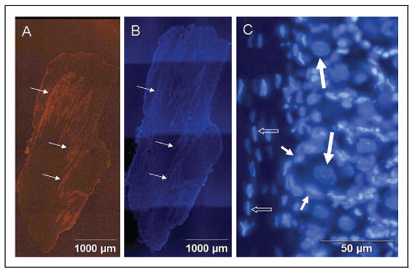
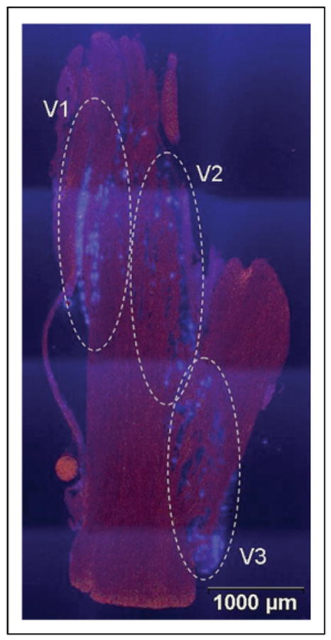
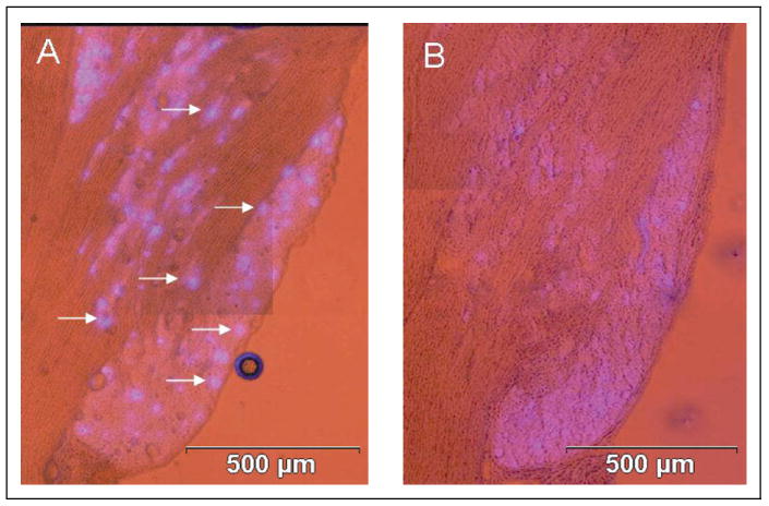
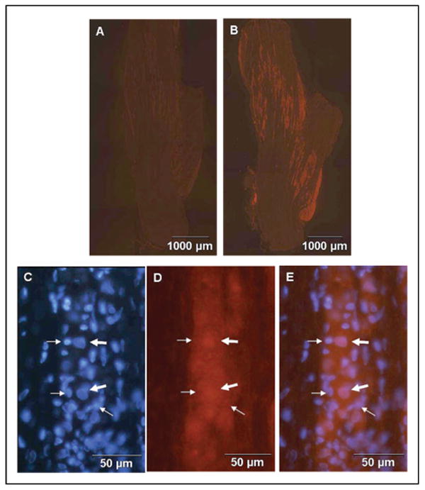
Results: In unstimulated control animals, True Blue was detected primarily in neuronal cell bodies localized in clusters within the ganglion corresponding to the V3 region (TMJ capsule), V2 region (whisker pad), or V1 region (eyebrow and eye). However, True Blue was detected in both neuronal cell bodies and adjacent glia in the V3 region of the ganglion obtained from animals injected with capsaicin. Dye movement into the surrounding glia correlated with the time after capsaicin injection. Chemical stimulation of V3 trigeminal nerves was found to increase the expression of the inflammatory proteins S100B and p38 in both neurons and glia within the V3 region. Unexpectedly, increased levels of these proteins were also observed in the V2 and V1 regions of the ganglion. CGRP and the vesicle docking protein SNAP-25 were colocalized in many neuronal cell bodies and processes. Decreased CGRP levels in the ganglion were observed 2 hours following capsaicin stimulation. Using protein microarray analysis, CGRP was shown to differentially regulate cytokine secretion from cultured trigeminal ganglion glia.

Conclusions: We demonstrated that activation of trigeminal neurons leads to changes in adjacent glia that involve communication through gap junctions and paracrine signaling. This is the first evidence, to our knowledge, of neuron-glia signaling via gap junctions within the trigeminal ganglion. Based on our findings, it is likely that neuronal-glial communication via gap junctions and paracrine signaling are involved in the development of peripheral sensitization within the trigeminal ganglion and, thus, are likely to play an important role in the initiation of migraine. Furthermore, we propose that propagation of inflammatory signals within the ganglion may help to explain commonly reported symptoms of comorbid conditions associated with migraine.


Conflict of interest statement

Conflict of Interest: None

Figures

Fig 1
Fig 1
Expression of TRPV1 and arrangement of neuronal and glial cells in adult rat trigeminal ganglion. (A) Staining of a 50 μm section of the entire trigeminal ganglion for expression of TRPV1 is shown. Neuronal cells are organized into bands or clusters (arrows). (B) Staining of the same section with the fluorescent dye DAPI is shown. All neuronal and glial cell nuclei are visible. (C) Neuronal and glial cell nuclei stained with DAPI are shown at higher magnification. Neuronal cell bodies (large arrows) are completely surrounded by satellite glial cells (small arrows). Schwann cells (small open arrows) are observed in association with nerve fibers.
Fig 2
Fig 2
Localization of neuronal cell bodies in trigeminal ganglion that provide sensory innervation of the TMJ capsule (V3), whisker pad (V2), and eye and eyebrow (V1). The entire ganglion is shown. The fluorescent dye True Blue was detected in neuronal cell bodies corresponding to the V3, V2, and V1 regions of the ganglion as identified.
Fig 3
Fig 3
Capsaicin stimulation of sensory neurons causes spreading of dye in trigeminal ganglion. The cellular localization pattern of True Blue in the V3 region of trigeminal ganglion under basal and stimulatory conditions is shown. (A) The dye was primarily detected in a few neuronal cell bodies (arrows) in a punctuate pattern in ganglion obtained from unstimulated control animals. (B) In contrast, the dye was detected in a diffuse pattern 2 hours after capsaicin stimulation.
Fig 4
Fig 4
Evidence of neuronal–glial gap junctions in trigeminal ganglion in response to capsaicin stimulation. Nerve cell bodies within the V3 region of the ganglion retrogradely labeled with True Blue were obtained from untreated animals (A) or animals injected in the TMJ capsule with capsaicin for 15 (B), 30 (C), 60 (D), or 120 (E) minutes prior to harvesting the ganglia. The corresponding boxed regions on the sections are shown at higher magnification in the right panels (a–e). While True Blue was detected primarily in the cytosol of neuronal cell bodies (thick arrows) of ganglion from control animals (A and a), more dye was detected in the adjacent glial satellite cells (thin arrows) with increasing time following capsaicin injection. At 2 hours, most of the dye is found in the cytosol of adjacent satellite cells (E and e).
Fig 5
Fig 5
Expression of S100B in trigeminal ganglion neurons is increased in response to capsaicin. A section of a ganglion from untreated animals or animals injected with capsaicin stained for S100B is shown. (A) In control ganglion, S100B staining was barely detectable. (B) In contrast, S100B staining was readily detectable in the V3, V2, and V1 regions of the ganglion following capsaicin stimulation for 120 minutes. At higher magnification, increased S100B expression was observed in both neuronal (thick arrows) and satellite glial cells (thin arrows) in response to capsaicin when compared to control (C–E). The same section was stained for DAPI (C) and S100B (D). A merged image is shown in E.
Fig 6
Fig 6
Expression of active p38 in neuronal and satellite glial cells is increased in response to NO/proton stimulation. Trigeminal ganglion from untreated animals or animals injected with 10 mm of the NO donor SNP in pH 5.5 medium were stained for the active, phosphorylated form of p38 MAP kinase. In control ganglion, p38 staining was barely detectable (A). In contrast, p38 staining was readily detectable in the V3, V2, and V1 regions of the ganglion 2 hours after stimulation with NO/protons (B). At higher magnification, elevated levels of p38 were detected in both neuronal (thick arrows) and satellite glial cells (thin arrow) in response to NO/protons (D). Retrograde labeling of the same section shown in panel B is shown in panel C. A single True Blue labeled neuron with surrounding satellite cells is indicated.
Fig 7
Fig 7
TNF-α increases expression of active p38 in neuronal and glial cells. Trigeminal ganglion from untreated animals or animals injected with 50 ng/mL TNF-α stained for the active, phosphorylated form of p38 MAP kinase are shown. In control ganglion, p38 staining was barely detectable (A). In contrast, p38 staining was readily detectable in the V3, V2, and V1 regions of the ganglion 2 hours after TNF-α injection (B). At higher magnification, increased p38 expression was observed in both neuronal (thick arrows) and glial cells (thin arrows) in response to TNF-α when compared to control (C–E). The same section was stained for DAPI (C) and p38 (D). A merged image is shown in E.
Fig 8
Fig 8
Expression of CGRP and SNAP-25 in trigeminal ganglion neurons. The expression of CGRP (A) and SNAP-25 (B) are shown in the entire trigeminal ganglion obtained from an untreated adult animal. At higher magnification, neuronal processes (arrows) expressing both CGRP (C) and SNAP-25 (D) that extend from neuronal cell bodies in one cluster to another cluster are observed. Neuronal processes expressing SNAP-25 are also observed within neuronal clusters (E).
Fig 9
Fig 9
Capsaicin stimulation of CGRP release from trigeminal ganglion neurons. The amount of CGRP was determined by radioimmunoassay in trigeminal ganglion obtained from control, untreated animals (CON, n = 13) or animals treated for 2 hours with DMSO (VEH, n = 4). The amount of CGRP is reported as pmol/μg of total protein. *P < .001 when compared to control- or vehicle-treated animals.
Fig 10
Fig 10
Primary cultures of trigeminal ganglion glial cells. Cells from d 3 trigeminal ganglia cultures stained with antibodies directed against glial fibrillary acidic protein (A). The same culture was costained with DAPI, which identifies nuclei of all cells (B).
Fig 11
Fig 11
CGRP regulation of cytokine release from cultured glial cells. Media was collected from primary trigeminal glial cell cultures left untreated (Control) or treated overnight with 100 nm CGRP and assayed for cytokine expression. Densometric analysis (n = 2 for each condition) was performed. The effect of CGRP on the relative level of regulated cytokines is reported.

Comment in

Similar articles

Cited by

References

    1. Stewart W, Lipton R, Celentano D, Reed M. Prevalence of migraine headache in the United States. Relation to age, income, race, and other sociodemographic factors. JAMA. 1991;267:64–69. - PubMed
    1. Pietrobon D. Migraine: New molecular mechanisms. Neuroscientist. 2005;11:373–386. - PubMed
    1. Appelgren A, Appelgren B, Kopp S, Lundeberg T, Theodorsson E. Neuropeptides in the arthritic TMJ and symptoms and signs from the stomatognathic system with special considerations to rheumatoid arthritis. J Orofac Pain. 1995;9:215–225. - PubMed
    1. Lazarov N. Comparative analysis of the chemical neuroanatomy of the mammalian trigeminal ganglion and mesencephalic trigeminal nucleus. Prog Neurobiol. 2002;66:19–59. - PubMed
    1. Hargreaves R, Shepheard S. Pathophysiology of migraine—New insights. Can J Neurol Sci. 1999;26:S12–S19. - PubMed

Publication types

MeSH terms