

The underside of the cerebral cortex: layer V/VI spiny inverted neurons
- PMID: 17635629
- PMCID: PMC2375765
- DOI: 10.1111/j.1469-7580.2007.00779.x
The underside of the cerebral cortex: layer V/VI spiny inverted neurons
Abstract
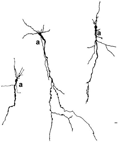
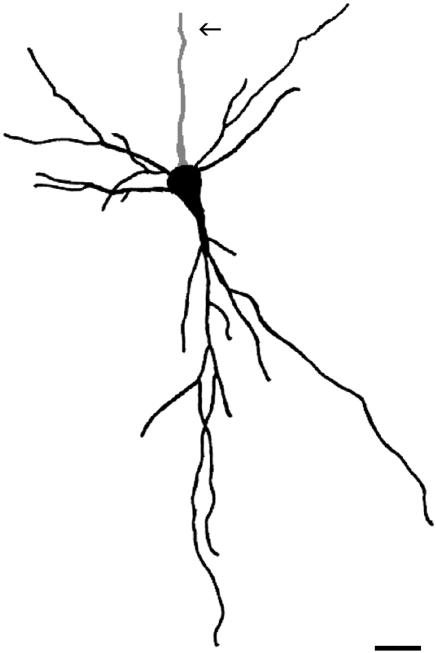
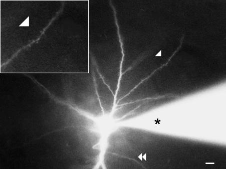
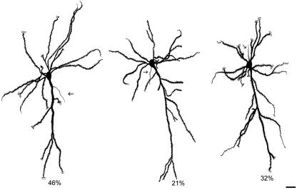
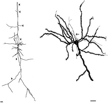
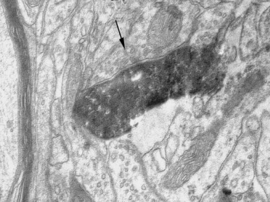
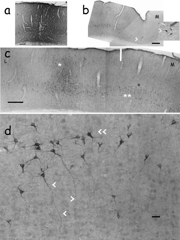
This paper presents an account of past and current research on spiny inverted neurons--alternatively also known as 'inverted pyramidal neurons'--in rats, rabbits and cats. In our laboratory, we have studied these cells with a battery of techniques suited for light and electron microscopy, including Nissl staining, Golgi impregnation, dye intracellular filling and axon retrograde track-tracing. Our results show that spiny inverted neurons make up less than 8.5 and 5.5% of all cortical neurons in the primary and secondary rabbit visual cortex, respectively. Infragranular spiny inverted neurons constitute 15 and 8.5% of infragranular neurons in the same animal and areas. Spiny inverted neurons congregate at layers V-VI in all studied species. Studies have also revealed that spiny inverted neurons are excitatory neurons which furnish axons for various cortico-cortical, cortico-claustral and cortico-striatal projections, but not for non-telencephalic centres such as the lateral and medial geniculate nuclei, the colliculi or the pons. As a group, each subset of inverted cells contributing to a given projection is located below the pyramidal neurons whose axons furnish the same centre. Spiny inverted neurons are particularly conspicuous as a source of the backward cortico-cortical projection to primary visual cortex and from this to the claustrum. Indeed, they constitute up to 82% of the infragranular cells that furnish these projections. Spiny inverted neurons may be classified into three subtypes according to the point of origin of the axon on the cell: the somatic basal pole which faces the cortical outer surface, the somatic flank and the reverse apical dendrite. As seen with electron microscopy, the axon initial segments of these subtypes are distinct from one another, not only in length and thickness, but also in the number of received synaptic boutons. All of these anatomical features together may support a synaptic-input integration which is peculiar to spiny inverted neurons. In this way, two differently qualified streams of axonal output may coexist in a projection which arises from a particular infragranular point within a given cortical area; one stream would be furnished by the typical pyramidal neurons, whereas spiny inverted neurons would constitute the other source of distinct information flow.
Figures

Similar articles
-
Morphological Characterization of a Cortico-cortical relay in the cat sensorimotor cortex.Cereb Cortex. 1997 Mar;7(2):100-9. doi: 10.1093/cercor/7.2.100. Cereb Cortex. 1997. PMID: 9087819
-
Morphometric analysis of the columnar innervation domain of neurons connecting layer 4 and layer 2/3 of juvenile rat barrel cortex.Cereb Cortex. 2003 Oct;13(10):1051-63. doi: 10.1093/cercor/13.10.1051. Cereb Cortex. 2003. PMID: 12967922
-
Columnar organization of dendrites and axons of single and synaptically coupled excitatory spiny neurons in layer 4 of the rat barrel cortex.J Neurosci. 2000 Jul 15;20(14):5300-11. doi: 10.1523/JNEUROSCI.20-14-05300.2000. J Neurosci. 2000. PMID: 10884314 Free PMC article.
-
The neocortex. An overview of its evolutionary development, structural organization and synaptology.Anat Embryol (Berl). 1994 Oct;190(4):307-37. doi: 10.1007/BF00187291. Anat Embryol (Berl). 1994. PMID: 7840420 Review.
-
Numerical relationships between geniculocortical afferents and pyramidal cell modules in cat primary visual cortex.Cereb Cortex. 1993 Jan-Feb;3(1):69-78. doi: 10.1093/cercor/3.1.69. Cereb Cortex. 1993. PMID: 8439740 Review.
Cited by
-
Axon topography of layer 6 spiny cells to orientation map in the primary visual cortex of the cat (area 18).Brain Struct Funct. 2017 Apr;222(3):1401-1426. doi: 10.1007/s00429-016-1284-z. Epub 2016 Aug 18. Brain Struct Funct. 2017. PMID: 27539451 Free PMC article.
-
Sensory deprivation differentially impacts the dendritic development of pyramidal versus non-pyramidal neurons in layer 6 of mouse barrel cortex.Brain Struct Funct. 2012 Apr;217(2):435-46. doi: 10.1007/s00429-011-0342-9. Epub 2011 Aug 23. Brain Struct Funct. 2012. PMID: 21861159 Free PMC article.
-
Adenosinergic Modulation of Layer 6 Microcircuitry in the Medial Prefrontal Cortex Is Specific to Presynaptic Cell Type.J Neurosci. 2024 Apr 10;44(15):e1606232023. doi: 10.1523/JNEUROSCI.1606-23.2023. J Neurosci. 2024. PMID: 38429106 Free PMC article.
-
Physiology and morphology of inverted pyramidal neurons in the rodent neocortex.Neuroscience. 2013 Sep 17;248:165-79. doi: 10.1016/j.neuroscience.2013.06.004. Epub 2013 Jun 14. Neuroscience. 2013. PMID: 23769893 Free PMC article.
-
A simplified morphological classification scheme for pyramidal cells in six layers of primary somatosensory cortex of juvenile rats.IBRO Rep. 2018 Oct 11;5:74-90. doi: 10.1016/j.ibror.2018.10.001. eCollection 2018 Dec. IBRO Rep. 2018. PMID: 30450442 Free PMC article.
References
-
- Albus K, Doñate-Oliver F, Sanides D, Fries W. The distribution of pontine projection cells in visual and association cortex of the cat: an experimental study with horseradish peroxidase. J Comp Neurol. 1981;201:175–189. - PubMed
-
- Arimatsu Y, Kojima M, Ishida M. Area- and lamina-specific organization of a neuronal subpopulation defined by expression of latexin in the rat cerebral cortex. Neuroscience. 1999;88:93–105. - PubMed
-
- Belichenko PV, Dahlstrom A, Von Essen C, Lindstrom S, Nordborg C, Sourander P. Atypical pyramidal cells in epileptic human cortex: CLSM and 3D reconstructions. Neuroreport. 1992;3:765–768. - PubMed
-
- Blanco-Santiago RI. University of the Basque Country; Desarrollo de las células invertidas corticales en el conejo. Doctoral dissertation.
Publication types
MeSH terms
LinkOut - more resources
Full Text Sources
Research Materials
Miscellaneous
