Review
. 2007 May-Jun;11(3):398-415.
doi: 10.1111/j.1582-4934.2007.00055.x.

Molecular and cellular key players in human islet transplantation

Affiliations
Review

Molecular and cellular key players in human islet transplantation

M Hermann et al. J Cell Mol Med. 2007 May-Jun.

Abstract

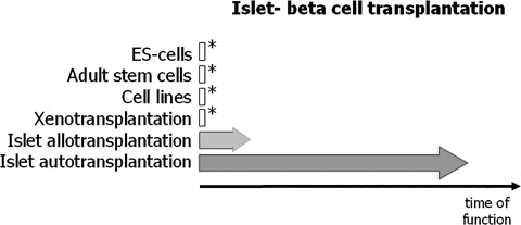
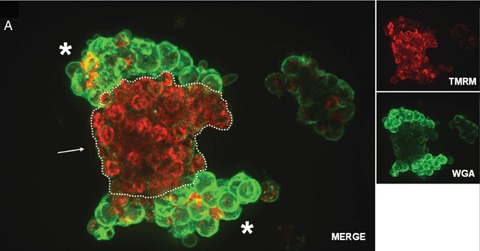
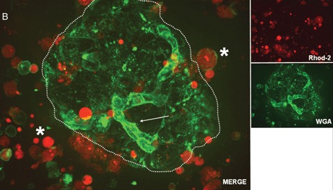
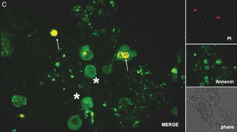
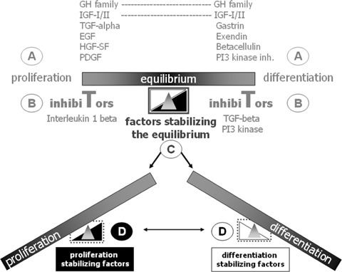
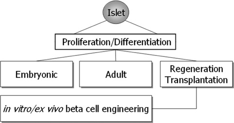
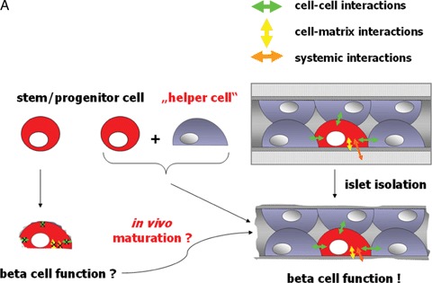
Human islet transplantation could represent an attractive alternative to insulin injections for the treatment of diabetes type 1. However, such an approach requires a better understanding of the molecular and cellular switches controlling ?-cell function in general as well as after transplantation into the liver. Although much research has been done into the suitability of stem or progenitor cells to generate a limitless supply of human ?-cells, a reproducible and efficient protocol for the differentiation of such cells into stably insulin-secreting ?-cells suitable for transplantation has yet to be reported. Fueled by recent findings showing that mature ?-cells are able to regenerate, many efforts have been undertaken to expand this cell pool. Unfortunately, also these approaches had problems to yield sufficiently differentiated human islet cells. The aim of this review is to summarize recent findings describing some of the molecular and cellular key players of islet biology. A more complete understanding of their orchestration and the use of new methods such as real time confocal imaging for the assessment of islet quality may yield the necessary advancements for more successful human islet transplantation.


Figures

1
1
Schematic representation of the progress/success in islet or β-cell transplantation. Asterisks symbolize the experimental state of the approach. The approach using embryonal stem cells still awaits ethical decisions. Similar to the approach aiming at the establishment of cell lines capable to substitute islet β-cells also embryonal stem cells bear the danger of oncologic transformations due to the many necessary in vitro population doublings. Another possibility is the use of xeno-islets, such as procine islets, for transplantation. Although having the advantage of being a sustainable islet source, many obstacles such as the antigenicity (due to the β-galactosyl antigen which is present in most non-human primates) and infection with zoonoses (such as the pig endogenous retroviruses) still remain [23]. The biological proof of principle showing a long-lasting and stable insulin production of islets transplanted into the liver has already been shown in autotransplantations [29]. Although the current immunosuppressive therapies have improved the outcome of islet allotransplantations, most patients once they reach 5 years post-transplantation do not sustain insulin independence [24].
2A
2A
Confocal real time microscopy of an entrapped human islet stained with tetramethylrhodamine methyl ester perchlorate (TMRM; red) and Alexa Fluor labeled wheat germ agglutinin (WGA; green). Vital mitochondria with an intact potential appear in red due to the TMRM staining. While islet (dotted line) mitochondria show a significantly higher vitality (see arrow), compared with the surrounding exocrine cells, the glycoconjugates, stained viaWGA, at the membrane surfaces are present in a significantly higher amount in the exocrine cells (see asterisks) compared with the islet cells.
2B
2B
Confocal real time microscopy of a human islet surrounded by stressed single cells in suspension after islet isolation procedure. The cell surfaces are stained with WGA.While endothelial cells (see arrow) show a strong staining of the cell surface after addition of WGA, islet cells (dotted circle) are only weakly stained. Note the strong positivity for cell permeant acetoxymethylester (Rhod-2) in the stressed single cells and the high cell vitality documented by the nearly absent staining for Rhod-2 positive cells in the islet.
2C
2C
Confocal real time microscopy of a human islet isolation stained with propidium iodide (PI; red; see arrows) and the FITC labeled apoptosis marker annexin V (green; see asterisks). Confocal microscopy was performed with a microlens-enhanced Nipkow disk-based confocal system mounted on an inverse microscope. All pictures were acquired with a 40 x water immersion objective.
3
3
Schematic model proposing four different molecular control levels of pancreatic β-cell proliferation and differentiation. For a more detailed description of the extrinsic factors influencing β-cell proliferation and differentiation see Soria, 2001 [43]. Pancreatic β-cell growth and differentiation requires precise control of the molecular mechanisms leading to either entry or exit of a replicative or differentiated state. A, B, C and D symbolize different control levels. Developmental state, age, metabolic factors, injuries or diseases such as diabetes affect both proliferation as well as differentiation of β-cells. Local signals coming either from surrounding cells or the extracellular matrix balance the proliferation and differentiation status of β-cells and thereby also their function. As impressively documented by islet autotransplantation, β-cells can handle with “big changes” i.e. even the transplantation into another organ such as the liver. The questions are how big the dependence of the β-cells is concerning an intact cytoarchitecture of the transplanted islet and how much help coming from the neighbouring cells, is needed (see also Figure 4)? Answering these questions is fundamental for further cell therapeutic approaches aiming at an expansion of the available pool of human β-cells.
4
4
Human islet β-cell proliferation and differentiation takes place either under embryonic or adult conditions. Besides these two physiological conditions a third one addressing regeneration or even transplantation of islets into the liver can be distinguished. In all three conditions, a correct balance between proliferation and differentiation is pivotal in order to ensure proper β-cell function. This balance is facing again a different situation when β-cells are maintained and propagated in vitro/ex vivo with the goal to gain an unlimited source of transplantable β-cells for human islet transplantation. It is this fourth situation that will benefit from a better understanding of the three other ones. Besides this, knowledge gained from the in vitro experiments with β-cells will round up our knowledge of in vivoβ-cell physiology.
5
5
(A) Under normal conditions β-cell function is influenced by cell-cell, cell-matrix and systemic interactions. Even after islet transplantation β-cell survival as well as function are maintained and stable for decades (as seen in autotransplantation of islets). Even after transplantation into the liver, islet cyto- architecture is maintained. Therefore, we hypothesize that β-cells either generated in vitro out of stem cells or ex vivo through expansion of progenitor cells might enface the problem of not being fully functional due to the lack of proper cell-cell or cell-matrix interactions. (B) While epithelial cells in the exocrine compartment (shown in brown colour at the left) produce their own basement membrane (see arrow), β-cells in the Islets of Langerhans (appear in red due to dithizone staining) do not (see arrow) [83]. Their basement membrane is produced from the underlying endothelial cells, a fact underlining the above-mentioned hypothesis.
5
5
(A) Under normal conditions β-cell function is influenced by cell-cell, cell-matrix and systemic interactions. Even after islet transplantation β-cell survival as well as function are maintained and stable for decades (as seen in autotransplantation of islets). Even after transplantation into the liver, islet cyto- architecture is maintained. Therefore, we hypothesize that β-cells either generated in vitro out of stem cells or ex vivo through expansion of progenitor cells might enface the problem of not being fully functional due to the lack of proper cell-cell or cell-matrix interactions. (B) While epithelial cells in the exocrine compartment (shown in brown colour at the left) produce their own basement membrane (see arrow), β-cells in the Islets of Langerhans (appear in red due to dithizone staining) do not (see arrow) [83]. Their basement membrane is produced from the underlying endothelial cells, a fact underlining the above-mentioned hypothesis.

References

    1. Humes HD. Cell therapy: leveraging nature's therapeutic potential. J Am Soc Nephrol. 2003;14:2211–3. - PubMed
    1. Dove A. Cell-based therapies go live. Nat Biotechnol. 2002;20:339–43. - PubMed
    1. Brower V. Human ES cells: Can you build a business around them? Nat Biotechnol. 1999;17:139–42. - PubMed
    1. Terada N, Hamazaki T, Oka M, Hoki M, Mastalerz DM, Nakano Y, Meyer EM, Morel L, Petersen BE, Scott EW. Bone marrow cells adopt the phenotype of other cells by spontaneous cell fusion. Nature. 2002;416:542–5. - PubMed
    1. Vassilopoulos G, Russell DW. Cell fusion: An alternative to stem cell plasticity and its therapeutic implications. Curr Opin Genet Dev. 2003;13:480–5. - PubMed

MeSH terms