Review
. 2007 Jul;5 Suppl 1(Suppl 1):102-15.
doi: 10.1111/j.1538-7836.2007.02516.x.

Serpins in thrombosis, hemostasis and fibrinolysis

Affiliations
Review

Serpins in thrombosis, hemostasis and fibrinolysis

J C Rau et al. J Thromb Haemost. 2007 Jul.

Abstract

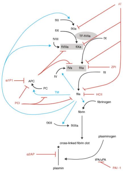
Hemostasis and fibrinolysis, the biological processes that maintain proper blood flow, are the consequence of a complex series of cascading enzymatic reactions. Serine proteases involved in these processes are regulated by feedback loops, local cofactor molecules, and serine protease inhibitors (serpins). The delicate balance between proteolytic and inhibitory reactions in hemostasis and fibrinolysis, described by the coagulation, protein C and fibrinolytic pathways, can be disrupted, resulting in the pathological conditions of thrombosis or abnormal bleeding. Medicine capitalizes on the importance of serpins, using therapeutics to manipulate the serpin-protease reactions for the treatment and prevention of thrombosis and hemorrhage. Therefore, investigation of serpins, their cofactors, and their structure-function relationships is imperative for the development of state-of-the-art pharmaceuticals for the selective fine-tuning of hemostasis and fibrinolysis. This review describes key serpins important in the regulation of these pathways: antithrombin, heparin cofactor II, protein Z-dependent protease inhibitor, alpha(1)-protease inhibitor, protein C inhibitor, alpha(2)-antiplasmin and plasminogen activator inhibitor-1. We focus on the biological function, the important structural elements, their known non-hemostatic roles, the pathologies related to deficiencies or dysfunction, and the therapeutic roles of specific serpins.


Figures

Fig. 1
Fig. 1
Serpin regulation of coagulation, protein C and fibrinolytic pathways. Serpins and inhibitory functions are shown in red, thrombin activity is shown in cyan. Prothrombinase and tenase complexes are shown in gray boxes. Coagulation is initiated by the exposure of tissue factor to factor VIIa shown in a gray oval. The symbol * indicates degradation. Necessary cofactors, Ca++, phospholipids, proteins S and Z, vitronectin and GAGs are not shown to maintain the simplicity of the schematic.
Fig. 2
Fig. 2
Serpin structure and mechanism of protease inhibition. (A) The shared serpin fold is illustrated by the structure of the prototypical native serpin α1PI. The ‘classic’ orientation shown on the left places the RSL (yellow) on top and the main β-sheet A (red) to the front. Sheets B and C are blue and orange, respectively, and helices A, D and H are colored green, cyan and magenta. The accessibility of the RSL is illustrated by rotating the molecule by 110° to the left along the long axis. It shows how the P1-P1′ (rods) scissile bond is exposed for proteolytic attack. Also clearer in this orientation are helices D and H, which are the heparin binding helices. (B) The serpin mechanism of protease inhibition is minimally expressed as a two-step process. In the first step, native serpin (ribbon with the P1 and P1′ residues as magenta balls) interacts reversibly with a protease (surface representation, colored according to temperature factors from blue to red) to form the Michaelis complex (middle). After formation of the acyl-enzyme intermediate the protease is flung to the opposite pole of the serpin and its catalytic architecture is destroyed, and consequently there is a loss of ordered structure (notice the smaller size and increase in temperature factors).
Fig. 3
Fig. 3
Native and complexed serpin structures. (A) The native structures of important hemostatic and fibrinolytic serpins are shown as ribbon diagrams, colored essentially as in Fig. 2. The monomeric structure of antithrombin is shown in the left panel, and is similar to that of heparin cofactor II (HCII) with the partial insertion of the N-terminal portion of the reactive site loop (RSL). A modeled position for the N-terminal tail of HCII is shown in magenta although its true position is not known. For AT, HCII and plasminogen activator inhibitor-1 the heparin binding helix (helix D) is shown in cyan, but for protein C inhibitor (PCI) heparin binds to helix H (blue). The increased size and flexibility of the RSL of PCI is also evident from this depiction. (B) Some important serpin complexes are shown. Using S195A proteases it was possible to obtain the structures of the AT Michaelis complexes with thrombin (magenta) and FXa (magenta) with their activating synthetic heparins (SR123781 and fondaparinux, rods). Similarly, the HCII-thrombin (blue) complex was also solved. The somatomedin (SMB) domain of VN (magenta) binds to s1A and helix E to prevent the latent transition through expansion of sheet A.

Similar articles

Cited by

References

    1. Bode W. The structure of thrombin: a janus-headed proteinase. Semin Thromb Hemost. 2006;32(Suppl 1):16–31. - PubMed
    1. Hoffman M, Monroe DM. Coagulation 2006: a modern view of hemostasis. Hematol Oncol Clin North Am. 2007;21:1–11. - PubMed
    1. Von Hundelshausen P, Weber C. Platelets as immune cells: bridging inflammation and cardiovascular disease. Circ Res. 2007;100:27–40. - PubMed
    1. Leger AJ, Covic L, Kuliopulos A. Protease-activated receptors in cardiovascular diseases. Circulation. 2006;114:1070–7. - PubMed
    1. Harker LA, Hanson SR, Runge MS. Thrombin hypothesis of thrombus generation and vascular lesion formation. Am J Cardiol. 1995;75:12B–7B. - PubMed

Publication types