Comparative Study
. 2007 Oct;75(10):4817-25.
doi: 10.1128/IAI.00532-07. Epub 2007 Jul 16.

Human pathogenic Borrelia spielmanii sp. nov. resists complement-mediated killing by direct binding of immune regulators factor H and factor H-like protein 1

Affiliations
Comparative Study

Human pathogenic Borrelia spielmanii sp. nov. resists complement-mediated killing by direct binding of immune regulators factor H and factor H-like protein 1

Pia Herzberger et al. Infect Immun. 2007 Oct.

Abstract

Borrelia spielmanii sp. nov. has recently been shown to be a novel human pathogenic genospecies that causes Lyme disease in Europe. In order to elucidate the immune evasion mechanisms of B. spielmanii, we compared the abilities of isolates obtained from Lyme disease patients and tick isolate PC-Eq17 to escape from complement-mediated bacteriolysis. Using a growth inhibition assay, we show that four B. spielmanii isolates, including PC-Eq17, are serum resistant, whereas a single isolate, PMew, was more sensitive to complement-mediated lysis. All isolates activated complement in vitro, as demonstrated by covalent attachment of C3 fragments; however, deposition of the later activation products C6 and C5b-9 was restricted to the moderately serum-resistant isolate PMew and the serum-sensitive B. garinii isolate G1. Furthermore, serum adsorption experiments revealed that all B. spielmanii isolates acquired the host alternative pathway regulators factor H and factor H-like protein (FHL-1) from human serum. Both complement regulators retained their factor I-mediated C3b inactivation activities when bound to spirochetes. In addition, two distinct factor H and FHL-1 binding proteins, BsCRASP-1 and BsCRASP-2, were identified, which we estimated to be approximately 23 to 25 kDa in mass. A further factor H binding protein, BsCRASP-3, was found exclusively in the tick isolate, PC-Eq17. This is the first report describing an immune evasion mechanism utilized by B. spielmanii sp. nov., and it demonstrates the capture of human immune regulators to resist complement-mediated killing.


Figures
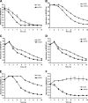
FIG. 1.
FIG. 1.
Serum susceptibility among B. spielmanii isolates. A growth inhibition assay was used to investigate the susceptibility to human serum of B. spielmanii isolates A14S (A), PC-Eq17 (B), PMai (C), PHap (D), PMew (E), and the serum-sensitive B. garinii isolate G1 (F). Spirochetes were incubated in either 50% NHS or 50% hiNHS over a cultivation period of 10 days at 33°C. Color changes were monitored by measurement of the absorbance at 562/630 nm. All experiments were performed three times, and each test was done five times with very similar results. For clarity, only data from representative experiments are shown. The error bars represent standard errors of the mean.
FIG. 2.
FIG. 2.
Deposition of complement components C3, C6, and C5b-9 on the surface of B. spielmanii. Complement components deposited on B. spielmanii isolates A14S, PC-Eq17, and PMew, as well as the serum-sensitive B. garinii isolate G1, were detected by indirect immunofluorescence microscopy. Spirochetes were incubated with either 25% NHS or hiNHS for 30 min at 37°C with gentle agitation, and bound C3, C6, and C5b-9 were analyzed with specific antibodies against each component and appropriate Alexa 488-conjugated secondary antibodies. For visualization of the spirochetes in a given microscopic field, the DNA-binding dye DAPI was used. The spirochetes were observed at a magnification of ×1,000. The data were recorded with a DS-5Mc charge-coupled device camera (Nikon) mounted on an Olympus CX40 fluorescence microscope. The images shown are representative of at least 20 microscope fields.
FIG. 3.
FIG. 3.
Binding of complement regulators factor H and FHL-1 by different B. spielmanii isolates. B. spielmanii isolates A14S, PC-Eq17, PMai, PHap, and PMew incubated in NHS-EDTA were extensively washed with 0.15 M NaCl, 0.03 M phosphate, 0.02% sodium azide, pH 7.2, containing 0.05% Tween 20, and bound proteins were eluted using 0.1 M glycine (pH 2.0). Both the last wash (w) and the eluate (e) fractions obtained from each strain were separated under nonreducing conditions in a 12.5% SDS-PAGE gel, transferred to nitrocellulose, and probed with either MAb VIG8 specific for SCR20 of factor H or MAb B22 for SCR5 of factor H and FHL-1.
FIG. 4.
FIG. 4.
Detection of factor H/FHL-1 on the surfaces of intact cells. Serum-resistant isolates A14S and PC-Eq17 and serum-sensitive B. garinii isolate G1 were incubated with NHS-EDTA. Bound proteins were detected by immunofluorescence microscopy after incubation with MAb B22 for factor H and FHL-1 (FH). For counterstaining, the DNA-binding dye DAPI was used to identify cells in a given microscopic field. The spirochetes were observed at a magnification of ×1,000. The data were recorded with a DS-5Mc charge-coupled device camera (Nikon) mounted on an Olympus CX40 fluorescence microscope. The images shown are representative of at least 20 microscope fields.
FIG. 5.
FIG. 5.
Analysis of functional activities of factor H and FHL-1 bound to B. spielmanii. The cofactor activities of factor H and FHL-1 bound to spirochetes were analyzed by measuring factor I-mediated conversion of C3b to iC3b. B. spielmanii isolates PC-Eq17, A14S, PMai, PHap, and PMew were incubated with either factor H (A) or purified FHL-1 (3 μg/ml each) (B) for 60 min at room temperature. For control purposes, the cells were incubated without factor H. After extensive washing with PBS, C3b (Calbiochem, Darmstadt, Germany; 10 μg/ml) and factor I (FI) (Calbiochem, Germany; 50 μg/ml) were added, and the mixture was incubated for 30 min at 37°C. Subsequently, the probes were boiled for 5 min, subjected to 12.5% SDS-PAGE, and transferred onto a nitrocellulose membrane. The various C3b degradation products were visualized by Western blotting using a polyclonal goat anti-human C3 antiserum (Calbiochem). As a positive control, purified factor H or FHL-1 (50 ng each) was added to the reaction mixture, and as a negative control, C3b and factor I were incubated in the absence of complement regulators.
FIG. 6.
FIG. 6.
Identification of factor H and FHL-1 binding proteins expressed within B. spielmanii isolates. Protein extracts (15 μg each) obtained from B. burgdorferi sensu stricto LW2, B. afzelii FEM1-D15, B. garinii G1, and B. spielmanii PC-Eq17, A14S, PMai, PHap, and PMew were separated by 10% Tris-Tricine SDS-PAGE and transferred to nitrocellulose. The membranes were incubated with either NHS as a source for factor H (A) or FHL-1 (B), and binding of the proteins was detected with MAb VIG8 specific for SCR20 of factor H or polyclonal serum specific for SCR1 to 4 of FHL-1. For detection of FlaB as a control, MAb L41 1C11 was applied. The identified CRASP proteins are indicated on the right, and the mobilities of the marker proteins are indicated on the left.
FIG. 7.
FIG. 7.
Protease treatment affects surface expression of native BsCRASP-1 and BsCRASP-2 and binding to factor H and FHL-1. (A) B. spielmanii A14S cells were incubated with the indicated concentrations of proteinase K or trypsin. After 2 h of incubation, the cells were lysed by sonication, and each protein lysate was subjected to 10% Tris-Tricine SDS-PAGE. BsCRASP-1 and BsCRASP-2 were identified using recombinant FHL-1 and polyclonal antibody αSCR1 to 4 (dilution 1/1,000) specific for the N terminus of FHL-1/factor H by ligand affinity analysis. (B) Flagellin (FlaB) was detected with MAb L41 1C11 (dilution 1/1,000) by Western blotting. (C) Part of a Coomassie-stained 10% Tris-Tricine SDS-polyacrylamide gel is shown to demonstrate the susceptibility of OspA and OspB to proteolytic degradation.

References

    1. Alitalo, A., T. Meri, L. Rämö, T. S. Jokiranta, T. Heikkilä, I. J. Seppälä, J. Oksi, M. Viljanen, and S. Meri. 2001. Complement evasion by Borrelia burgdorferi: serum-resistant strains promote C3b inactivation. Infect. Immun. 69:3685-3691. - PMC - PubMed
    1. Alitalo, A., T. Meri, P. Comstedt, L. Jeffery, J. Tornberg, T. Strandin, H. Lankinen, S. Bergström, M. Cinco, S. R. Vuppala, D. R. Akins, and S. Meri. 2005. Expression of complement factor H binding immunoevasion proteins in Borrelia garinii isolated from patients with neuroborreliosis. Eur. J. Immunol. 35:3043-3053. - PubMed
    1. Blackmore, T. K., V. A. Fischetti, T. A. Sadlon, H. M. Ward, and D. L. Gordon. 1998. M protein of the group A streptococcus binds to the seventh short consensus repeat of human complement factor H. Infect. Immun. 66:1427-1431. - PMC - PubMed
    1. Brade, V., I. Kleber, and G. Acker. 1992. Differences of two Borrelia burgdorferi strains in complement activation and serum resistance. Immunobiology 185:453-465. - PubMed
    1. Breitner-Ruddock, S., R. Würzner, J. Schulze, and V. Brade. 1997. Heterogeneity in the complement-dependent bacteriolysis within the species of Borrelia burgdorferi. Med. Microbiol. Immunol. 185:253-260. - PubMed

Publication types

MeSH terms

LinkOut - more resources