. 2007 Jul 16;178(2):297-308.
doi: 10.1083/jcb.200701078.

Type I gamma phosphatidylinositol phosphate kinase is required for EGF-stimulated directional cell migration

Affiliations

Type I gamma phosphatidylinositol phosphate kinase is required for EGF-stimulated directional cell migration

Yue Sun et al. J Cell Biol. .

Abstract

Phosphatidylinositol 4,5-bisphosphate (PI4,5P(2)) modulates a plethora of cytoskeletal interactions that control the dynamics of actin assembly and, ultimately, cell migration. We show that the type Igamma phosphatidylinositol phosphate kinase 661 (PIPKIgamma661), an enzyme that generates PI4,5P(2), is required for growth factor but not G protein-coupled receptor-stimulated directional migration. By generating PI4,5P(2) and regulating talin assembly, PIPKIgamma661 modulates nascent adhesion formation at the leading edge to facilitate cell migration. The epidermal growth factor (EGF) receptor directly phosphorylates PIPKIgamma661 at tyrosine 634, and this event is required for EGF-induced migration. This phosphorylation regulates the interaction between PIPKIgamma661 and phospholipase Cgamma1 (PLCgamma1, an enzyme previously shown to be involved in the regulation of EGF-stimulated migration). Our results suggest that phosphorylation events regulating specific PIPKIgamma661 interactions are required for growth factor-induced migration. These interactions in turn define the spatial and temporal generation of PI4,5P(2) and derived messengers required for directional migration.


Figures
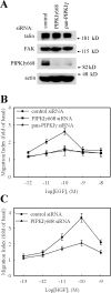
Figure 1.
Figure 1.
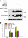
Knockdown of PIPKIγ attenuated EGF-stimulated cell migration. HeLa cells were transfected with control siRNA, pan-PIPKIγ siRNA, or PIPKIγ668-specific siRNA separately as indicated. (A) Expression of PIPKIγ668, talin, FAK, and actin were detected by their specific antibodies. (B) EGF (10−12 to 10−8 M) was used to stimulate cell migration. (C) HGF (10−13 to 10−9 M) was used to stimulate cell migration. Quantifications are means ± SEM of three separate experiments.
Figure 2.
Figure 2.
The specificity of PIPKIγ in regulating cell migration. HeLa cells were transfected with control siRNA or PIPKIγ668-specific siRNA separately as indicated, and LPA (A) or SDF1α (B) was used to stimulate cell migration. (C) HeLa cells were transfected with control siRNA or PIPKIγ668- or PIPKIα-specific siRNA separately as indicated, and EGF was used to stimulate cell migration. Expression of PIPKIγ668, PIPKIα, and actin was detected by their specific antibodies. Quantifications are expressed as means ± SEM of three separate experiments (*, P < 0.01 compared with control siRNA–transfected HeLa cells).
Figure 3.
Figure 3.
EGF-induced cell migration requires PIPKIγ661 kinase activity and its talin binding ability. Parental HeLa cells or HeLa tet-off stable cell lines expressing HA-tagged wild-type PIPKIγ661, PIPKIγ635, PIPKIγKD, PIPKIγY644F, or PIPKIγS645F were transfected with control siRNA or PIPKIγ668-specific siRNA separately as indicated. (A) The expression of PIPKIγ in these cell lines was detected by anti–pan-PIPKIγ antibody or anti-HA antibody separately. As the loading control, the amount of actin was detected by anti-actin antibody. (B) EGF was used to stimulate the migration of these cell lines. Quantifications are expressed as means ± SEM of three separate experiments (*, P < 0.01 compared with control siRNA–transfected HeLa cells).
Figure 4.
Figure 4.
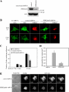
PIPKIγ is required for EGF-induced talin assembly to adhesions. (A) The expression of PIPKIγ668 in control shRNA– or PIPKIγ668-specific shRNA–transfected HeLa cells was detected. (B) Control shRNA or PIPKIγ668-specific shRNA–transfected HeLa cells were stimulated with 10−10 M EGF for 15 min. Cells were fixed and stained with anti-talin antibody. (C) EGF-induced talin assembly to adhesions was quantified. (D) Control shRNA or PIPKIγ668-specific shRNA were cotransfected with GFP-talin into HeLa cells. A micropipette filled with EGF was put near the cell to stimulate GFP-talin recruitment. GFP-talin assembly kinetics was quantified. Rate constants for assembly of individual adhesions were calculated as described in Materials and methods. Quantifications are expressed as means ± SEM (*, P < 0.01 compared with control siRNA–transfected HeLa cells). (E) GFP-talin assembly to adhesions at different time point of micropipette stimulation was shown. See Videos 1 and 2 (available at http://www.jcb.org/cgi/content/full/jcb.200701078/DC1). Bars, 10 μm.
Figure 5.
Figure 5.
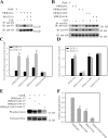
PIPKIγ661 is tyrosine phosphorylated by EGF stimulation. (A) HeLa cells were transfected with β-gal or HA-PIPKIγ661 separately, pretreated with or without EGFR-specific inhibitor PD153035, and stimulated with 10−9 M EGF for 5 min. Cells were used in immunoprecipitation with anti-HA antibody, and the tyrosine phosphorylation was detected by anti-phosphotyrosine antibody. (B) HA-tagged PIPKIγ661, PIPKIγY644F, PIPKIγY649F, or PIPKIγY634F were transfected into HeLa cells separately and stimulated with 10−9 M EGF for 5 min. Cells were subjected into immunoprecipitation with anti-HA antibody, and the tyrosine phosphorylation was detected. The amount of talin in the immunoprecipitation complex was detected by anti-talin antibody. (C) Tyrosine phosphorylation of HA-tagged PIPKIγ661, PIPKIγY644F, PIPKIγY649F, or PIPKIγY634F with or without EGF stimulation was quantified. (D) Talin association with HA-tagged PIPKIγ661, PIPKIγY644F, PIPKIγY649F, or PIPKIγY634F with or without EGF stimulation was quantified. (E) Reconstituted c-tail of wild-type PIPKIγ661, PIPKIγY644F, PIPKIγY634F, or PIPKIγY634F/Y644F was used as substrate of purified EGFR in the in vitro kinase assay. (F) The relative phosphorylation of wild-type PIPKIγ661, PIPKIγY644F, PIPKIγY634F, or PIPKIγY634F/Y644F was quantified. The phosphorylation of wild-type PIPKIγ661 was set as 100% (*, P < 0.01 compared with wild-type PIPKIγ661). Quantifications are expressed as means ± SEM of three separate experiments.
Figure 6.
Figure 6.
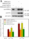
Expression of PIPKIγY634F could not rescue EGF-stimulated cell migration in PIPKIγ668 knockdown HeLa cells. Parental HeLa cells or HeLa tet-off stable cell lines expressing HA-tagged wild-type PIPKIγ661 or PIPKIγY634F were transfected with control or PIPKIγ668 siRNA separately as indicated. (A) The expression of PIPKIγ in these cell lines was detected by anti-PIPKIγ antibody or anti-HA antibody. As the loading control, the amount of actin was detected by anti-actin antibody. (B) EGF was used to stimulate the migration of these cell lines. Quantifications are expressed as means ± SEM of three separate experiments (*, P < 0.01 compared with control siRNA–transfected HeLa cells).
Figure 7.
Figure 7.
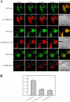
The effect of PIPKIγ661, PIPKIγY634F, PIPKIγKD, or PIPKIγS645F on EGF-induced talin assembly into adhesions. (A) HA-tagged PIPKIγ661, PIPKIγY634F, PIPKIγKD, or PIPKIγS645F stably expressing HeLa cells were stimulated with 10−10 M EGF for 15 min. Cells were fixed and stained with anti-HA and anti-talin antibodies. (B) EGF-induced talin assembly to adhesions in these cells was quantified. Quantifications are expressed as means ± SEM (*, P < 0.01 compared with wild-type PIPKIγ661). Bar, 10 μm.
Figure 8.
Figure 8.
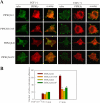
Expression of PIPKIγY634F decreased EGF-induced talin assembly to nascent adhesions at the leading edge. (A) GFP-talin was cotransfected with mc-PIPKIγ661, mc-PIPKIγY634F, or mc-PIPKIγKD into HeLa cells. A micropipette filled with EGF was put near the cell to stimulate talin recruitment. See Videos 3–8 (available at http://www.jcb.org/cgi/content/full/jcb.200701078/DC1). Bar, 10 μm. (B) GFP- talin assembly kinetics in mc-PIPKIγ661–, mc-PIPKIγY634F–, or mc-PIPKIγKD–expressing HeLa cells were quantified. Quantifications are expressed as means ± SEM (*, P < 0.01 compared with mc-PIPKIγ661–expressing HeLa cells).
Figure 9.
Figure 9.
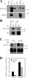
PLCγ differentially associates with PIPKIγ661 and PIPKIγY634F. (A) HeLa cells were put into immunoprecipitation assay with the antibodies indicated. The amount of endogenous PIPKIγ or PLCγ1 in the immunoprecipitation complex was detected by anti-PIPKIγ antibody or anti-PLCγ1 antibody separately. (B) HeLa cells were transfected with HA-PIPKIγ661 or HA-PIPKIγY634F. Cells were put into immunoprecipitation assay with anti-HA antibody, and the amount of PLCγ1 in the immunoprecipitation complex was detected. (C) HeLa cells transfected with HA-PIPKIγ661 or HA-PIPKIγY634F were stimulated with 10−9 M EGF for 5 min. Cells were subjected into immunoprecipitation with anti-HA antibody, and the amount of PLCγ1 in the immunoprecipitation complex was detected. (D) The effect of EGF stimulation on PLCγ1 association with wild-type PIPKIγ661 or PIPKIγY634F was quantified. Quantifications are expressed as means ± SEM of three separate experiments.
Figure 10.
Figure 10.
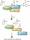
Model of how PIPKIγ661 is involved in EGF-induced migration. By associating with PIPKIγ661, PLCγ1 could hydrolyze the PI4,5P2 produced by PIPKIγ661; the hydrolysis would diminish the PI4,5P2 required to regulate talin assembly into adhesions. EGF-induced phosphorylation of PIPKIγ661 at Y634 causes a disassembly of the PLCγ1–PIPKIγ661 complex, and this could enhance PI4,5P2 accumulation and thus enhance talin assembly into adhesions. By modulating talin assembly, PIPKIγ661 regulates adhesion formation at the leading edge and facilitates EGF-induced migration.

Similar articles

Cited by

References

    1. Anderson, R.A., and V.T. Marchesi. 1985. Regulation of the association of membrane skeletal protein 4.1 with glycophorin by a polyphosphoinositide. Nature. 318:295–298. - PubMed
    1. Anderson, R.A., I.V. Boronenkov, S.D. Doughman, J. Kunz, and J.C. Loijens. 1999. Phosphatidylinositol phosphate kinases, a multifaceted family of signaling enzymes. J. Biol. Chem. 274:9907–9910. - PubMed
    1. Bairstow, S.F., K. Ling, X. Su, A.J. Firestone, C. Carbonara, and R.A. Anderson. 2006. Type Igamma661 phosphatidylinositol phosphate kinase directly interacts with AP2 and regulates endocytosis. J. Biol. Chem. 281:20632–20642. - PubMed
    1. Bershadsky, A.D., and A.H. Futerman. 1994. Disruption of the Golgi apparatus by brefeldin A blocks cell polarization and inhibits directed cell migration. Proc. Natl. Acad. Sci. USA. 91:5686–5689. - PMC - PubMed
    1. Borm, B., R.P. Requardt, V. Herzog, and G. Kirfel. 2005. Membrane ruffles in cell migration: indicators of inefficient lamellipodia adhesion and compartments of actin filament reorganization. Exp. Cell Res. 302:83–95. - PubMed

Publication types

MeSH terms