. 2007 Aug 6;204(8):1837-47.
doi: 10.1084/jem.20070406. Epub 2007 Jul 16.

IL-25 augments type 2 immune responses by enhancing the expansion and functions of TSLP-DC-activated Th2 memory cells

Affiliations

IL-25 augments type 2 immune responses by enhancing the expansion and functions of TSLP-DC-activated Th2 memory cells

Yui-Hsi Wang et al. J Exp Med. .

Abstract

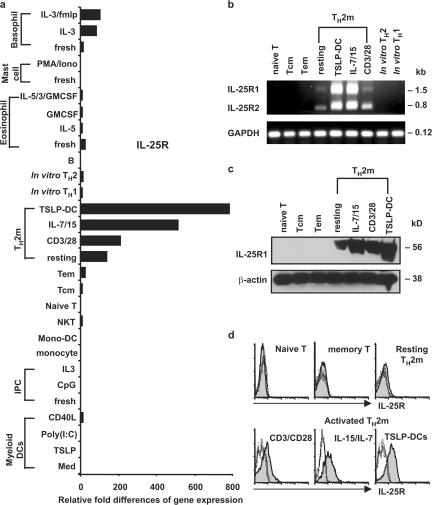
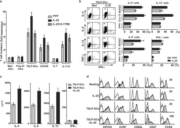
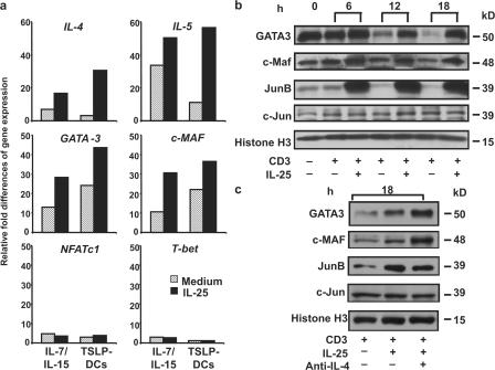
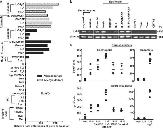
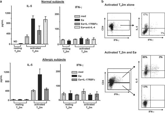
Interleukin (IL) 25 (IL-17E), a distinct member of the IL-17 cytokine family, plays important roles in evoking T helper type 2 (Th2) cell-mediated inflammation that features the infiltrations of eosinophils and Th2 memory cells. However, the cellular sources, target cells, and underlying mechanisms remain elusive in humans. We demonstrate that human Th2 memory cells expressing distinctive levels of IL-25 receptor (R) are one of the responding cell types. IL-25 promotes cell expansion and Th2 cytokine production when Th2 central memory cells are stimulated with thymic stromal lymphopoietin (TSLP)-activated dendritic cells (DCs), homeostatic cytokines, or T cell receptor for antigen triggering. The enhanced functions of Th2 memory cells induced by IL-25 are associated with sustained expression of GATA-3, c-MAF, and JunB in an IL-4-independent manner. Although keratinocytes, mast cells, eosinophils, and basophils express IL-25 transcripts, activated eosinophils and basophils from normal and atopic subjects were found to secrete bioactive IL-25 protein, which augments the functions of Th2 memory cells. Elevated expression of IL-25 and IL-25R transcripts was observed in asthmatic lung tissues and atopic dermatitis skin lesions, linking their possible roles with exacerbated allergic disorders. Our results provide a plausible explanation that IL-25 produced by innate effector eosinophils and basophils may augment the allergic inflammation by enhancing the maintenance and functions of adaptive Th2 memory cells.


Figures

Figure 1.
Figure 1.
Distinctive expression of IL-25R by human Th2 memory cells. (a) Expression of IL-25R was measured by quantitative PCR on a panel of human cDNA templates made from various cell types, as described in Materials and methods. Different T cell subsets were isolated as previously described (reference 20). (b) Expression of IL-25R isoforms by activated Th2 memory cells was demonstrated by RT-PCR using primers, as described in Materials and methods. Sorted resting T cell subsets, or activated Th2 memory cells stimulated by autologous TSLP-DCs, the homeostatic cytokine IL-7/15, and anti-CD3/CD28 for 7 d were isolated for cell lysate preparation (c) or flow cytometry analysis (d). Quantified cell lysates were subjected to Western blot analysis using biotinylated goat anti–human IL-17RB polyclonal antibody (c). Surface expression of IL-25R of activated Th2 memory cells or resting T cell subsets were examined using flow cytometry, as described in Materials and methods (d). Shaded histograms represent IL-25 R staining; open histograms represent the isotype control. Relative fold differences in IL-25R gene expression between samples listed in panel a are indicated on the x axis. Data are from one of three independent experiments.
Figure 2.
Figure 2.
IL-25 enhances the proliferation and Th2 polarization of stimulated Th2 memory cells. Sorted Th2 memory T cells were cultured with the cytokines IL-7 only, IL-15 plus IL-7, anti-CD3/CD28, or autologous CD11c+ DCs activated by various stimuli (x axis) at a DC/T cell ratio of 1:2 for 7 d in the presence or absence of IL-25. The neutralizing recombinant protein IL-17RB-Fc was used in culture. Their proliferative responses were compared, and graphic bars represent the times of expansion by dividing the final cell number by the initial T cell number in six individual experiments (a). To characterize the effect of IL-25 on Th2 memory cells, sorted cells were cultured in medium only or expanded by IL-15 plus IL-7, anti-CD3/CD28, or TSLP-DCs for 7 d in the presence or absence of IL-25. The proliferated cells were collected and restimulated with PMA plus ionomycin for analysis of intracellular cytokine production (b), or Th2 memory cells expanded by TSLP-DCs in the presence or absence of IL-25 for 7 d were collected and restimulated with anti-CD3/CD28 for 24 h before the measurements of cytokines in the culture supernatants by ELISA (c). For phenotypic analyses, sorted Th2 memory cells cultured in IL-25 alone, or expanded by TSLP-DCs, or TSLP-DCs plus IL-25 were examined using flow cytometry (d). Shaded histograms represent staining of Th2 memory T cells with the markers indicated below histograms; open histograms represent the isotype control. Numbers within the quadrants indicate the percentage of expanded cells that stained positive for each respective cytokine. The results in panels a, b, c, and d are for separate experiments. Data represent the mean ± SD of five experiments.
Figure 3.
Figure 3.
IL-25 induces elevated cytokine production by TSLP-DC–activated Th2 memory cells. Freshly isolated resting or activated Th2 memory cells expanded by TSLP-DCs for 7 d were collected, washed, and stimulated directly with IL-25 or cultured in medium only for 24 h in the presence or absence of neutralizing soluble receptor IL-17RB-FC or anti-IL-4 mAbs. Cultured supernatants were collected for ELISA analyses. Data represent the mean ± SD of five experiments.
Figure 4.
Figure 4.
IL-25 up-regulates the expression of GATA3, c-MAF, and JunB. Sorted Th2 memory T cells were cultured with the cytokine IL-15 plus IL-7 or TSLP-DCs for 7 d in the presence or absence of IL-25. Expression levels of the indicated genes that are involved in Th2 cell differentiation were measured by real-time PCR, as described in Materials and methods. (a) Fold differences in the changes of expression of each gene between samples are marked on the y axes. Th2 memory cells expanded by TSLP-DCs for 7 d were collected (0 h; b) and restimulated with anti-CD3 mAb for different time courses (b) or 18 h (c) in the presence or absence of IL-25 (b and c) or anti-IL-4 mAbs (c) before collection and were subjected to Western blot analysis. Data are from one of three independent experiments.
Figure 5.
Figure 5.
Activated eosinophils and basophils are the producers of IL-25. Expression of IL-25 transcript was measured by quantitative PCR on a panel of human cDNA libraries made from various cell types (a) or examined by RT-PCR analysis (b), as described in Materials and methods. Purified eosinophils and basophils obtained from normal or atopic subjects were cultured for 3 d with the indicated cytokines or stimuli before the RNA isolation for gene expression analysis (a) or harvesting of supernatants for the measurement of IL-25 using ELISA analyses (b). Relative fold differences in IL-25 gene expression between samples listed in panel a are indicated on the x axis. Data in panel a are from one of three independent experiments. Data in panel c are from five normal or four atopic subjects, respectively, and represent the mean ± SD of tested samples (c). White lines indicate that intervening lanes have been spliced out.
Figure 6.
Figure 6.
Eosinophil-secreted IL-25 promotes cytokine production by TSLP-DC–activated Th2 memory cells. Resting or TSLP-DC–activated Th2 memory cells purified from normal (a and b) or atopic subjects (a) were cultured in medium only or co-cultured with autologous eosinophils for 3 d in the presence or absence of anti–IL-4 Abs or IL-17RB-Fc recombinant proteins (a). Co-cultured supernatants were harvested for the measurement of Th2 cytokine production using ELISA analyses (a). Activated Th2 memory cells cultured in medium alone or co-cultured with eosinophils for 3 d were collected and restimulated with PMA plus ionomycin before being stained with CD4 and the indicated intracellular cytokines. Numbers within the quadrants indicate the percentage of cells that stained positive for each respective cytokine. Data are from five normal or four atopic subjects and represent the mean ± SD of tested samples.
Figure 7.
Figure 7.
Expression of IL-25 and IL-25R transcripts by asthmatic and atopic subjects. Expression of IL-25 and IL-25R transcripts were measured by quantitative PCR on cDNA templates obtained from a bronchial biopsy of asthmatic or normal subjects, and a skin biopsy, including psoriasis vulgaris, cutaneous lupus erythematosus, prurigo nodularis, atopic dermatitis, and normal subjects. Relative fold differences in gene expression between samples are indicated on the x axis.

Similar articles

Cited by

References

    1. Cohn, L., J.A. Elias, and G.L. Chupp. 2004. Asthma: mechanisms of disease persistence and progression. Annu. Rev. Immunol. 22:789–815. - PubMed
    1. Wills-Karp, M. 1999. Immunologic basis of antigen-induced airway hyper responsiveness. Annu. Rev. Immunol. 17:255–281. - PubMed
    1. Gleich, G.J., C.R. Adolphson, and K.M. Leiferman. 1993. The biology of the eosinophilic leukocyte. Annu. Rev. Med. 44:85–101. - PubMed
    1. Rothenberg, M.E. 1998. Eosinophilia. N. Engl. J. Med. 338:1592–1600. - PubMed
    1. Pan, G., D. French, W. Mao, M. Maruoka, P. Risser, J. Lee, J. Foster, S. Aggarwal, K. Nicholes, S. Guillet, et al. 2001. Forced expression of murine IL-17E induces growth retardation, jaundice, a Th2-biased response, and multiorgan inflammation in mice. J. Immunol. 167:6559–6567. - PubMed

Publication types