. 2007 Aug 1;120(Pt 15):2683-93.
doi: 10.1242/jcs.006346. Epub 2007 Jul 17.

The neuronal Arf GAP centaurin alpha1 modulates dendritic differentiation

Affiliations

The neuronal Arf GAP centaurin alpha1 modulates dendritic differentiation

Carlene D Moore et al. J Cell Sci. .

Abstract

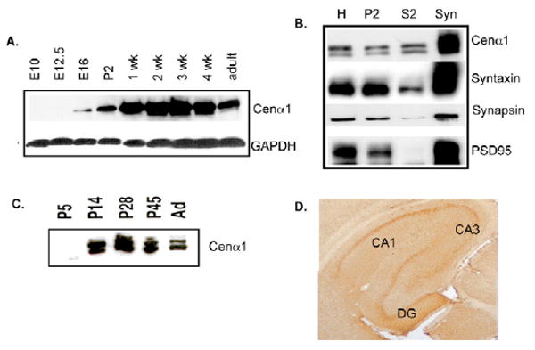
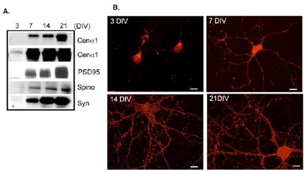
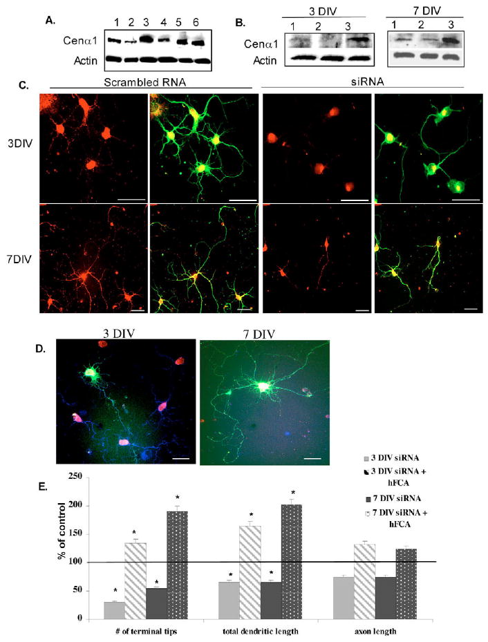
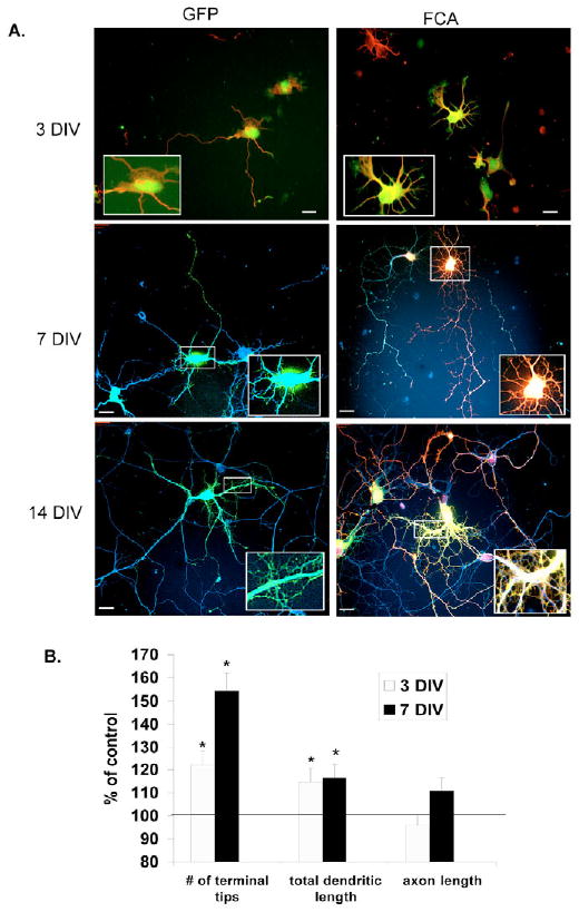
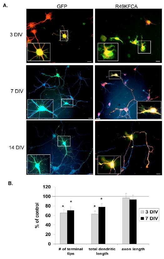
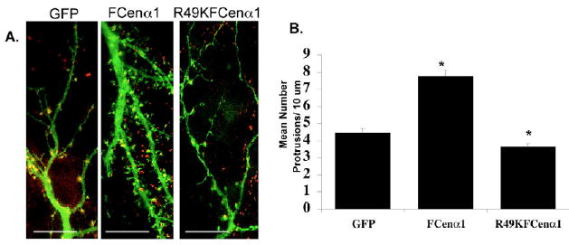
Centaurin alpha1 is an Arf GTPase-activating protein (GAP) that is highly expressed in the nervous system. In the current study, we show that endogenous centaurin alpha1 protein is localized in the synaptosome fraction, with peak expression in early postnatal development. In cultured dissociated hippocampal neurons, centaurin alpha1 localizes to dendrites, dendritic spines and the postsynaptic region. siRNA-mediated knockdown of centaurin alpha1 levels or overexpression of a GAP-inactive mutant of centaurin alpha1 leads to inhibition of dendritic branching, dendritic filopodia and spine-like protrusions in dissociated hippocampal neurons. Overexpression of wild-type centaurin alpha1 in cultured hippocampal neurons in early development enhances dendritic branching, and increases dendritic filopodia and lamellipodia. Both filopodia and lamellipodia have been implicated in dendritic branching and spine formation. Following synaptogenesis in cultured neurons, wild-type centaurin alpha1 expression increases dendritic filopodia and spine-like protrusions. Expression of a GAP-inactive mutant diminishes spine density in CA1 pyramidal neurons within cultured organotypic hippocampal slice cultures. These data support the conclusion that centaurin alpha1 functions through GAP-dependent Arf regulation of dendritic branching and spines that underlie normal dendritic differentiation and development.


Figures

Fig. 1
Fig. 1
Developmental expression and localization of endogenous centaurin α1 in the rodent brain. (A) Immunoblot analysis of centaurin α1 protein from lysates (25 μg total lysate per lane) from whole brain at different developmental stages from E10 to P42 (=adult). (B) Immunoblot analysis of centaurin α1 and synaptic markers in fractionated P23 whole rat brain. H, whole brain homogenate; P2, crude microsomes and synaptosomes; S2, supernatant; Syn, purified synaptosomes; 20 μg of each fraction was loaded per lane. (C) Immunoblot analysis of centaurin α1 in the synaptosome fraction prepared from the developing brain. 10 μg of each fraction was loaded per lane. (D) Immunohistochemistry with anti-centaurin α1 antibody in hippocampus from P28 rat. DG, dentate gyrus.
Fig. 2
Fig. 2
Expression of endogenous centaurin α1 in primary cultured dissociated hippocampal neurons. (A) Immunoblot showing developmental expression of centaurin α1 and the specific synaptic proteins PDS95, spinophilin (Spino) and synaptotagmin (Syn) in primary cultured neurons at 3, 7, 14 and 21 DIV. 20 μg of total lysate was loaded per lane. (B) Indirect immunofluorescence shows endogenous centaurin α1 localization (red) and levels at 3, 7, 14 and 21 DIV. Bars, 10 μm.
Fig. 3
Fig. 3
Localization of endogenous centaurin α1 in primary cultured dissociated hippocampal neurons. Indirect immunofluorescence was used to visualize endogenous centaurin α1 localization (red) compared with microtubule associated protein 2 (MAP-2), tau, synaptophysin (syn), PSD-95 and spinophilin (Spin) in 14 DIV neurons. Arrows indicate areas of juxtaposition; arrowheads indicate areas of colocalization. Bars, 10 μm.
Fig. 4
Fig. 4
Effects of knockdown of centaurin α1 protein by siRNA in dissociated cultured hippocampal neurons. Hippocampal neurons were transfected after dissociation (0 DIV) and cultured for 3, 7 and 14 DIV. (A,B) Immunoblot analysis of endogenous centaurin α1 in hippocampal cultures. Total cell lysates (12.5 μg lysate per lane) were probed with anti-centaurin α1 and an anti-actin antibodies, which were used to assess actin levels for calculation of the knockdown percentage. (A) Lane 1, scrambled (Scr) siRNA 3 DIV; lane 2, rat centaurin α1 (rCena1) siRNA 3 DIV; lane 3, Scr siRNA 7 DIV; lane 4, rCena1 siRNA 7 DIV; lane 5, Scr siRNA 14 DIV; lane 6, rCena1 siRNA 14 DIV. (B) Lane 1, Scr siRNA; lane 2, rCena1 siRNA; lane 3, rCena1 siRNA plus human Flag-centaurin α1. (C) Indirect immunofluorescence showing endogenous centaurin α1 (red) compared with β-tubulin (green) to visualize neuronal morphology at 3 and 7 DIV. Double-stranded scrambled RNA control is shown in left-hand panels and double-stranded siRNA to rat centaurin α1 in the right-hand panels. Bars, 20 μm. (D) Rescue by expression of human centaurin α1. Neurons were transfected with rat centaurin α1 siRNA, GFP and human centaurin α1. Indirect immunofluorescence showing endogenous plus heterologous centaurin α1-(red), β-tubulin (blue) to visualize neuronal morphology and GFP (green) to label neurons co-expressing human Flag centaurin α1 at 3 and 7 DIV. Bars, 20 μm. (E) Quantification of the effects of siRNA knockdown of centaurin α1 at 3 and 7 DIV and rescue with human centaurin α1 compared with scrambled siRNA control. The data were quantified from three independent experiments (n=30 at 3 DIV, n=25 at 7 DIV cells for each condition). Data represent mean ± s.e. *P<0.05.
Fig. 5
Fig. 5
Effects of overexpression of wild-type centaurin α1 in developing dissociated hippocampal neurons. Hippocampal neurons were transfected by Nucleofection after dissociation (0 DIV) and cultured for 3, 7 and 14 DIV. (A) Fluorescence (GFP, green) and indirect immunofluorescence (β-tubulin; red, 3 DIV; blue, 7 and 14 DIV; Flag-tagged centuarin α1, red) were used to visualize transfected neurons and neuronal morphology. GFP control (left panels). GFP plus Flag-centaurin α1 (right panels). Insets show higher magnification. Bars, 20 μm. (B) Quantification of the effects of expression of FLAG-centaurin α1 at 3 and 7 DIV compared with GFP control. The data was quantified from three independent experiments (n=50 cells from 3 DIV and n=30 cells from 7 DIV, respectively, for each condition). Data represent mean ± s.e. *P<0.05.
Fig. 6
Fig. 6
Effects of overexpression of a GAP-inactive mutant centaurin α1 on dendritic differentiation. Hippocampal neurons were transfected after dissociation (0 DIV) and cultured for 3-14 DIV. (A) Fluorescence (GFP, green) and indirect immunofluorescence (β-tubulin, red) were used to visualize transfected neurons and neuronal morphology. Expression of GFP control (left panels) compared with GFP plus Flag-R49K centaurin α1 (right panel). Boxed areas show a higher magnification. Bars, 20 um. (B) Quantification of the effects of overexpression of Flag-R49Kcentaurin α1 for 3 and 7 DIV compared with GFP control. The data was quantified from three independent experiments (n=50 cells at 3 DIV and 30 cells at 7 DIV). Data represent mean ± s.e. *P<0.05.
Fig. 7
Fig. 7
Effects of overexpression of wild-type and GAP-inactive centaurin α1 on dendritic protrusions in dissociated cultured hippocampal neurons. Dissociated hippocampal neurons were transfected at 1 DIV and cultured for 13 DIV. (A) Fluorescence (GFP, green) and indirect immunofluorescence (spinophilin or neurabin, red) were used to visualize transfected neurons and morphological effects. Expression of GFP control (left panels) compared with GFP plus Flag-centaurin α1 (middle panel) and GFP plus Flag-R49Kcentaurin α1 (right panel). Bars, 10 μm. (B) Quantification of the number of dendritic protrusions per 10 μm dendrite length in neurons expressing GFP, GFP plus Flag-centaurin α1 and GFP plus Flag-R49K centaurin α1. Data represent mean ± s.e. *P<0.05.
Fig. 8
Fig. 8
Centaurin α1 localizes to dendritic spines and regulates dendritic spine density in CA1 neurons in organotypic hippocampal slice cultures. (A) Representative dendritic segments from CA1 pyramidal cells in organotypic hippocampal slice cultures transfected with eYFP and Flag-centaurin α1. Centaurin α1 was visible in the dendritic shaft and spines (arrows). Bar, 25 μm. (B) Representative dendritic segments of CA1 pyramidal neurons from organotypic hippocampal slice cultures transfected with eYFP and either dsRed, Flag-centaurin α1 or Flag-R49K centaurin α1. Bars, 2 μm. (C) Quantification of spine density in each condition. Data represent mean ± s.e. *P<0.00016.

Similar articles

Cited by

References

    1. Aggensteiner M, Reiser G. Expression of the brain-specific membrane adapter protein p42IP4/centaurin a, a Ins(1,3,4,5)P4/PtdIns(3,4,5)P3 binding protein, in developing rat brain. Brain Res Dev Brain Res. 2003;142:77–87. - PubMed
    1. Albertinazzi C, Za L, Paris S, de Curtis I. ADP-ribosylation factor 6 and a functional PIX/p95-APP1 complex are required for Rac1B-mediated neurite outgrowth. Mol Biol Cell. 2003;14:1295–1307. - PMC - PubMed
    1. Allen P, Ouimet C, Greengard P. Spinophilin, a novel protein phosphatase 1 binding protein localized to dendritic spines. Proc Natl Acad Sci USA. 1997;94:9956–9961. - PMC - PubMed
    1. Alonso M, Medina J, Pozzo-Miller L. ERK1/2 activation is necessary for BDNF to increase dendritic spine density in hippocampal CA1 pyramidal neurons. Learn Mem. 2004;11:172–178. - PMC - PubMed
    1. Binder L, Frankfurter A, Rebhun L. The distribution of tau in the mammalian central nervous system. J Cell Biol. 1985;101:1371–1378. - PMC - PubMed

Publication types

MeSH terms