. 2007 Aug 1;120(Pt 15):2594-608.
doi: 10.1242/jcs.03472. Epub 2007 Jul 17.

An in vivo model of apoptosis: linking cell behaviours and caspase substrates in embryos lacking DIAP1

Affiliations

An in vivo model of apoptosis: linking cell behaviours and caspase substrates in embryos lacking DIAP1

Dhianjali Chandraratna et al. J Cell Sci. .

Abstract

The apoptotic phenotype is characterised by dynamic changes in cell behaviours such as cell rounding and blebbing, followed by chromatin condensation and cell fragmentation. Whereas the biochemical pathways leading to caspase activation have been actively studied, much less is known about how caspase activity changes cell behaviours during apoptosis. Here, we address this question using early Drosophila melanogaster embryos lacking DIAP1. Reflecting its central role in the inhibition of apoptosis, loss of DIAP1 causes massive caspase activation. We generated DIAP1-depleted embryos by either using homozygous null mutants for thread, the gene coding DIAP1, or by ectopically expressing in early embryos the RGH protein Reaper, which inhibits DIAP1. We show that (1) all cells in embryos lacking DIAP1 follow synchronously the stereotypic temporal sequence of behaviours described for apoptotic mammalian cells and (2) these cell behaviours specifically require caspase activity and are not merely a consequence of cellular stress. Next, we analyse the dynamic changes in the localisation of actomyosin, Discs large, Bazooka and DE-cadherin in the course of apoptosis. We show that early changes in Bazooka and Discs large correlate with early processing of these proteins by caspases. DE-cadherin and Myosin light chain do not appear to be cleaved, but their altered localisation can be explained by cleavage of known regulators. This illustrates how embryos lacking DIAP1 can be used to characterise apoptotic changes in the context of an embryo, thus providing an unprecedented in vivo model in which thousands of cells initiate apoptosis simultaneously.


Figures

Fig. 1
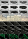
Fig. 1. Cells round up and disperse swiftly in th5 mutant embryos.
(A-C) Stage 10 embryos stained for Engrailed. In wild-type (A), the germ-band is fully extended and the Engrailed-expressing cells are arranged in segmental stripes in the ectoderm. In hypomorphic th4 mutants (B), germ-band extension fails halfway, and the Engrailed-expressing cells in the dorsal-most part of the ectoderm are found dispersed. In th5-null mutants (C), germ-band extension fails completely, and all Engrailed-expressing cells are dispersed. The pale regions correspond to fragments of the yolk cell emerging at the surface of the embryo. (D-I) Stage 8 embryos showing distribution of Even-skipped (Eve), Twist (Twi) and Forkhead (Fkh) to mark the ectoderm, mesoderm and endoderm, respectively. In wild-type embryos (D), Eve-expressing cells are arranged into pair-rule stripes. These cells are found completely dispersed in th5 embryos (E). (F) Ventral view showing Twist-expressing cells at the ventral midline and underneath, where the mesoderm has invaginated. In th5 embryos (G), the mesoderm invagination collapses and the Twist-expressing cells reappear at the surface of the embryo. Fkh-expressing cells mark the anterior (arrow) and posterior midgut rudiment in wild type. Note that at stage 8, the posterior Fkh domain has already invaginated (arrowhead) (H). In th5 embryos, the Fkh-expressing cells reappear at the surface of the embryo (I). (J-M) Kinetics of cell dispersal in live embryos, observed either by labelling nuclei using HistoneYFP (J,K, see supplementary material Movie 1), or in unlabelled embryos filmed under Nomarski optics (L,M, see supplementary material Movie 2). Times are indicated from the end of cellularisation (abbreviated as C). In both movies, no defects are detected until the beginning of germ-band extension, at which point the morphogenetic folds of the embryos start to regress (C+21-minute frame in K and C+18-minute frame in M), then the embryos contract abruptly and the cells round up and disperse in a few minutes (C+26-minute frame in K and C+25-minute frame in M). Note that in K, the nuclei within the field of view at C+26 minutes are entirely different from those within the field of view at C+21 minutes, showing that the nuclei have dispersed extensively. In J and K, anterior, posterior, ventral and dorsal sides are labelled, and the arrow indicates the posterior transverse furrow. In L and M, the arrow marks the cephalic furrow, and the arrowhead indicates the posterior midgut invagination.
Fig. 2
Fig. 2. Progression of the apoptotic phenotype in th5 embryos.
(A-D) Wild-type or homozygous th5 embryos were hand-selected at the end of cellularisation and fixed after ageing for another 30 minutes (T1), 1 hour (T2) or 1.5 hours (T3). Fixed embryos are stained for either Engrailed (A), activated Drice (B,C) or TUNEL (D), and photographed using Nomarski optics. (A) T1 shows a th5 embryo that is still morphologically normal: it has initiated germ-band extension and exhibits regular Engrailed stripes. T2 shows a th5 embryo that has completed cell dispersal: the cells have lost their columnar shape and adopted a rounded shape, and as a consequence these appear larger (compare close-ups for T1 and T2). The Engrailed cells have scattered and the yolk cell has fragmented and emerged at the surface of the embryo. The embryo in T3 shows a striking change in cell and nuclear shapes (the latter revealed by the nuclear localisation of the Engrailed protein). The nuclei have condensed and the cell bodies are smaller and more numerous, indicating that the cells have fragmented (compare close-ups for T2 and T3). (B) No staining for the activated form of Drice is detected in wild-type embryos before stage 10 (T3) at which two nuclei are found positive in the amnioserosa (arrow). This is consistent with the fact that there is no apoptosis in early wild-type embryos and that the amnioserosa is the first tissue in which apoptosis is detected at stage 10-11 (Abrams et al., 1993). Similarly, no staining was detected in early embryos stained for TUNEL (not shown). (C) T1: just prior to cell dispersal, discrete cells already show detectable levels of activated Drice. Activated Drice staining patterns appear random, except in the head, where an anterior spot is always detected. T2: once cell dispersal is complete, activated Drice is detected in every cell (unstained areas are yolk fragments). T3: the nuclei have condensed (compare close-ups for T2 and T3). (D) TUNEL staining is not detected in any th5 homozygous embryos prior to cell dispersal (T1). However, once cell dispersal is completed (T2), all nuclei are positive for TUNEL. In T3, nuclei condensation is clearly seen (compare close-ups for T2 and T3). (E) Stills from a time-lapse movie (supplementary material Movie 3) of a th5 homozygous embryo in which cell membranes have been labelled with SrcGFP and imaged by confocal microscopy. Times are indicated from the end of cellularisation. The arrow in the first frame indicates the cephalic furrow. In the following frames, arrowheads label examples of the blebs observed in this movie. The embryo appears normal at first (C+10 minutes), then the cephalic furrow starts to regress, signalling the onset of cell shape changes. The embryo contracts abruptly, the cells undergo extreme cell shape changes and small membranous blebs start to form (C+25 minutes). The cells reach a more regular, rounded shape and the blebs form dynamically at the cell surface (C+40 minutes). Later, cells start fragmenting and blebbing decreases in intensity (C+55 minutes). Towards the end of the movie (C+65 minutes), most cells are fragmenting, and blebbing is detectable in only a fraction of the cell bodies. The timing of cell fragmentation in this movie matches the timing of cell fragmentation in the fixed samples (T3). (F) Summary of the sequence of apoptotic phenotypes in th5 embryos. Note that two hours after the end of cellularisation (T4), Engrailed expression is not detectable anymore in fixed th5 embryos (data not shown).
Fig. 3
Fig. 3. Embryos overexpressing Reaper and Ricin exhibit distinct cell shape and cytoskeletal changes.
(A-C) Frames from time-lapse movies taken under Nomarski optics showing a wild-type embryo (A), an embryo overexpressing Reaper (B, see supplementary material Movie 4), and an embryo overexpressing Ricin (C, see supplementary material Movie 5). Times are indicated from the end of cellularisation. In reaperOVER embryos, the cells round up shortly after the end of cellularisation, precluding any morphogenetic movements (C+10 minutes). When all cells have rounded up, the cells are found in multiple layers at the surface of the embryo (C+40 minutes). In ricinOVER embryos, the embryo development is also arrested shortly after the end of cellularisation, before the start of morphogenetic movements. But in contrast to reaperOVER embryos, cells in ricinOVER embryos still appear attached basally to the yolk cell and do not round up. (D-G) Close-up of fixed embryos taken under Nomarski optics. In wild-type embryos, the epithelium is made up of a single layer of columnar cells arranged on top of the yolk cell (y). In reaperOVER embryos, cells round up abruptly at the end of cellularisation (arrowhead), losing both lateral and basal contacts (E) and rearrange to form multiple layers at the surface of the embryo (F). In ricinOVER embryos, development also arrests at the end of cellularisation, but the cells stay columnar in shape and in a single layer (G). (H-J) Wild-type, reaperOVER (supplementary material Movie 6) and ricinOVER (supplementary material Movie 7) embryos labelled with DEcadGFP to visualise the adherens junctions. In wild-type, DEcadGFP is enriched in an apical and basal junction towards the end of cellularisation (arrowheads in C–10-minute frame). Later on, the basal staining disappears and most of the signal is concentrated in the apical junction (C+10 minutes). As germ-band extension proceeds, the cells become less columnar but DEcadGFP is still clearly localised in an apical junction. In reaperOVER embryos, DEcadGFP localisation is normal up to the beginning of germ-band extension (frames C–10 minutes and C+10 minutes). When the embryo starts contracting, DEcadGFP is still localised at the cell apices (C+20 minutes) and then becomes delocalised in dots once cells have rounded up (C+35 minutes). By contrast, DEcadGFP is not delocalised in arrested ricinOVER embryos and remains associated with the apical cortex (Frame C+35 minutes in J). Apical (K-M) and sagital (N-P) views of wild-type, reaperOVER and ricinOVER embryos labelled with phalloidin. Actin is dramatically delocalised in reaperOVER embryos (L,O), and concentrates in a dense spot on one side of the cell. By contrast, actin is still cortical in ricinOVER embryos (M,P). Moreover, the basal actin at the cellularisation front is still present (P). Note that in the ricinOVER embryo shown in P, the actin rings at the basal side of the cell have closed, whereas actin rings in WT embryos are still open at late cellularisation (N). This suggests that in arrested ricinOVER embryos, actin ring closure proceeds in the absence of translation.
Fig. 4
Fig. 4. Behaviour of the actomyosin cytoskeleton in th5 embryos.
(A-D) Actin-phalloidin staining of WT and th5 mutant embryos. (A) In WT embryos, actin is found at the apical cell cortex, as well as in small discrete dots. In th5 embryos undergoing cell rounding up (B), a subset of the cells show an increased staining with a fibrous appearance. Shortly after cells have rounded up, some cells (right-side of C) maintain a wild-type-looking cortical staining associated with the presence of small dots. The only difference is the change from a columnar cell shape to a rounded cell shape. In the same embryo (left-side of C), some cells show a dramatic delocalisation of actin, with a very faint cortical staining, and dense accumulation of actin in one dense spot at the cortex. In older embryos (D), all cells have delocalised their actin. (E) Embryos lacking DIAP1 were collected 1 hour after cellularisation and extracts were analysed by western blotting using an antibody against the phosphorylated form of Myosin light chain (three independent extracts of 40 embryos each were loaded for each genotype). The blot was reprobed with an antibody against tubulin as a loading control (boxed bands). Band intensities were quantified and the ratio of phospho-MLC to tubulin is indicated in the graph. Shaded boxes show average values across the three samples for each genotype, whereas dotted boxes show the minimum and maximum values found for each genotype. (F,G) Stills from time-lapse supplementary material Movies 8 and 9 of WT and th5 mutant embryos labelled with SqhGFP. (F) Wild-type embryos undergoing germ-band extension exhibit a planar polarisation of SqhGFP, with cables running parallel to the dorsoventral (D/V) axis (C+20-50 minutes). In th5 embryos (G), SqhGFP planar polarisation is initially normal (C+20 minutes), and then an enrichment of Myosin II is seen in the cables compared with wild-type embryos (C+33 minutes). This enrichment culminates when the embryo contracts, and large regions of the embryo show a strong accumulation of Myosin II in cables as well as in fibrous aggregates at the cell cortices (C+38 minutes). When cells have rounded up (C+50 minutes), Myosin II is found in small dots that are likely to correspond to the membraneous blebs observed in the srcGFP movie (supplementary material Movie 3). Larger dots are also seen, which are likely to be remnants of the basal actomyosin II rings present in wild-type embryos between ectodermal cells and the yolk cell.
Fig. 5
Fig. 5. Epithelial depolarisation in th5 embryos.
(A-D) Staining for Baz in WT and th5 mutant embryos. (A) In WT, Baz localises at the cell apex and is enriched at the dorsoventral membranes of intercalating cells in the embryo trunk (head is in the top-left corner). In th5 embryos immediately after cell rounding (B), Baz ‘fibres’ are still intact, although their relative position at the cell cortex is altered, presumably because cells are changing from a elongated columnar shape to a rounded shape. (C) Close-up of th5 embryo undergoing cell dispersal. In the trunk region (right-part of the panel), Baz staining has now lost its fibrous appearance and is found at the cortex of the round cells, with a slight enrichment on one side, presumably reflecting the previous position of the apical domain. In this embryo, the head region (left-part of the panel) still exhibits epithelial characteristics, with Baz localised at the cell apices. (D) th5 embryo at the cell fragmentation stage. Baz staining is completely delocalised and is found in the cytoplasm of fragmenting cells as well as in smaller nuclear cell fragments. (E,F) Stills from time-lapse movies of WT and th5 mutant embryos labelled with DEcadGFP. No initial difference in DEcadGFP localisation can be detected between WT (E) and th5 embryos (F) after cephalic furrow formation (frames C+0-10 minutes). Then the apposing cells in the cephalic furrow become misaligned in th5 embryos compared with WT (C+16 minutes). Whereas germ-band extension proceeds in WT (C+16-64 minutes), the th5 embryo contracts, DEcadGFP appears to move apically (arrowhead in C+24-minute frame), the cell apices contract (arrowhead in C+48-minute frame) and then DEcadGFP is delocalised in dots (C+64 minutes). (G-I) Dlg staining of WT and th5 mutant embryos. (G) In WT, Dlg labels the lateral membrane. In th5 embryos undergoing cell dispersal (H), the cells that have already rounded up have lost all Dlg localisation, whereas Dlg staining is normal in the cells that still retain epithelial characteristics. In older th5 mutant embryos (I), Dlg localisation is lost in all cells. (J) Schema showing the localisation of Baz (apical membrane), DE-cadherin (adherens junctions) and Dlg (lateral membrane) in the early epidermis of Drosophila (see also Tepass et al., 2001). Note that in the early embryos looked at in this study the cells are either still connected to the yolk cell basally, by a cytoplasmic bridge or have just separated from the yolk cell through a cytokinesis-like process.
Fig. 6
Fig. 6. Processing of Dlg and Baz in extracts of embryos lacking DIAP1.
Embryos lacking DIAP1 were collected either 1, 2 or 3 hours after cellularisation, and extracts were analysed on western blots using antibodies against Dlg (A,B), Baz (C) and DE-cadherin (D). Each blot was reprobed with an antibody against tubulin as a loading control (boxed bands). (A) Two isoforms are detected for Dlg in the WT. In th5 homozygous embryos, the amount of these isoforms markedly decreased and three shorter bands are detected (marked with asterisks), which are likely to be products of caspase cleavage of Dlg. Two of these products are still detectable in the th5 embryo extract 3 hours after the end of cellularisation. (B) Embryos overexpressing Reaper were collected 2 hours after the end of cellularisation on the basis of their phenotype and compared with wild-type-looking embryos from the same experiment, as well as th5 embryos. The same two Dlg isoforms are observed in wt, ReaperOVER and th5 extracts, with an apparent MW of 120 and 100 kDa, respectively. Shorter bands of identical sizes are found in both th5 and Reaper embryos, indicating that they are both caspase digestion products. The apparent MWs of the putative caspase products are 85 kDa, 35 kDa (gels A and B) and 20 kDa (gel A). (C) A major band of apparent MW of 200 kDa is detected for Baz. The N-terminal antibody used for this experiment also recognises several smaller bands as previously reported (Wodarz et al., 1999). In th5 homozygous embryos, one main cleavage product is detected (apparent MW of 100 kDa) in addition to two minor ones (apparent MW of 80 kDa). (D) One major band of ~120 kDa is detected for DE-cadherin in WT embryos. A minor band of 180 kDa is also found, which is thought to correspond to the precursor of DE-cadherin (Oda et al., 1994). The minor band is reduced in th5 embryos, but the levels of the major band remain the same. Moreover, we could not detect any cleavage products in th5 homozygous embryos. Together, these findings indicate that DE-cadherin is not processed by caspases in DIAP1 mutants.

Similar articles

Cited by

References

    1. Abrams JM, White K, Fessler LI, Steller H. Programmed cell death during Drosophila embryogenesis. Development. 1993;117:29–43. - PubMed
    1. Adrain C, Creagh EM, Cullen SP, Martin SJ. Caspase-dependent inactivation of proteasome function during programmed cell death in Drosophila and man. J Biol Chem. 2004;279:36923–36930. - PubMed
    1. Adrain C, Brumatti G, Martin SJ. Apoptosomes: protease activation platforms to die from. Trends Biochem Sci. 2006;31:243–247. - PubMed
    1. Allen MJ, O’Kane CJ, Moffat KG. Cell ablation using wild-type and cold-sensitive ricin-A chain in Drosophila embryonic mesoderm. Genesis. 2002;34:132–134. - PubMed
    1. Bellaiche Y, Gho M, Kaltschmidt JA, Brand AH, Schweisguth F. Frizzled regulates localization of cell-fate determinants and mitotic spindle rotation during asymmetric cell division. Nat Cell Biol. 2001;3:50–57. - PubMed

Publication types

MeSH terms

LinkOut - more resources