Comparative Study
. 2007 Sep;27(18):6420-32.
doi: 10.1128/MCB.01946-06. Epub 2007 Jul 16.

The mitochondrial respiratory chain controls intracellular calcium signaling and NFAT activity essential for heart formation in Xenopus laevis

Affiliations
Comparative Study

The mitochondrial respiratory chain controls intracellular calcium signaling and NFAT activity essential for heart formation in Xenopus laevis

Yong Chen et al. Mol Cell Biol. 2007 Sep.

Abstract

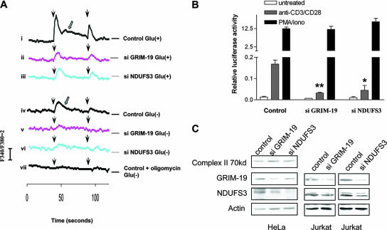
The mitochondrial respiratory chain (MRC) plays crucial roles in cellular energy production. However, its function in early embryonic development remains largely unknown. To address this issue, GRIM-19, a newly identified MRC complex I subunit, was knocked down in Xenopus laevis embryos. A severe deficiency in heart formation was observed, and the deficiency could be rescued by reintroducing human GRIM-19 mRNA. The mechanism involved was further investigated. We found that the activity of NFAT, a transcription factor family that contributes to early organ development, was downregulated in GRIM-19 knockdown embryos. Furthermore, the expression of a constitutively active form of mouse NFATc4 in these embryos rescued the heart developmental defects. NFAT activity is controlled by a calcium-dependent protein phosphatase, calcineurin, which suggests that calcium signaling may be disrupted by GRIM-19 knockdown. Indeed, both the calcium response and calcium-induced NFAT activity were impaired in the GRIM-19 or NDUFS3 (another complex I subunit) knockdown cell lines. We also showed that NFAT can rescue expression of Nkx2.5, which is one of the key genes for early heart development. Our data demonstrated the essential role of MRC in heart formation and revealed the signal transduction and gene expression cascade involved in this process.


Figures

FIG. 1.
FIG. 1.
Cloning and expression pattern of XGRIM-19 in X. laevis. (A) Comparison of GRIM-19 amino acid sequences between X. laevis, X. tropicalis, mouse, and human. Amino acid numbers are indicated, and the nonconserved amino acids are highlighted with boxes. (B) Whole-mount in situ hybridization of XGRIM-19 in Xenopus embryos. XGRIM-19 mRNA was detected in the animal cap and the marginal zone of embryos at stage 10, as indicated in panel i. During organogenesis, XGRIM-19 mRNA was strongly expressed in the central nervous system (CNS) and eyes at stage 25 (ii), skeletal muscle and kidney at stage 35 (iii), and jaw muscle and heart (iv) at stage 42, as indicated. (C) XGRIM-19 mRNA in the different developmental stages was detected by RT-PCR with 28S and 18S RNA as loading controls. (D) XGRIM-19 protein level was detected by Western blot analysis (WB) using anti-mouse GRIM-19 antibody. The blot was reprobed with antiactin as a control.
FIG. 2.
FIG. 2.
KD efficiency of XGRIM-19 and its effect on complex I activity. (A) XGRIM-19 MOs inhibit translation of XGRIM-19 mRNA in vitro. The pcDNA3-XGRIM-19 construct was transcribed and translated in vitro in the presence of XGRIM-19 MO1, MO2, or the control MO (C) at the indicated concentrations (Conc). (B) XGRIM-19 MO1 inhibits XGRIM-19 protein expression in vivo. Xenopus embryos were injected with control MO (lanes 1 and 4) or XGRIM-19 MO1 (lanes 2 and 5) at stage 1. Embryos were harvested at the indicated stages, and total lysates were prepared for Western blot analysis with anti-GRIM-19 antibody. For the rescue experiment, the hG19 mRNA was injected into two dorsal cells at stage 3 after injection with MO1 at stage 1 (lanes 3 and 6). (C) KD of GRIM-19 impairs complex I activity. The representative activities of complex I and complex II were assayed in the control (C) and XGRIM-19 KD (MO1) embryos at stage 28 by in-gel OXPHOS assay (left panel). Enzymatic activities of complex I and II in the in-gel assay were quantified by densitometry. The graph depicts the average of complex I or II activity from three independent experiments with standard deviations (right panel). **, statistical significance at a P value of <0.01, as determined by paired t test analysis.
FIG. 3.
FIG. 3.
KD of XGRIM-19 causes heart defect in Xenopus embryos. (A and B) Statistical data showing heart defects in control, XGRIM-19 MO1, or XGRIM-19 MO1 with hG19 mRNA rescue (A) and 0.5 μM rotenone or DMSO-treated WT Xenopus embryos (B). Numbers (n) of treated embryos are indicated. Heart development was examined under the microscope at stage 45 and grouped as “normal,” “abnormal,” “no heart,” or “dilated,” as indicated by different colors. Percentages of embryos displaying different phenotypes are presented as bars (**, P < 0.01). (C to F) Histological analysis of heart morphology in control (C), XGRIM-19 MO1-injected (E and F), and human GRIM-19 mRNA-rescued (D) embryos at stage 45. The numbers of embryos examined in each group were 4, 4, and 3, respectively. Transverse sections of the heart were examined, and only one set of embryos is represented. Black arrows indicate the atria, green arrows indicate ventricles, “T” indicates the heart tube, and the arrowhead indicates heart valves.
FIG. 4.
FIG. 4.
Depletion of XGRIM-19 downregulates expression of several cardiac genes and NFAT activity. (A) Whole-mount in situ hybridization showing the expression of Nkx2.5, MLC2, cardiac actin, BMP4, and GATA4 in the control (left panels) or the XGRIM-19 MO1 (right panels) embryos at stages 28 (lateral views). The cardiac actin expression in the heart region is indicated by white arrows and in the somite by black arrows. (B) Various gene expression levels in the embryos injected with control MO (C) or XGRIM-19 MO1 at different stages, as detected by RT-PCR. ODC, ornithine decarboxylase. (C) Three sets of images representing normal (++), reduced (+), or absence of (−) gene expression. Statistical data on heart-specific gene expression in control and XGRIM-19 KD embryos are summarized in Table 1.
FIG. 5.
FIG. 5.
Depletion of XGRIM-19 inhibits NFAT activity. (A) Whole embryos injected with control or MO1 together with pNFAT-TA-luc reporter and control Renilla luciferase construct were harvested from different stages (as indicated), and a dual luciferase assay was performed. The average levels of luciferase activity are shown in the graph, with error bars representing the standard deviations of the means from three experiments. Activities in control and GRIM-19 KD show significant difference at stage 18 (**, P < 0.01), but not at stage 22 and 28, likely due to large variations. (B) The NFAT activity from the tissue of cardiac region (indicated by an arrow in the upper right panel) was measured at stage 28 using the same method as that described for panel A (P < 0.05).
FIG. 6.
FIG. 6.
Rescue of heart deficiency by CA-NFATc4 in XGRIM-19 KD Xenopus embryos. (A) Comparison of NFATc4 and CA-NFATc4 activity in MCF-7 cells. MCF-7 cells were cotransfected with the pNFAT-TA-luc construct and either pcDNA3-NFATc4 or pcDNA3-CA-NFATc4. Cells were either left untreated or were treated with ionomycin to increase intracellular calcium levels. Dual luciferase assays were performed in triplicate, and the average luciferase activity from three independent experiments is shown, with error bars representing the standard deviations of the means. (B) CA-NFATc4 partially rescues the heart defect in XGRIM-19 KD embryos. Embryos were injected with control MO, XGRIM-19 MO1, or XGRIM-19 MO1 and CA-NFATc4 mRNA, as indicated. The percentages of embryos displaying heart phenotypes of normal, abnormal, and no heart are indicated as bars, and the number of embryos is shown on top (n) (**, P < 0.01). (C) Embryos were injected with control MO, XGRIM-19 MO1, or XGRIM-19 MO1 and CA-NFATc4 mRNA. Nkx2.5 mRNA was detected by in situ hybridization in these embryos at stage 28. (D) The percentages of embryos displaying an absence of (−), weak (+), or normal (++) Nkx2.5 expression are indicated as bars, and the number of embryos is shown on top (n) (**, P < 0.01; *, P < 0.05). (E) Gene expression in embryos injected with control MO, XGRIM-19 MO1, or XGRIM-19 MO1 and CA-NFATc4 mRNA. Total embryo lysate at stage 28 was harvested and analyzed by Western blotting with antibodies against GRIM-19, NFATc4, VDAC, Hsp60, and actin, as indicated on the left.
FIG. 7.
FIG. 7.
Ultrastructure of heart and skeletal muscle from Xenopus embryos at stage 45. Embryos injected by control MO, GRIM-19 MO1, or GRIM-19 MO1 plus CA-NFAT were examined with a transmission electron microscope. XGRIM-19 KD causes severely perturbed sarcomeres in heart muscle (white arrowhead, top middle panel). Mitochondrial proliferation (white arrow, middle panel in middle row) and morphological abnormalities in skeletal muscle were observed (white arrow, bottom middle panel). CA-NFATc4 can rescue the sarcomere formation in heart muscle (top right panel). Lipid droplets (LD) are commonly visible in XGRIM-19 KD muscles. Scale bars, 0.5 μM.
FIG. 8.
FIG. 8.
Inhibition of calcineurin-NFAT activity results in early heart defect in Xenopus embryos. (A) Statistical data showing heart defects in the Xenopus embryos treated with solvent (control) or 2 μM or 5 μM of FK506 and CsA. The number (n) of treated embryos is indicated. Heart development was examined under the microscope at stage 42 and grouped as “normal,” “abnormal,” “no heart,” or “dilated” heart, indicated by different colors. The percentages of embryos displaying different phenotype are presented as bars (**, P < 0.01). (B) Transverse sections represent the normal heart in control or the heart tube in the Fk506/CsA-treated embryos at stage 42. Black arrow, atrium; green arrow, ventricle; arrowhead, heat tube.
FIG. 9.
FIG. 9.
GRIM-19 KD compromises intracellular calcium mobilization and NFAT activity. (A) GRIM-19 and NDUFS3 KD HeLa cells and control cells expressing nonspecific siRNA (si) were stimulated with 2.5 μM His (arrowhead) in sample medium containing 5 mM glucose (i to iii) or 2 mM pyruvate (iv to vii). As a control, 25 μM oligomycin was added to medium to inhibit complex V activity (vii). Calcium mobilization was measured using a luminescence spectrophotometer. The Fura 340/380 ratio indicates the change in [Ca2+]i. The open arrow indicates Ca2+ influx. (B) Control, GRIM-19, and NDUFS3 KD Jurkat cells were transiently transfected with pNFAT-TA-luc and stimulated with either anti-CD3/CD28 or ionomycin/PMA. Endogenous NFAT activity was analyzed by luciferase assay. The difference between NFAT activity in the KD cell lines in comparison to that in the control is statistically significant (**, P < 0.01; *, P < 0.05). (C) Western blots showing the KD efficiency of GRIM-19 and NDUFS3 in HeLa and Jurkat cells.

Similar articles

Cited by

References

    1. Angell, J. E., D. J. Lindner, P. S. Shapiro, E. R. Hofmann, and D. V. Kalvakolanu. 2000. Identification of GRIM-19, a novel cell death-regulatory gene induced by the interferon-beta and retinoic acid combination, using a genetic approach. J. Biol. Chem. 275:33416-33426. - PubMed
    1. Berridge, M. J., M. D. Bootman, and H. L. Roderick. 2003. Calcium signalling: dynamics, homeostasis and remodelling. Nat. Rev. Mol. Cell Biol. 4:517-529. - PubMed
    1. Bezprozvanny, I., and B. E. Ehrlich. 1993. ATP modulates the function of inositol 1,4,5-trisphosphate-gated channels at two sites. Neuron 10:1175-1184. - PubMed
    1. Bushdid, P. B., H. Osinska, R. R. Waclaw, J. D. Molkentin, and K. E. Yutzey. 2003. NFATc3 and NFATc4 are required for cardiac development and mitochondrial function. Circ. Res. 92:1305-1313. - PubMed
    1. Carafoli, E. 2002. Calcium signaling: a tale for all seasons. Proc. Natl. Acad. Sci. USA 99:1115-1122. - PMC - PubMed

Publication types

MeSH terms

LinkOut - more resources