. 2007 Oct;27(19):6889-902.
doi: 10.1128/MCB.00762-07. Epub 2007 Jul 16.

Epigenetic regulation of protein-coding and microRNA genes by the Gfi1-interacting tumor suppressor PRDM5

Affiliations

Epigenetic regulation of protein-coding and microRNA genes by the Gfi1-interacting tumor suppressor PRDM5

Zhijun Duan et al. Mol Cell Biol. 2007 Oct.

Abstract

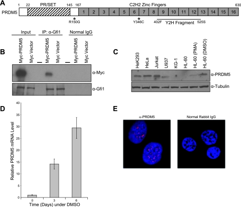
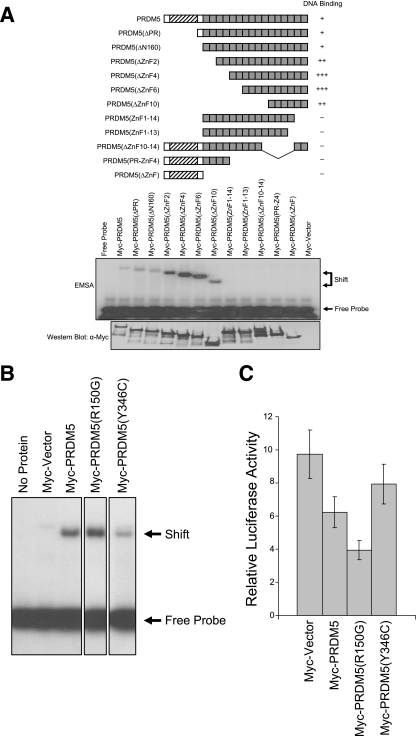
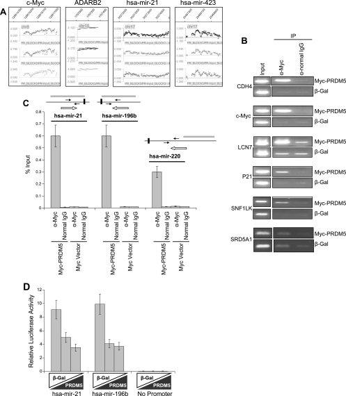
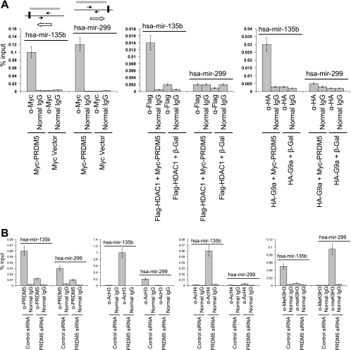
Gfi1 transcriptionally governs hematopoiesis, and its mutations produce neutropenia. In an effort to identify Gfi1-interacting proteins and also to generate new candidate genes causing neutropenia, we performed a yeast two-hybrid screen with Gfi1. Among other Gfi1-interacting proteins, we identified a previously uncharacterized member of the PR domain-containing family of tumor suppressors, PRDM5. PRDM5 has 16 zinc fingers, and we show that it acts as a sequence-specific, DNA binding transcription factor that targets hematopoiesis-associated protein-coding and microRNA genes, including many that are also targets of Gfi1. PRDM5 epigenetically regulates transcription similarly to Gfi1: it recruits the histone methyltransferase G9a and class I histone deacetylases to its target gene promoters and demonstrates repressor activity on synthetic reporters; on endogenous target genes, however, it functions as an activator, in addition to a repressor. Interestingly, genes that PRDM5 activates, as opposed to those it represses, are also targets of Gfi1, suggesting a competitive mechanism through which two repressors could cooperate in order to become transcriptional activators. In neutropenic patients, we identified PRDM5 protein sequence variants perturbing transcriptional function, suggesting a potentially important role in hematopoiesis.


Figures

FIG. 1.
FIG. 1.
PRDM5 structure, Gfi1 interaction, neutropenic sequence variants, and subcellular localization. (A) PRDM5 schematic representation. Asterisks denote variants found in neutropenic individuals. Zinc finger fragment recovered in yeast two-hybrid (Y2H) screen with zinc fingers of Gfi1 is indicated. Numbers on top of schematic correspond to protein sequence. (B) Interaction of PRDM5 with Gfi1 in cotransfected HeLa cells. Coimmunoprecipitation assays were performed using an antibody against Gfi1 or normal goat IgG as a negative control with HeLa cells cotransfected with the indicated plasmids. Dash indicates empty lane. (C) Western blot analysis of PRDM5 in human cell lines. PMA, phorbol myristate acetate; DMSO, dimethyl sulfoxide. (D) Up-regulation of PRDM5 during dimethyl sulfoxide (DMSO)-induced granulocytic differentiation of HL-60 cells, as measured by quantitative real-time RT-PCR with GAPDH as internal control. (E) Confocal microscopy of indirect immunofluorescent staining of HeLa cells with PRDM5 antibody (red) and control IgG. Nuclei counterstained with DAPI are blue. α, anti.
FIG. 2.
FIG. 2.
DNA sequence-specific transcriptional repressor activity of PRDM5 and its disruption by neutropenia-associated sequence variants. (A) Deletion of PRDM5 to determine domains responsible for DNA binding via gel-shift assay. Hatching and shading are as indicated in Fig. 1A. EMSA, electrophoretic mobility shift assay. The number of plus signs indicates the relative strength of binding. −, no binding. (B) Effects of neutropenia-associated PRDM5 sequence variants on DNA binding in gel-shift assay. R150G shows slightly more DNA binding, Y346C less DNA binding. (An amount of 0.5 μl of 50 μl total in vitro TnT transcription/translation system product of each protein was used.) The figure shows a single gel image, where intervening lanes have been cropped for the purpose of clarity of presentation. α, anti. (C) PRDM5 transcriptional repressor activity disrupted by neutropenia-associated sequence variants. Reporter assay in HeLa cells transiently transfected with 100 ng of a construct containing a dimer of the PRDM5 DNA binding consensus sequence fused to a simian virus 40 promoter driving luciferase expression along with 50 ng of the indicated form of PRDM5 expression vector. Note that the R150G mutation increased repressor activity of PRDM5, while Y346C diminished activity, consistent with effects on DNA binding.
FIG. 3.
FIG. 3.
PRDM5 target gene identification. (A) Examples of ChIP-chip hybridization signals, with vertical axis representing log2 ratio of ChIP to input, showing results for three probe replicates as visualized using NimbleGen SignalMap software. (B) Confirmation of ChIP-chip results by semiquantitative PCR in transfected HEK293 cells. SRD5A1 (excluded as target by ChIP-chip) serves as negative control. Cells were transfected with either Myc-PRDM5 expression constructs or equivalent quantities of vector expressing β-gal. (C) Confirmation of ChIP-chip results for miRNA target genes via quantitative real-time PCR in transfected HEK293 cells. Paired arrows indicate location of PCR primers relative to mature RNA (horizontal black bars). Gray bars demonstrate enriched regions in ChIP-chip assays. Open arrows show transcription direction. (D) PRDM5 repression of hsa-mir-21 and hsa-mir-196b gene promoters, demonstrated by transient reporter assays in HeLa cells transfected with 0, 50, or 150 ng of PRDM5 expression vector (black triangles) and 100 ng of luciferase reporter construct. In each titration, the total quantity of expression vector was held constant at 150 ng with compensatory quantities of vector expressing β-gal (white triangles). α, anti.
FIG. 4.
FIG. 4.
Stable suppression of PRDM5 in HEK293 cells by RNAi. (A) Quantitative real-time RT-PCR assays demonstrating reduction of PRDM5 by two PRDM5 RNAi retroviral constructs, V2HS_29874 (shRNA1) and V2HS_29875 (shRNA2), compared to control vector RHS1703 and empty vector pSM2c. Pools correspond to different polyclonal populations selected for vector-conferred puromycin resistance. (B) Western blot assays demonstrating PRDM5 suppression at protein level. α, anti.
FIG. 5.
FIG. 5.
Fluorescence-activated cell sorter analysis of cell cycle profile following PRDM5 suppression in HEK293 cells compared to controls. FL2-H: FL2-PI corresponds to the height of the fluorescence signal for propidium iodide staining of DNA content. (Repetition of the experiment showed cells in G1, S, and G2/M stages with control RNAi and PRDM5 RNAi at 46%, 18%, and 36% and 63%, 13%, and 24%, respectively [data not shown].)
FIG. 6.
FIG. 6.
Confirmation of target gene deregulation following PRDM5 silencing in HEK293 cells. (A) Spot check of PRDM5 targets shown to be deregulated by microarray confirmed here by results of quantitative real-time RT-PCR with GAPDH and β-actin as internal controls. (B) PRDM5 miRNA target gene expression profiling following PRDM5 silencing, measured by TaqMan assays with RNU6B as internal control and RNU66 as negative control.
FIG. 7.
FIG. 7.
PRDM5 recruitment of G9a HMTase. (A) Immunoprecipitated PRDM5 complex possesses HMTase activity. Vectors expressing Myc-PRDM5, or β-gal as a negative control, were transfected into HeLa cells, and cell extracts were immunoprecipitated with Myc or nonspecific antibody and then incubated with histones and S-[3H]adenosylmethionine and resolved by sodium dodecyl sulfate-polyacrylamide gel electrophoresis with Coomassie staining. Autoradiography demonstrates transfer of tritiated methyl to histone H3. (B) The PRDM5 complex methylates H3-K9, but not H3-K4. A synthetic peptide corresponding to the amino-terminal 20 residues of H3, as well as one in which the K4 was premethylated, are both methylated, but premethylation of K9 blocks its function as a substrate. (C) Coexpression of G9a, but not Suv39H1, in HeLa cells increases HMTase activity of immunoprecipitated PRDM5 complex. (Western blots were performed on cell extract to confirm expected expression.) Asterisks denote equivalent electrophoretic migration distance for H3 in different panels. α, anti.
FIG. 8.
FIG. 8.
In vivo association of PRDM5 with HDAC1 and G9a. (A) When coexpressing Myc-tagged PRDM5 with HA-tagged G9a (left two panels) and Flag-tagged HDAC1 (middle right panel) in transfected HEK293 cells, PRDM5 could be coimmunoprecipitated with G9a by antibodies against either HA epitope (left panel) or Myc epitope (middle left panel) and with HDAC1 by HDAC1 antibody (middle right panel), respectively. Transfected Myc-PRDM5 also coimmunoprecipitated with endogenous HDAC1 in HEK293 cells (right panel). (B) Endogenous PRDM5 forms an immunoprecipitable complex with endogenous G9a and HDAC1 in HEK293 cells. Antibody against PRDM5 coimmunoprecipitates G9a (left panel) and HDAC1 (right panel). α, anti.
FIG. 9.
FIG. 9.
PRDM5 recruitment of G9a and HDAC1 to its miRNA target promoters and histone modification pattern changes associated with its silencing in HEK293 cells. (A) HEK293 cells were cotransfected with the noted combinations of plasmids and subjected to ChIP with antibodies against the indicated epitope tags or control IgG followed by semiquantitative PCR. Left panel, occupancy of Myc-PRDM5 on the promoters of hsa-mir-135b and hsa-mir-299 genes. Middle panel, PRDM5 recruitment of HDAC1 to hsa-mir-135b promoter, but not hsa-mir-299 promoter. Right panel, PRDM5 recruitment of G9a to hsa-mir-135b promoter, but not hsa-mir-299 promoter. Paired arrows indicate location of PCR primers relative to mature RNA (horizontal black bars). Gray bars demonstrate enriched regions in ChIP-chip assays. Open arrows show transcription direction. (B) Histone modification pattern changes associated with silencing of PRDM5 in HEK293 cells. Left panel, presence of endogenous PRDM5 on promoters of hsa-mir-135b and hsa-mir-299, demonstrated by diminished binding with RNAi depletion. Left middle panel, PRDM5 depletion results in acetylated histone H3 becoming increased in hsa-mir-135b and decreased in hsa-mir-299 promoters. Right middle panel, PRDM5 depletion results in acetylated histone H4 becoming increased in hsa-mir-135b and not significantly changed on H4 in hsa-mir-299 promoters. Right panel, depletion of PRDM5 results in K9-methylated histone H3 becoming decreased in hsa-mir-135b and increased in hsa-mir-299 promoters. α, anti.

References

    1. Abuin, A., G. M. Hansen, and B. Zambrowicz. 2007. Gene trap mutagenesis. Handb. Exp. Pharmacol. 178:129-147. - PubMed
    1. Ancelin, K., U. C. Lange, P. Hajkova, R. Schneider, A. J. Bannister, T. Kouzarides, and M. A. Surani. 2006. Blimp1 associates with Prmt5 and directs histone arginine methylation in mouse germ cells. Nat. Cell Biol. 8:623-630. - PubMed
    1. Bieda, M., X. Xu, M. A. Singer, R. Green, and P. J. Farnham. 2006. Unbiased location analysis of E2F1-binding sites suggests a widespread role for E2F1 in the human genome. Genome Res. 16:595-605. - PMC - PubMed
    1. Canote, R., Y. Du, T. Carling, F. Tian, Z. Peng, and S. Huang. 2002. The tumor suppressor gene RIZ in cancer gene therapy (review). Oncol. Rep. 9:57-60. - PubMed
    1. Carling, T., K. C. Kim, X. H. Yang, J. Gu, X. K. Zhang, and S. Huang. 2004. A histone methyltransferase is required for maximal response to female sex hormones. Mol. Cell. Biol. 24:7032-7042. - PMC - PubMed

Publication types

MeSH terms