. 2007 Sep;27(18):6433-45.
doi: 10.1128/MCB.00135-07. Epub 2007 Jul 16.

Phosphorylation of Slx4 by Mec1 and Tel1 regulates the single-strand annealing mode of DNA repair in budding yeast

Affiliations

Phosphorylation of Slx4 by Mec1 and Tel1 regulates the single-strand annealing mode of DNA repair in budding yeast

Sonja Flott et al. Mol Cell Biol. 2007 Sep.

Abstract

Budding yeast (Saccharomyces cerevisiae) Slx4 is essential for cell viability in the absence of the Sgs1 helicase and for recovery from DNA damage. Here we report that cells lacking Slx4 have difficulties in completing DNA synthesis during recovery from replisome stalling induced by the DNA alkylating agent methyl methanesulfonate (MMS). Although DNA synthesis restarts during recovery, cells are left with unreplicated gaps in the genome despite an increase in translesion synthesis. In this light, epistasis experiments show that SLX4 interacts with genes involved in error-free bypass of DNA lesions. Slx4 associates physically, in a mutually exclusive manner, with two structure-specific endonucleases, Rad1 and Slx1, but neither of these enzymes is required for Slx4 to promote resistance to MMS. However, Rad1-dependent DNA repair by single-strand annealing (SSA) requires Slx4. Strikingly, phosphorylation of Slx4 by the Mec1 and Tel1 kinases appears to be essential for SSA but not for cell viability in the absence of Sgs1 or for cellular resistance to MMS. These results indicate that Slx4 has multiple functions in responding to DNA damage and that a subset of these are regulated by Mec1/Tel1-dependent phosphorylation.


Figures

FIG. 1.
FIG. 1.
Analysis of DNA replication during recovery from MMS-induced replisome stalling in slx4Δ cells. (A) Strains BY4741 (wild type), SFY008 (slx4Δ), SFY009 (slx1Δ), and SFY010 (sgs1Δ) were grown to mid-log phase, arrested in G1 by the presence of α-factor, and then released from G1 arrest into YPD. After 10 min, MMS (0.05%) was added for 45 min. Cells were then filtered, washed extensively, and incubated in YPD at 30°C for the times indicated. Samples for PFGE were taken before treatment with MMS (lanes 1, 6, 11, and 16), at 0 (lanes 2, 7, 12, and 17), 3 (lanes 3, 8, 13, and 18), 4 (lanes 4, 9, 14, and 19), and 5 (lanes 5, 10, 15, and 20) hours after cells were washed free of MMS. After PFGE, chromosomes were visualized by staining with ethidium bromide. (B) Same experiment as in panel A, except that samples were subjected to FACS analysis after staining with propidium iodide. (C) Wild-type (PP108) and slx4Δ (SFY063) cells were arrested in G1 with α-factor and were released into S phase in the presence of BrdU and MMS (0.05%). After 60 min, cells were washed and resuspended for the indicated times in fresh medium containing BrdU. Chromosomal DNA was stretched on silanized coverslips, and replicated tracks were detected with an anti-BrdU antibody (green). DNA fibers were counterstained with an antiguanosine antibody (red). Merged images (BrdU and DNA) and BrdU signals alone are shown for representative DNA fibers, with arrowheads pointing to unreplicated gaps. (D) Box plots of the length distribution of BrdU tracks. The Mann-Whitney rank-sum test was used to show that the differences between wild-type and slx4Δ cells are significant (P < 0.001). (E) For each time point, DNA fibers and BrdU tracks were measured and the percentage of replication was determined for wild-type cells (filled circles) and slx4Δ cells (open circles). Mean fiber length, which is indicative of the persistence of DSBs or fragile replication intermediates, was significantly shorter in slx4Δ mutants. Mutant cells also show a 3.2-fold increase of DNA fibers with unreplicated gaps relative to wild type after 130 min of recovery.
FIG. 1.
FIG. 1.
Analysis of DNA replication during recovery from MMS-induced replisome stalling in slx4Δ cells. (A) Strains BY4741 (wild type), SFY008 (slx4Δ), SFY009 (slx1Δ), and SFY010 (sgs1Δ) were grown to mid-log phase, arrested in G1 by the presence of α-factor, and then released from G1 arrest into YPD. After 10 min, MMS (0.05%) was added for 45 min. Cells were then filtered, washed extensively, and incubated in YPD at 30°C for the times indicated. Samples for PFGE were taken before treatment with MMS (lanes 1, 6, 11, and 16), at 0 (lanes 2, 7, 12, and 17), 3 (lanes 3, 8, 13, and 18), 4 (lanes 4, 9, 14, and 19), and 5 (lanes 5, 10, 15, and 20) hours after cells were washed free of MMS. After PFGE, chromosomes were visualized by staining with ethidium bromide. (B) Same experiment as in panel A, except that samples were subjected to FACS analysis after staining with propidium iodide. (C) Wild-type (PP108) and slx4Δ (SFY063) cells were arrested in G1 with α-factor and were released into S phase in the presence of BrdU and MMS (0.05%). After 60 min, cells were washed and resuspended for the indicated times in fresh medium containing BrdU. Chromosomal DNA was stretched on silanized coverslips, and replicated tracks were detected with an anti-BrdU antibody (green). DNA fibers were counterstained with an antiguanosine antibody (red). Merged images (BrdU and DNA) and BrdU signals alone are shown for representative DNA fibers, with arrowheads pointing to unreplicated gaps. (D) Box plots of the length distribution of BrdU tracks. The Mann-Whitney rank-sum test was used to show that the differences between wild-type and slx4Δ cells are significant (P < 0.001). (E) For each time point, DNA fibers and BrdU tracks were measured and the percentage of replication was determined for wild-type cells (filled circles) and slx4Δ cells (open circles). Mean fiber length, which is indicative of the persistence of DSBs or fragile replication intermediates, was significantly shorter in slx4Δ mutants. Mutant cells also show a 3.2-fold increase of DNA fibers with unreplicated gaps relative to wild type after 130 min of recovery.
FIG. 2.
FIG. 2.
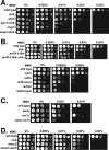
SLX4 interacts with genes involved in DNA damage bypass. Strains BY4741 (wild type), SFY008 (slx4Δ), SFY022 (apn1Δ), SFY023 (apn1Δ slx4Δ), SFY020 (mag1Δ), SFY021 (mag1Δ slx4Δ), SFY036 (pol30k164rΔ), SFY037 (pol30k164rΔ slx4Δ), SFY026 (rad6Δ), SFY027 (rad6Δ slx4Δ), SFY028 (rad18Δ), SFY029 (rad18Δ slx4Δ), SFY034 (rev3Δ), SFY035 (rev3Δ slx4Δ), SFY030 (mms2Δ), and SFY031 (mms2Δ slx4Δ) were grown to saturation in liquid culture, and 10-fold serial dilutions were spotted on YPD agar plates with or without the indicated amounts of MMS. Plates were incubated for 3 days at 30°C.
FIG. 3.
FIG. 3.
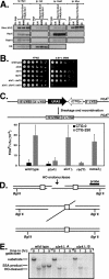
Slx4 interacts with Rad1-Rad10 and is required for SSA. (A) Cells expressing Myc13-Slx4 and HA6-Slx4 (in which PEP4 was disrupted) or cells expressing Slx4 were grown to mid-exponential phase in liquid culture and left untreated or incubated with MMS (0.02%) for 30 min or exposed to ionizing radiation (IR; 180 Gy) and left to recover for 30 min, respectively. Native lysates were prepared, and Myc-Slx4 immunoprecipitates or HA-Slx1 or anti-Rad1 precipitates were subjected to SDS-PAGE and Western blot analysis with the antibodies indicated. (B) Same experiment as described for Fig. 2, except that different (indicated) strains were used. (C) Top: a schematic diagram of the assay to measure SSA according to reference is shown. Bottom: yeast strains SFY047 (CTG-0), SFY049 (CTG-250), SFY048 (slx4Δ CTG-0), SFY050 (slx4Δ CTG-250), SFY064 (rad1Δ CTG-250), and SFY066 (mms2Δ CTG-250) were grown to mid-log phase and plated on YPD agar and on plates containing FOA, according to the methods described in reference . Frequency of URA3 marker loss was determined as described in Materials and Methods. Average frequencies of FOA-resistant colonies per CFU and the standard deviations are shown. (D) HO endonuclease cleaves at its recognition site between 205-bp repeats of the ura3 sequence (boxes). The probe used for blotting is a HindIII-BamHI fragment as indicated. (E) The kinetics of SSA was monitored as described previously (43) at the indicated times after HO induction by Southern blot hybridization of BglII-digested genomic DNA using the probe indicated in panel D. The uncut SSA product and HO-cut bands are 8.3, 5.5, and 4.8 kb, respectively. Two different SLX4-disrupted strains are shown.
FIG. 4.
FIG. 4.
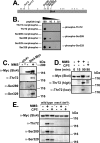
Identification of Ser/Thr residues in Slx4 phosphorylated by Mec1 and Tel1 after DNA damage. (A) Schematic diagram of Slx4. Sites phosphorylated by DNA-PK are indicated. Asterisks denote all S/T-Q motifs. (B) Different amounts of phosphopeptide (“phospho”) corresponding to Slx4 Thr72, Ser289, and Ser329 and the corresponding non-phospho peptides (“nonphospho”) were spotted onto nitrocellulose, and Western blot analysis was carried out with the relevant phospho-specific antibody. (C to E) The yeast strains indicated were grown to mid-exponential phase in liquid culture and left untreated or incubated with MMS (0.02%) for 90 min (D and F) or with MMS (0.02%) or CPT (5 μg/ml) for 15 or 90 min, respectively (E). Native lysates were prepared, and Myc-Slx4 immunoprecipitates were subjected to SDS-PAGE and Western blot analysis with antibodies against Myc, phospho-Thr72, phospho-Ser289, or phospho-Ser329. Thr72 (high), long exposure time; Thr72 (low), short exposure time.
FIG. 5.
FIG. 5.
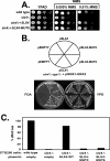
Phosphorylation of Slx4 promotes SSA. (A) Wild-type cells (strain HP30) transformed with pRS413 (empty vector) or slx4Δ cells (SFY018) transformed with empty vector or plasmids pSLX4 or pSLX4-MUT6 were grown to saturation in liquid culture. Tenfold serial dilutions, starting from an OD600 of 0.6, were spotted on YPD agar or onto YPD agar plates containing MMS. Cells were then incubated at 30°C for 3 days. (B) sgs1Δ slx4Δ [pSGS1-URA3] cells (strain NJY561) (30) were transformed with the following HIS3 plasmids: empty plasmid (pRS413), pSLX4, or pSLX4-MUT6. Cells were then restreaked on YPD agar or on agar containing 5-FOA. Plates were incubated for 3 days at 30°C. (C) Same experiment as in Fig. 3C except that strains SFY049 (CTG-250) transformed with pRS413 (empty vector) and SFY050 (slx4Δ CTG-250) transformed with pRS413 (empty vector), pSLX4, pSLX4-MUT3, or pSLX4-MUT6 were used.
FIG. 6.
FIG. 6.
Model for cellular roles of Slx4. Slx4 exists in at least two mutually exclusive, functionally distinct Slx4-containing complexes. Esc4-Slx4-Slx1 protects cells in the absence of Sgs1 but is not required for SSA. The Slx4-Rad1-Rad10 complex, on the other hand, promotes SSA. The two Slx4-containing complexes appear to be distinct in that deletion of RAD1 is not synthetically lethal with deletion of SGS1 (4), and Rad1 is not required for resistance to, or for recovery from, MMS (Fig. 3B and data not shown). Similarly, Slx1 is not required for recovery from MMS (Fig. 1A) or for SSA (Fig. 3C). It is clear that Slx4 is required for recovery from MMS-induced replisome stalling, possibly by regulating error-free bypass, but it is not yet clear which, if any, of the known Slx4-interacting proteins is responsible. Since SLX4 is epistatic with ESC4 in terms of MMS hypersensitivity, it is likely that this subset of the Esc4-Slx4-Slx1 complex is important. Alternatively, one or more as-yet-unidentified Slx4-interacting proteins may help promote resistance to MMS. Mec1 and Tel1 together phosphorylate Slx4, and this is required for efficient SSA but not for viability in the absence of Sgs1, for cellular resistance to MMS, or for completion of DNA replication when replisomes stall. The precise molecular basis for the modulation of Slx4 function by phosphorylation is not yet known.

References

    1. Bardwell, L., A. J. Cooper, and E. C. Friedberg. 1992. Stable and specific association between the yeast recombination and DNA repair proteins RAD1 and RAD10 in vitro. Mol. Cell Biol. 12:3041-3049. - PMC - PubMed
    1. Bastin-Shanower, S. A., W. M. Fricke, J. R. Mullen, and S. J. Brill. 2003. The mechanism of Mus81-Mms4 cleavage site selection distinguishes it from the homologous endonuclease Rad1-Rad10. Mol. Cell. Biol. 23:3487-3496. - PMC - PubMed
    1. Beranek, D. T. 1990. Distribution of methyl and ethyl adducts following alkylation with monofunctional alkylating agents. Mutat. Res. 231:11-30. - PubMed
    1. Brusky, J., Y. Zhu, and W. Xiao. 2000. UBC13, a DNA-damage-inducible gene, is a member of the error-free postreplication repair pathway in Saccharomyces cerevisiae. Curr. Genet. 37:168-174. - PubMed
    1. Chang, M., M. Bellaoui, C. Boone, and G. W. Brown. 2002. A genome-wide screen for methyl methanesulfonate-sensitive mutants reveals genes required for S phase progression in the presence of DNA damage. Proc. Natl. Acad. Sci. USA 99:16934-16939. - PMC - PubMed

Publication types

MeSH terms

Substances