Apoptosis regulation by Bcl-x(L) modulation of mammalian inositol 1,4,5-trisphosphate receptor channel isoform gating
- PMID: 17636122
- PMCID: PMC1941509
- DOI: 10.1073/pnas.0702489104
Apoptosis regulation by Bcl-x(L) modulation of mammalian inositol 1,4,5-trisphosphate receptor channel isoform gating
Abstract
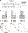
Members of the Bcl-2 family of proteins regulate apoptosis, with some of their physiological effects mediated by their modulation of endoplasmic reticulum (ER) Ca(2+) homeostasis. Antiapoptotic Bcl-x(L) binds to the inositol trisphosphate receptor (InsP(3)R) Ca(2+) release channel to enhance Ca(2+)- and InsP(3)-dependent regulation of channel gating, resulting in reduced ER [Ca(2+)], increased oscillations of cytoplasmic Ca(2+) concentration ([Ca(2+)](i)), and apoptosis resistance. However, it is controversial which InsP(3)R isoforms mediate these effects and whether reduced ER [Ca(2+)] or enhanced [Ca(2+)](i) signaling is most relevant for apoptosis protection. DT40 cell lines engineered to express each of the three mammalian InsP(3)R isoforms individually displayed enhanced apoptosis sensitivity compared with cells lacking InsP(3)R. In contrast, coexpression of each isoform with Bcl-x(L) conferred enhanced apoptosis resistance. In single-channel recordings of channel gating in native ER membranes, Bcl-x(L) increased the apparent sensitivity of all three InsP(3)R isoforms to subsaturating levels of InsP(3). Expression of Bcl-x(L) reduced ER [Ca(2+)] in type 3 but not type 1 or 2 InsP(3)R-expressing cells. In contrast, Bcl-x(L) enhanced spontaneous [Ca(2+)](i) signaling in all three InsP(3)R isoform-expressing cell lines. These results demonstrate a redundancy among InsP(3)R isoforms in their ability to sensitize cells to apoptotic insults and to interact with Bcl-x(L) to modulate their activities that result in enhanced apoptosis resistance. Furthermore, these data suggest that modulation of ER [Ca(2+)] is not a specific requirement for ER-dependent antiapoptotic effects of Bcl-x(L). Rather, apoptosis protection is conferred by enhanced spontaneous [Ca(2+)](i) signaling by Bcl-x(L) interaction with all isoforms of the InsP(3)R.
Conflict of interest statement
The authors declare no conflict of interest.
Figures


Similar articles
-
Apoptosis protection by Mcl-1 and Bcl-2 modulation of inositol 1,4,5-trisphosphate receptor-dependent Ca2+ signaling.J Biol Chem. 2010 Apr 30;285(18):13678-84. doi: 10.1074/jbc.M109.096040. Epub 2010 Feb 26. J Biol Chem. 2010. PMID: 20189983 Free PMC article.
-
The endoplasmic reticulum gateway to apoptosis by Bcl-X(L) modulation of the InsP3R.Nat Cell Biol. 2005 Oct;7(10):1021-8. doi: 10.1038/ncb1302. Epub 2005 Sep 18. Nat Cell Biol. 2005. PMID: 16179951 Free PMC article.
-
Single-channel properties in endoplasmic reticulum membrane of recombinant type 3 inositol trisphosphate receptor.J Gen Physiol. 2000 Mar;115(3):241-56. doi: 10.1085/jgp.115.3.241. J Gen Physiol. 2000. PMID: 10694253 Free PMC article.
-
Inositol 1,4,5-trisphosphate receptor-isoform diversity in cell death and survival.Biochim Biophys Acta. 2014 Oct;1843(10):2164-83. doi: 10.1016/j.bbamcr.2014.03.007. Epub 2014 Mar 15. Biochim Biophys Acta. 2014. PMID: 24642269 Review.
-
A dual role for the anti-apoptotic Bcl-2 protein in cancer: mitochondria versus endoplasmic reticulum.Biochim Biophys Acta. 2014 Oct;1843(10):2240-52. doi: 10.1016/j.bbamcr.2014.04.017. Epub 2014 Apr 21. Biochim Biophys Acta. 2014. PMID: 24768714 Review.
Cited by
-
Ca(2+) channels on the move.Biochemistry. 2009 Dec 29;48(51):12062-80. doi: 10.1021/bi901739t. Biochemistry. 2009. PMID: 19928968 Free PMC article. Review.
-
Mitochondria-Associated Membranes As Networking Platforms and Regulators of Cancer Cell Fate.Front Oncol. 2017 Aug 18;7:174. doi: 10.3389/fonc.2017.00174. eCollection 2017. Front Oncol. 2017. PMID: 28868254 Free PMC article. Review.
-
Titanium dioxide (TiO2) nanoparticles preferentially induce cell death in transformed cells in a Bak/Bax-independent fashion.PLoS One. 2012;7(11):e50607. doi: 10.1371/journal.pone.0050607. Epub 2012 Nov 21. PLoS One. 2012. PMID: 23185639 Free PMC article.
-
Nanospaces between endoplasmic reticulum and mitochondria as control centres of pancreatic β-cell metabolism and survival.Protoplasma. 2012 Feb;249 Suppl 1:S49-58. doi: 10.1007/s00709-011-0349-3. Epub 2011 Nov 22. Protoplasma. 2012. PMID: 22105567 Review.
-
Cellular and molecular effects of hyperglycemia on ion channels in vascular smooth muscle.Cell Mol Life Sci. 2021 Jan;78(1):31-61. doi: 10.1007/s00018-020-03582-z. Epub 2020 Jun 27. Cell Mol Life Sci. 2021. PMID: 32594191 Free PMC article. Review.
References
Publication types
MeSH terms
Substances
Grants and funding
LinkOut - more resources
Full Text Sources
Research Materials
Miscellaneous

