

Structural basis of antigenic escape of a malaria vaccine candidate
- PMID: 17636123
- PMCID: PMC1941496
- DOI: 10.1073/pnas.0701464104
Structural basis of antigenic escape of a malaria vaccine candidate
Abstract
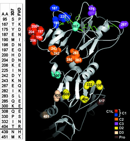
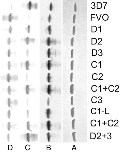
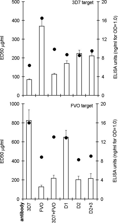
Antibodies against the malaria vaccine candidate apical membrane antigen-1 (AMA-1) can inhibit invasion of merozoites into RBC, but antigenic diversity can compromise vaccine efficacy. We hypothesize that polymorphic sites located within inhibitory epitopes function as antigenic escape residues (AER). By using an in vitro model of antigenic escape, the inhibitory contribution of 24 polymorphic sites of the 3D7 AMA-1 vaccine was determined. An AER cluster of 13 polymorphisms, located within domain 1, had the highest inhibitory contribution. Within this AER cluster, antibodies primarily targeted five polymorphic residues situated on an alpha-helical loop. A second important AER cluster was localized to domain 2. Domain 3 polymorphisms enhanced the inhibitory contribution of the domain 2 AER cluster. Importantly, the AER clusters could be split, such that chimeras containing domain 1 of FVO and domain 2 + 3 of 3D7 generated antisera that showed similarly high level inhibition of the two vaccine strains. Antibodies to this chimeric protein also inhibited unrelated strains of the parasite. Interstrain AER chimeras can be a way to incorporate inhibitory epitopes of two AMA-1 strains into a single protein. The AER clusters map in close proximity to conserved structural elements: the hydrophobic trough and the C-terminal proteolytic processing site. This finding led us to hypothesize that a conserved structural basis of antigenic escape from anti-AMA-1 exists. Genotyping high-impact AER may be useful for classifying AMA-1 strains into inhibition groups and to detect allelic effects of an AMA-1 vaccine in the field.
Conflict of interest statement
The authors declare no conflict of interest.
Figures

Similar articles
-
Overcoming antigenic diversity by enhancing the immunogenicity of conserved epitopes on the malaria vaccine candidate apical membrane antigen-1.PLoS Pathog. 2013;9(12):e1003840. doi: 10.1371/journal.ppat.1003840. Epub 2013 Dec 26. PLoS Pathog. 2013. PMID: 24385910 Free PMC article.
-
Immunogenicity of a recombinant malaria vaccine candidate, domain I+II of AMA-1 ectodomain, from Indian P. falciparum alleles.Vaccine. 2008 Aug 18;26(35):4526-35. doi: 10.1016/j.vaccine.2008.06.031. Epub 2008 Jun 30. Vaccine. 2008. PMID: 18590786
-
Identification of Plasmodium falciparum reticulocyte binding protein homologue 5-interacting protein, PfRipr, as a highly conserved blood-stage malaria vaccine candidate.Vaccine. 2016 Nov 4;34(46):5612-5622. doi: 10.1016/j.vaccine.2016.09.028. Epub 2016 Sep 28. Vaccine. 2016. PMID: 27692771
-
Apical membrane antigen 1: a malaria vaccine candidate in review.Trends Parasitol. 2008 Feb;24(2):74-84. doi: 10.1016/j.pt.2007.12.002. Epub 2008 Jan 15. Trends Parasitol. 2008. PMID: 18226584 Review.
-
Apical membrane antigen 1 as an anti-malarial drug target.Curr Top Med Chem. 2011;11(16):2039-47. doi: 10.2174/156802611796575885. Curr Top Med Chem. 2011. PMID: 21619512 Review.
Cited by
-
Assessing Prevalence and Transmission Rates of Malaria through Simultaneous Profiling of Antibody Responses against Plasmodium and Anopheles Antigens.J Clin Med. 2022 Mar 26;11(7):1839. doi: 10.3390/jcm11071839. J Clin Med. 2022. PMID: 35407447 Free PMC article.
-
Humoral immune response to mixed PfAMA1 alleles; multivalent PfAMA1 vaccines induce broad specificity.PLoS One. 2009 Dec 1;4(12):e8110. doi: 10.1371/journal.pone.0008110. PLoS One. 2009. PMID: 19956619 Free PMC article.
-
Development of quantitative suspension array assays for six immunoglobulin isotypes and subclasses to multiple Plasmodium falciparum antigens.J Immunol Methods. 2018 Apr;455:41-54. doi: 10.1016/j.jim.2018.01.009. Epub 2018 Jan 31. J Immunol Methods. 2018. PMID: 29397157 Free PMC article.
-
Microarray analyses reveal strain-specific antibody responses to Plasmodium falciparum apical membrane antigen 1 variants following natural infection and vaccination.Sci Rep. 2020 Mar 3;10(1):3952. doi: 10.1038/s41598-020-60551-z. Sci Rep. 2020. PMID: 32127565 Free PMC article.
-
Use of immunodampening to overcome diversity in the malarial vaccine candidate apical membrane antigen 1.Infect Immun. 2014 Nov;82(11):4707-17. doi: 10.1128/IAI.02061-14. Epub 2014 Aug 25. Infect Immun. 2014. PMID: 25156737 Free PMC article.
References
-
- Klenerman P, Zinkernagel RM. Nature. 1998;394:482–485. - PubMed
-
- Genton B, Al-Yaman F, Betuela I, Anders RF, Saul A, Baea K, Mellombo M, Taraika J, Brown GV, Pye D, et al. Vaccine. 2003;22:30–41. - PubMed
-
- Polhemus M, Magill AJ, Cummings JF, Kester KE, Ockenhouse CF, Lanar DE, Dutta S, Barbosa A, Soisson L, Diggs C, et al. Vaccine. 2007 in press. - PubMed
Publication types
MeSH terms
Substances
LinkOut - more resources
Full Text Sources
