. 2007 Jul 24;104(30):12482-7.
doi: 10.1073/pnas.0705533104. Epub 2007 Jul 16.

A viral adaptor protein modulating casein kinase II activity induces cytopathic effects in permissive cells

Affiliations

A viral adaptor protein modulating casein kinase II activity induces cytopathic effects in permissive cells

Jürg P F Nüesch et al. Proc Natl Acad Sci U S A. .

Abstract

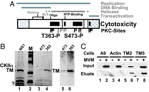
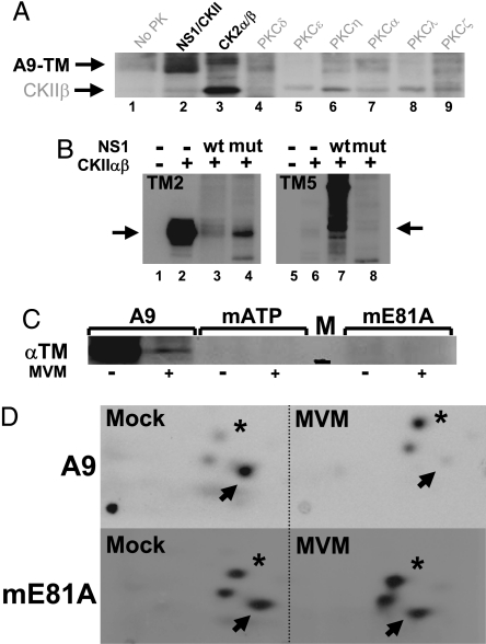
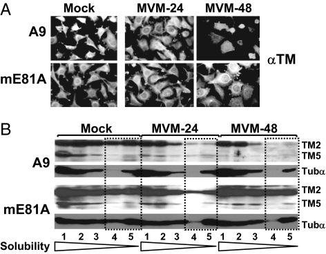
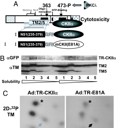
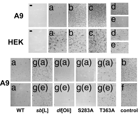
Autonomous parvoviruses induce severe morphological and physiological alterations in permissive host cells, eventually leading to cell lysis and release of progeny virions. Viral cytopathic effects (CPE) result from specific rearrangements and destruction of cytoskeletal micro- and intermediate filaments. We recently reported that inhibition of endogenous casein kinase II (CKII) protects target cells from parvovirus minute virus of mice (MVM)-induced CPE, pointing to this kinase as an effector of MVM toxicity. The present work shows that the parvoviral NS1 protein mediates CKII-dependent cytoskeletal alterations and cell death. NS1 can act as an adaptor molecule, linking the cellular protein kinase CKIIalpha to tropomyosin and thus modulating the substrate specificity of the kinase. This action results in an altered tropomyosin phosphorylation pattern both in vitro and in living cells. The capacity of NS1 to induce CPE was impaired by mutations abolishing binding with either CKIIalpha or tropomyosin. The cytotoxic adaptor function of NS1 was confirmed with fusion peptides, where the tropomyosin-binding domain of NS1 and CKIIalpha are physically linked. These adaptor peptides were able to mimic NS1 in its ability to induce death of transformed MVM-permissive cells.


Conflict of interest statement

The authors declare no conflict of interest.

Figures

Fig. 1.
Fig. 1.
Determinants of NS1-induced cytotoxicity. (A) Domain structure of NS1 (5). The common N terminus of NS1 and NS2, the region of homology with the SV40 large T-antigen, and the trans activation domain are represented, respectively, as dotted, cross-hatched, and checkered boxes. The nucleotide-binding (NTP-binding) site, the oligomerization region (Oligo), the nuclear localization signal (NLS), and nicking motifs (metal coordination site and linkage tyrosine) are positioned. Black bars denote NS1 regions mediating cytopathogenicity. Consensus PKC-phosphorylation sites that modulate (black P) or do not modulate (gray P) CPE are indicated, including 363-P and 473-P targeted for differential affinity chromatography. (.B Left) SDS/PAGE and Coomassie blue staining of proteins eluted from GST-NS1 affinity columns. NS1, proteins in A9-P3-DE1 binding to GST-NS1WT; 473, proteins lacking affinity for GST-NS1:S473A and binding to GST-NS1WT; 363, proteins lacking affinity for GST-NS1:T363A and binding to GST-NS1WT. M, molecular mass markers (97, 66, 45, 30, and 20.1 kDa (Rainbow marker, Amersham). Proteins overrepresented in either 474 or 363 are marked with dots. The doublet marked TM was cut from the gel and subjected to MS/MS analysis. (Right) Western blot analyses of both flow-through samples with TM-specific antibodies. (C) Interaction of NS1 with TM in MVM-infected cells. Binding of NS1 to cytoskeletal proteins was estimated on the basis of its affinity for GST-tagged TM2, TM5, and β-actin. Cell extracts from (MVM-infected) A9 cells (lanes 1 and 2) or from derivatives expressing GST-tagged cytoskeleton proteins (β-actin, lanes 3 and 4; TM2, lanes 5 and 6; TM5, lanes 7 and 8) were run through glutathione–Sepharose columns specifically retaining GST-tagged proteins. Binding partners (e.g., NS1) of GST-tagged proteins were eluted with 700 mM NaCl (eluate) and analyzed by Western blotting with anti-NS1C compared with total amounts (input).
Fig. 2.
Fig. 2.
In vitro and in vivo phosphorylation of A9 cell tropomyosin. (A and B) In vitro kinase assays of the indicated PK substrate combinations were performed in the presence of [γ-32P]ATP and analyzed by SDS/PAGE and autoradiography. (A) A9-TM purified by NS1 affinity (Fig. 1B lane 4), alone (no PK), or combined with similarly purified NS1–CKIIα (Fig. 1B lane 3) or with the indicated recombinant PK. Migrations of TM (A9-TM) and recombinant CKIIβ are indicated. (B) Phosphorylation of recombinant TM2 (Left) and TM5 (Right) without (lanes 1 and 5) or with CKIIαβ in the absence (lanes 2 and 6) or presence of WT (lanes 3 and 7) or mutant S473A (lanes 4 and 8) GST-NS1. The migration of monomeric TM is indicated by arrows. (C and D) MVM infection and functional CKIIα were tested for their impact on the in vivo phosphorylation of TM. A9 cells or derivatives expressing the dominant-negative CKII(mATP) or CKII(mE81A) were infected (or not) with MVM (30 pfu per cell) and subjected to metabolic 32P labeling. TM was isolated by immunoprecipation and analyzed by SDS/PAGE (C), with reference to a 30-kDa marker (black bar in lane M). Bands corresponding to phospholabeled TM were excised, and their tryptic phosphopeptide patterns were analyzed (D). Phosphopeptides characteristically overrepresented in mock- (arrow) or MVM (asterisk)-infected cells are marked.
Fig. 3.
Fig. 3.
Effect of CKIIα functional modifications on MVM-induced cytoskeleton alterations. A9 or A9:P38-CKII(mE81A) cells were infected (or not) with MVM (30 pfu per cell). (A) For immunofluorescence microscopy, cells were fixed with paraformaldehyde at the indicated time after infection, and the TM filament network was revealed by indirect fluorescence microscopy. (B) For biochemical characterization of cytoskeletal filaments, extracts were fractionated according to their differential solubility in detergent(s) as described in Materials and Methods,“ and the individual fractions (S1–S5) were analyzed for TM and tubulin α by Western blotting. Low-solubility fractions corresponding to rigid fiber structures are framed by dotted lines. The migration of TM2, TM5, and tubulin α (Tubα) are indicated.
Fig. 4.
Fig. 4.
TM phosphorylation by adaptor molecules mimicking NS1-mediated targeting of CKIIα. (A) Schematic representation of NS1 and adaptor molecules (I and II). NS1 is shown with its cellular binding partners CKIIα and TM. The semisynthetic adaptor molecules consist of the NS1 TR (amino acids 235–379), a spacer element (GFP), and either WT or mutant (mE81A) CKIIα. (B and C) In vivo phosphorylation of TM by adaptor proteins 1 and 2. A9 cells were infected with recombinant adenoviruses expressing functional (Ad:TR-CKIIα) or dominant-negative (Ad:TR-mE81A) adaptor molecules. Infected cells were subjected (C) or not (B) to metabolic labeling and processed for protein analyses. The expression of effector proteins (TR-CKIIα) and substrates (TM2, TM5) was measured after cell fractionations by Western blotting with anti-GFP or anti-TM antiserum, respectively (B). (C) Tryptic phosphopeptide pattern of TM isolated from radiolabeled cell extracts. Phosphopeptides characteristically overrepresented in mock- vs. MVM-infected A9 cells and conversely are indicated, respectively, by an arrow and an asterisk.
Fig. 5.
Fig. 5.
Cytotoxic activity of adaptor molecules targeting CKIIα activity. Colony-formation inhibition assays are shown. A variety of adaptor constructs were made expressing the TM-interaction domain of NS1 (TR) and either CKIIα- or a CKIIα-binding site (BS, DLEPDEELED) in physically linked or dissociated forms, as WT or mutant polypeptides cloned into pCR3.1 containing the neoR expression cassette. A9 or HEK293 cells transfected with these constructs were tested for the ability to inhibit colony formation in the presence of G418, as visualized by crystal violet staining. (a) TR connected to WT CKIIα. (b) TR linked to dominant-negative CKII(mE81A). (c) GFP-WT CKIIα. (d) TR connected to GFP (without PK). (e) TR connected by GFP to CKIIα-BS recruiting endogenous CKIIα. (f) GFP-BS (GFP with a C-terminal CKIIαBS). [g(a)] Mutated NS1 targeting region (mTR) connected to functional CKIIα. [g(b)] mTR linked to CKIIαBS. The impact of structural elements in TR was investigated by testing the effects of the following mutations: sb[L], methionine-for-leucine substitutions in a leucine (L) stretch; dl[Oli], deletion of the oligomerization signal; S283A and T363A, alanine substitutions at consensus PKC phosphorylation sites.

References

    1. Geletneky K, Herrero YCM, Rommelaere J, Schlehofer JR. J Vet Med. 2005;52:327–330. - PubMed
    1. Nüesch JP. In: Parvoviruses. Kerr JR, Cotmore S, Bloom ME, Linden RM, Parrish CR, editors. London: Edward Arnold; 2006. pp. 275–290.
    1. Corbau R, Duverger V, Rommelaere J, Nüesch JP. Virology. 2000;278:151–167. - PubMed
    1. Daeffler L, Horlein R, Rommelaere J, Nüesch JP. J Virol. 2003;77:12466–12478. - PMC - PubMed
    1. Nüesch JP, Rommelaere J. J Virol. 2006;80:4729–4739. - PMC - PubMed

LinkOut - more resources