. 2007 Jul 24;104(30):12389-94.
doi: 10.1073/pnas.0703787104. Epub 2007 Jul 16.

Isolation, characterization, and in vitro and in vivo differentiation of putative thecal stem cells

Affiliations

Isolation, characterization, and in vitro and in vivo differentiation of putative thecal stem cells

Arata Honda et al. Proc Natl Acad Sci U S A. .

Abstract

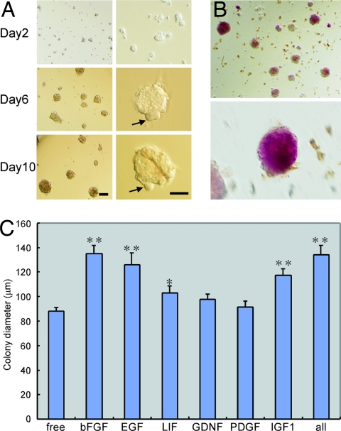
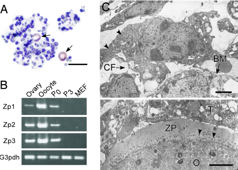
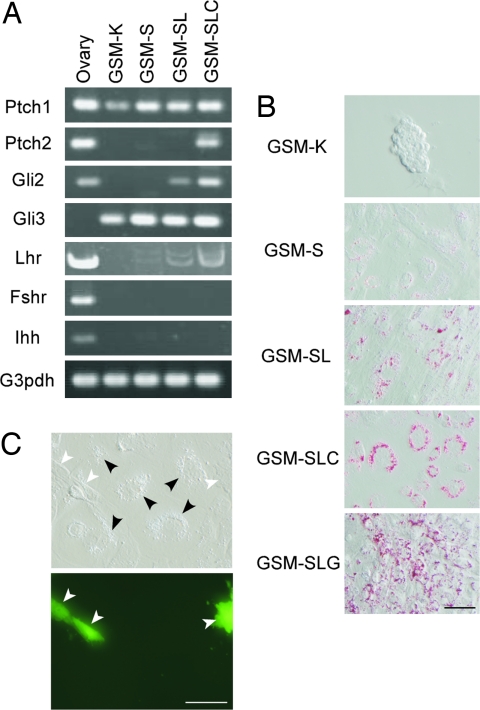
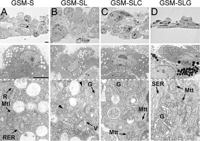
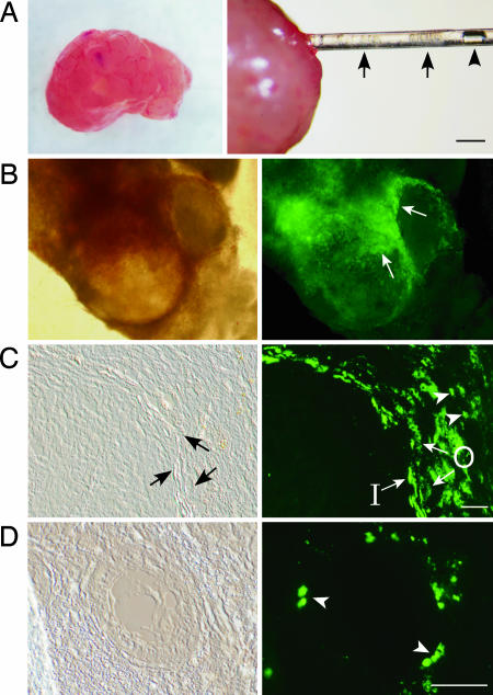
Although ovarian theca cells play an indispensable role in folliculogenesis by providing follicular structural integrity and steroid substrates for estrogen production, little information is available about their recruitment, growth, and differentiation because their immature forms have not been identified. We have isolated putative thecal stem cells with the ability to self-renew and differentiate in vivo and in vitro. They are similar to fibroblasts in morphology and proliferate in vitro as round colonies with a homogenous cell population. They were induced to differentiate into early precursors and steroidogenic cells in a stepwise manner after treatment with serum, luteinizing hormone, and paracrine factors from granulosa cells. At each differentiation step, these cells displayed appropriate gene expression and morphological markers and later secreted androstenedione. The fully mature morphology was achieved by coculture with isolated granulosa cells. When transplanted into the ovaries, the putative thecal stem cells colonized exclusively in the ovarian interstitium and the thecal layer of follicles as differentiated cells. Thus, thecal stem cells appear to be present in neonatal ovaries and can be isolated, purified, and induced to differentiate in vitro. Thecal stem cells could provide an invaluable in vitro experimental system to study interactions among the oocytes, granulosa cells, and theca cells during normal folliculogenesis and to study ovarian pathology caused by theca cell dysfunction.


Conflict of interest statement

The authors declare no conflict of interest.

Figures

Fig. 1.
Fig. 1.
Development of thecal stem cell colonies in vitro. (A) The size of the colonies increased rapidly during the first several days and gradually reached the maximum size of ≈120 μm in diameter. Many oocytes (≈20 μm in diameter) continued to protrude from the surface of the colonies (arrows). (Scale bars: Left, 100 μm; Right, 50 μm.) (B) The colonies are weakly positive for alkaline phosphatase staining. (C) Effect of growth factors on colony size during the first 14 days of culture. Single growth factors, or some combination thereof, were added to the basic GSM. *, P < 0.05; **, P < 0.01 [compared with growth-factor-free GSM (three replicates, one-way ANOVA)].
Fig. 2.
Fig. 2.
Cytological and gene expression analysis of thecal stem cell colonies. (A) Paraffin-embedded section of colonies stained for BrdU (blue) and MVH (brown). Small cells were generally positive for BrdU but not for MVH. Growing oocytes (arrows) were positive for MVH but not for BrdU, indicating that the oocytes came from preexisting cells in the colonies. (Scale bar: 50 μm.) (B) Gene expression analysis (RT-PCR) of the colonies. After the third passage (P3), oocytes disappeared from the colonies, as demonstrated by the absence of oocyte-specific markers (zona pellucida proteins Zp1, Zp2, and Zp3). MEF, mouse embryonic fibroblast. (C) Electron micrographs of the colonies. Each cell in the colonies had a relatively small cytoplasm that was rich in ribosomes and rough ER (arrowheads) (Upper). Cell-to-cell connections were sparse, and their intercellular space was filled with basement membrane (BM) and collagen fibers (CF) (Upper). A thecal stem cell (T) directly surrounds the zona pellucida (ZP) of an oocyte (O), but unlike typical granulosa cells, it does not protrude foot processes to the oocyte surface through the zona (Lower). Arrowheads indicate microvilli from the oocyte. (Scale bars: 2 μm.)
Fig. 3.
Fig. 3.
The stepwise differentiation of thecal stem cells as indicated by gene expression patterns and cytoplasmic lipid droplet development. (A) Gene expression analysis (RT-PCR) of differentiating theca cells under different culture conditions. The Lhr, Gli2, and Ptch2 genes were expressed as the thecal cells differentiated in vitro. The Fshr and Ihh genes are markers for granulosa cells. GSM-SL, GSM supplemented with LH, IGF1, and stem cell factor. (B) Cytoplasmic lipid droplets stained with Oil red O. Initially, very few lipid droplets were visible in the thecal stem cell colonies (GSM-K), but these increased in number progressively as the theca cells differentiated to their fully mature state (GSM-SLG). (Scale bar: 50 μm.) (C) Thecal stem cells were cocultured with isolated granulosa cells (GSM-SLG). To identify the cells differentiating from the thecal stem cells correctly, granulosa cells were prepared from EGFP-expressing mice. As expected, only fluorescence-negative cells had lipid droplets (granular inclusions under Hoffman optics) (black arrowheads). White arrowheads indicate EGFP-expressing granulosa cells. (Scale bar: 50 μm.)
Fig. 4.
Fig. 4.
Light and electron micrographs of cells that differentiated from thecal stem cells in vitro. The lower images are the higher magnifications of the rectangular areas of the middle images. Cells in GSM-S (A) retained the original undifferentiated features, such as the presence of rough ER (RER), ribosomes (R), and mitochondria with lamellar cristae (Mtl), whereas differentiating cells in GSM-SL (B) accumulated Golgi apparatuses (G), vacuoles (V), and mitochondria with cristae intermediate between the lamellar and tubular type (arrowheads). Cells in GSM conditioned with granulosa cells (C) and GSM-SLG (D) showed more differentiated steroidogenic features. Mtt, mitochondria with typical tubular cristae; SER, smooth ER; G, Golgi apparatuses; GSM-SL, GSM supplemented with LH, IGF1, and stem cell factor; GSM-SLC, GSM conditioned with granulosa cells. (Scale bars: 10 μm.)
Fig. 5.
Fig. 5.
Intraovarian transplantation of thecal stem cells (A) and ovaries 2 weeks after transplantation of thecal stem cells, which colonized exclusively in the ovarian interstitium and the thecal layer of follicles (B–D). (A) A host ovary removed from a mature female mouse (Left). The blood was wiped from the ovary on sterilized filter paper. Thecal stem cell colonies (arrows in Right) were transplanted into the ovary by a glass pipette. Arrowhead indicates an air bubble placed for controllable transfer. After transfer, the ovary was inserted into the empty ovarian bursa of another female mouse. (Scale bar: 2 μm.) (B) The donor thecal cells (EGFP-positive) surrounding two large follicles were clearly visible by fluorescence microscopy (arrows). (C) A frozen section of a large follicular area. The donor thecal stem cells differentiated into large cells and were located in both the inner (I) and outer (O) thecal layers. They were also present in the interstitial area of small cells (arrowheads). (Scale bar: 50 μm.) (D) A frozen section of a small primary follicle. A few small, probably less differentiated, theca cells were present around the follicle (arrowheads). (Scale bar: 50 μm.) (B–D Right) Corresponding fields observed by fluorescent microscopy are shown.

Similar articles

Cited by

References

    1. Magoffin DA. Int J Biochem Cell Biol. 2005;37:1344–1349. - PubMed
    1. Demeestere I, Centner J, Gervy C, Englert Y, Delbaere A. Reproduction (Bristol, UK) 2005;130:147–156. - PubMed
    1. Hirshfield AN. Biol Reprod. 1991;44:1157–1162. - PubMed
    1. Lo KC, Lei Z, Rao Ch V, Beck J, Lamb DJ. Endocrinology. 2004;145:4011–4015. - PubMed
    1. Ge RS, Dong Q, Sottas CM, Papadopoulos V, Zirkin BR, Hardy MP. Proc Natl Acad Sci USA. 2006;103:2719–2724. - PMC - PubMed

Publication types