. 2007 Jul 24;104(30):12359-64.
doi: 10.1073/pnas.0701180104. Epub 2007 Jul 16.

FRET imaging in living maize cells reveals that plasma membrane aquaporins interact to regulate their subcellular localization

Affiliations

FRET imaging in living maize cells reveals that plasma membrane aquaporins interact to regulate their subcellular localization

Enric Zelazny et al. Proc Natl Acad Sci U S A. .

Abstract

Zea mays plasma membrane intrinsic proteins (ZmPIPs) fall into two groups, ZmPIP1s and ZmPIP2s, that exhibit different water channel activities when expressed in Xenopus oocytes. ZmPIP1s are inactive, whereas ZmPIP2s induce a marked increase in the membrane osmotic water permeability coefficient, P(f). We previously showed that, in Xenopus oocytes, ZmPIP1;2 and ZmPIP2;1 interact to increase the cell P(f). Here, we report the localization and interaction of ZmPIP1s and ZmPIP2s in living maize cells. ZmPIPs were fused to monomeric yellow fluorescent protein and/or monomeric cyan fluorescent protein and expressed transiently in maize mesophyll protoplasts. When expressed alone, ZmPIP1 fusion proteins were retained in the endoplasmic reticulum, whereas ZmPIP2s were found in the plasma membrane. Interestingly, when coexpressed with ZmPIP2s, ZmPIP1s were relocalized to the plasma membrane. Using FRET/fluorescence lifetime imaging microscopy, we demonstrated that this relocalization results from interaction between ZmPIP1s and ZmPIP2s. Immunoprecipitation experiments provided additional evidence for the association of ZmPIP1;2 and ZmPIP2;1 in maize roots and suspension cells. These data suggest that PIP1-PIP2 interaction is required for in planta PIP1 trafficking to the plasma membrane to modulate plasma membrane permeability.


Conflict of interest statement

The authors declare no conflict of interest.

Figures
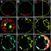
Fig. 1.
Fig. 1.
Subcellular localization of ZmPIPs fused to mYFP or mCFP in maize cells. (A–G) Maize mesophyll protoplasts transiently expressing mYFP::ZmPIP2;1 (A), mCFP::ZmPIP2;5 (B), ROP6::YFP (D), mYFP::ZmPIP1;2 (E), mYFP::ZmPIP1;1 (F), or mYFP::ZmPIP1;6 (G). In C, the protoplast plasma membrane was labeled with the FM4-64 steryl dye. (H–J) Coexpression of mCFP::ZmPIP1;2 with the ER marker, YFP::HDEL. (H) mCFP::ZmPIP1;2 fluorescence. (I) YFP::HDEL fluorescence. (J) Merged images. Chlorophyll autofluorescence (red) in chloroplasts is seen in A, B, E, and J. The diameters of the maize mesophyll protoplasts ranged from 25 to 35 μm.
Fig. 2.
Fig. 2.
The subcellular localization of mYFP::ZmPIP1s is modified by coexpression of mCFP::ZmPIP2s. Protoplasts coexpressing mCFP::ZmPIP2;1 and mYFP::ZmPIP1;1 (A–C); mCFP::ZmPIP2;1 and mYFP::ZmPIP1;2 (D–F); mCFP::ZmPIP2;1 and YFPm::ZmPIP1;6 (G–I); mCFP::ZmPIP2;5 and mYFP::ZmPIP1;1 (J–L); or mCFP::ZmPIP1;2 and mYFP::ZmPIP1;1 (M–O). The CFP channel (Top), the YFP channel (Middle), and the superimposed image for the two channels (Bottom) are shown. Chlorophyll autofluorescence (red) from chloroplasts can be seen in I and L. (C Inset) Magnified image showing unidentified structures in the vicinity of the plasma membrane containing only one type of fluorophore.
Fig. 3.
Fig. 3.
FRET/FLIM analyses of maize protoplasts transiently expressing different combinations of tagged ZmPIP1s and ZmPIP2s. (A–F) mCFP::ZmPIP2;1 alone (A) or coexpressed with mYFP::ZmPIP2;1 (B), ROP6::YFP (negative control) (C), mYFP::ZmPIP1;1 (D), mYFP::ZmPIP1;2 (E), or mYFP::ZmPIP1;6 (F). (G and H) mCFP::ZmPIP2;5 alone (G) or coexpressed with mYFP::ZmPIP1;1 (H). (I–K) mCFP::ZmPIP1;2 alone (I) or coexpressed with mYFP::ZmPIP1;2 (J) or mYFP::ZmPIP1;1 (K). The donor fluorescence lifetime τ was calculated as described in Methods and is indicated by a color code from red for τ values of ≈1 ns to blue for τ values of >2 ns.
Fig. 4.
Fig. 4.
Coimmunopurification of ZmPIP1;2 and ZmPIP2;1 from maize BMS cells and roots. Proteins from BMS suspension cell plasma membranes (A and B) or maize root plasma membranes (C and D) were solubilized and subjected to immunoprecipitation with anti-ZmPIP2;1 antiserum and Protein A Sepharose as described in Methods, and different fractions were analyzed by using anti-ZmPIP2;1/2;2 (A and C) or anti-ZmPIP1;2 (B and D) antibodies. Monomer and dimer forms of each protein are shown (at ≈29 kDa and 55 kDa, respectively). (A and B) Lanes: 1, purified plasma membranes; 2, solubilized proteins; 3, immunoprecipitated proteins; and 4, anti-ZmPIP2;1 antibodies (negative control). (C and D) Lanes: 1, solubilized proteins; 2, immunoprecipitated proteins; and 3, anti-ZmPIP2;1 antibodies. (A and C Upper) H+ ATPase (PMA) was used as a negative control for immunoprecipitation and interaction. The positions of the molecular mass markers are indicated on the y axis.

Similar articles

Cited by

References

    1. Lee JK, Kozono D, Remis J, Kitagawa Y, Agre P, Stroud RM. Proc Natl Acad Sci USA. 2005;102:18932–18937. - PMC - PubMed
    1. Ringler P, Borgnia MJ, Stahlberg H, Maloney PC, Agre P, Engel A. J Mol Biol. 1999;291:1181–1190. - PubMed
    1. Savage DF, Egea PF, Robles-Colmenares Y, O'Connell JD, III, Stroud RM. PLoS Biol. 2003;1:E72. - PMC - PubMed
    1. Murata K, Mitsuoka K, Hirai T, Walz T, Agre P, Heymann JB, Engel A, Fujiyoshi Y. Nature. 2000;407:599–605. - PubMed
    1. Sui H, Han BG, Lee JK, Walian P, Jap BK. Nature. 2001;414:872–878. - PubMed

Publication types

LinkOut - more resources