. 2007 Sep 14;282(37):27155-27164.
doi: 10.1074/jbc.M704408200. Epub 2007 Jul 18.

Anti-inflammatory effects of phosphatidylcholine

Affiliations

Anti-inflammatory effects of phosphatidylcholine

Irina Treede et al. J Biol Chem. .

Abstract

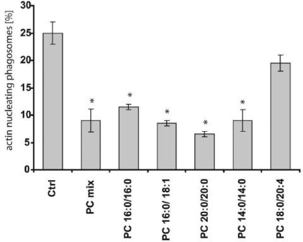
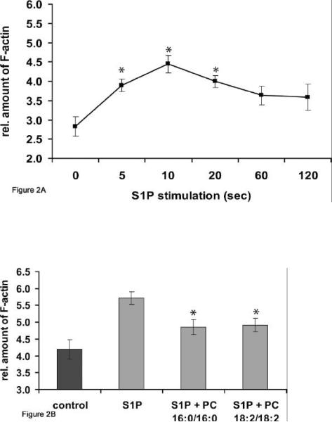
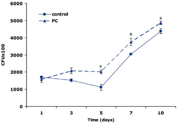
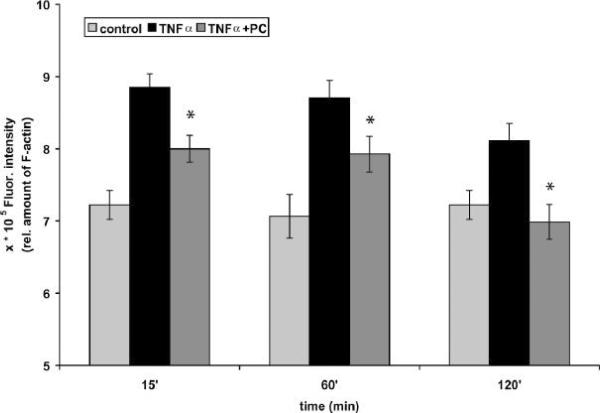
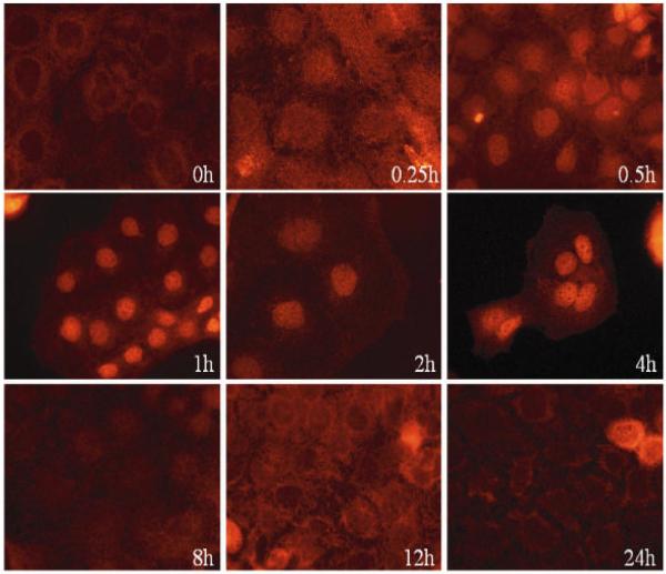
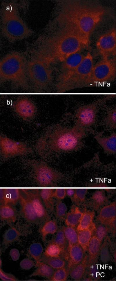
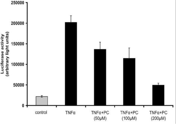
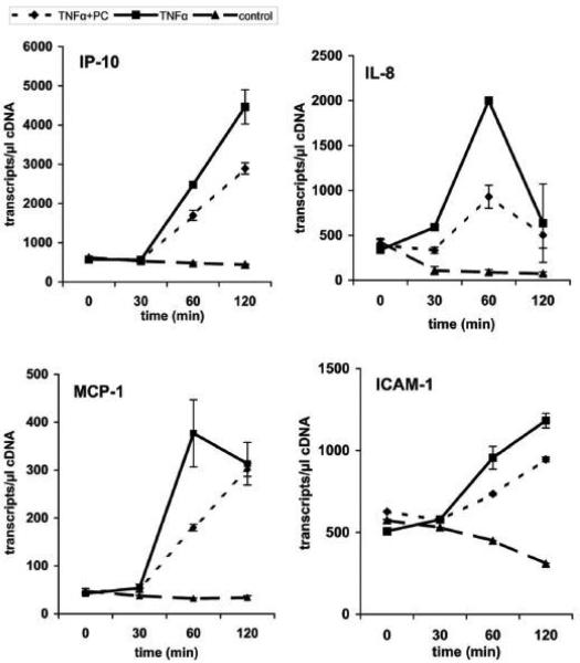
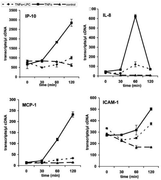
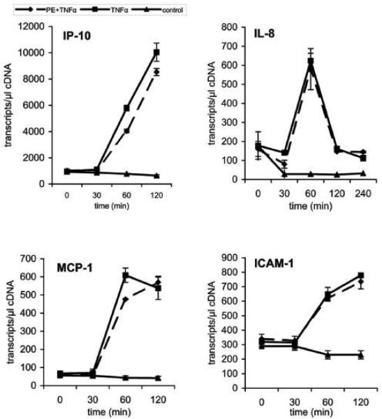
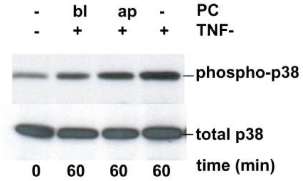
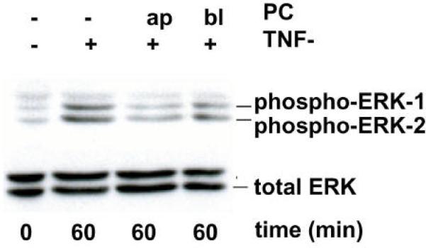
We recently showed that mucus from patients with ulcerative colitis, a chronic inflammatory disorder of the colon, is characterized by a low level of phosphatidylcholine (PC) while clinical studies reveal that therapeutic addition of PC using slow release preparations is beneficial. The positive role of PC in this disease is still elusive. Here we tested the hypothesis that exogenous application of PC has anti-inflammatory properties using three model systems. First, human Caco-2 cells were treated with tumor necrosis factor-alpha (TNF-alpha) to induce a pro-inflammatory response via activation of NF-kappaB. Second, latex bead phagosomes were analyzed for their ability to assemble actin in vitro, a process linked to pro-inflammatory signaling and correlating with the growth versus killing of mycobacteria in macrophages. The third system used was the rapid assembly of plasma membrane actin in macrophages in response to sphingosine 1-phosphate. TNF-alpha induced a pro-inflammatory response in Caco-2 cells, including 1) assembly of plasma membrane actin; 2) activation of both MAPKs ERK and p38; 3) transport of NF-kappaB subunits to the nucleus; and 4) subsequent up-regulation of the synthesis of pro-inflammatory gene products. Exogenous addition of most PCs tested significantly inhibited these processes. Other phospholipids like sphingomyelin or phosphatidylethanolamine showed no effects in these assays. PC also inhibited latex bead phagosome actin assembly, the killing of Mycobacterium tuberculosis in macrophages, and the sphingosine 1-phosphate-induced actin assembly in macrophages. TNF-alpha induces the activation of signaling molecules and the reorganization of the actin cytoskeleton in human intestinal cells. Exogenous application of PC blocks pro-inflammatory signaling in Caco-2 cells, in phagosomes in vitro and facilitates intracellular survival of mycobacteria. We provide further evidence that actin assembly by membranes is part of the pro-inflammatory response. Collectively, these results provide a molecular foundation for the clinical studies showing a beneficial effect of PC therapy in ulcerative colitis.


Figures

Figure 1
Figure 1
Effect of different PC species on actin assembly of isolated phagosomal membranes from J774 macrophages. Phagosomes containing latex beads were treated with a 10 μM preparation of different PC species. Polymerization of actin filaments was clearly inhibited by the different PC species tested in vitro. ATP alone activated the system (ctrl). Columns represent the mean percentages of phagosomes being positive for nucleating actin filaments. The percentages plus and minus standard deviation from three independent experiments are given. Asterisks indicate significant differences (p< 0.05).
Figure 2
Figure 2
Effect of PC 16:0/16:0 and PC 18:2/18:2 on S1P-induced actin assembly in RAW macrophages. The incubation of RAW macrophages with S1P (10nM) for either 5 or 10 seconds demonstrated a very quick F-actin-peak analyzed by a fluorometric assay after staining of F-actin with TRITC-phalloidin (A). RAW macrophages were pre-treated with a 200 μM preparation of either PC 18:2/18:2 (1, 2-dilinoleoyl-phosphatidylcholine) or PC 16:0/16:0 (1, 2-dipalmitoyl-glycero-3- phosphocholine) for 1 h at 37°C. The stimulation of RAW macrophages with S1P (10nM) with PC (S1P + PC 18:2/18:2 or S1P + PC 16:0/16:0) resulted in a significant reduction of actin assembly relative to the F-actin amount of cells stimulated with S1P alone (S1P) for 10 sec (B). Results were compared with relative F-actin amounts of untreated cells (control) within the same experiment using triplicate samples and are representative of three others carried out independently. Values are means ± SD (n=3) with a significance of *p<0.01.
Figure 3
Figure 3
Effect of PC on the intracellular survival of M. tuberculosis H37 RV in J774 macrophages. Cells were infected with bacteria and the effect of a 10 μM PC preparation (natural phosphatidylcholine from bovine brain) on the intracellular survival of bacteria was analyzed after 1 h of bacterial uptake. At indicated times intracellular bacteria were counted after lyses of infected macrophages. The figure clearly shows that the growths of M. tuberculosis was increased by PC-treatment (PC) compared to the intracellular survival of bacteria in untreated macrophages (control). Values are means ± SD (n=3) with a signific ance of *p<0.05.
Figure 4
Figure 4
PC 18:2/18:2 inhibits the TNF-a-induced F-actin assembly in Caco-2 cells. The incubation of cells with TNF-a (50 ng/mL) for 15, 60 and 120 min demonstrated an increased formation of F-actin in Caco-2 cells. Cells were then pre-treated with a 200 μM preparation of PC 18:2/18:2 (1, 2-dilinoleoyl-phosphatidylcholine) for 1 h at 37°C. For fluorometric analyses F-actin was stained with TRITC-phalloidin. The stimulation of Caco-2 cells with TNF-a after pre-treatment with PC (TNF-a + PC) resulted in a decrease of the F-actin amount of cells stimulated with TNF-a alone (TNF-a) for the indicated times. Results were compared to F-actin amounts of untreated cells (control) within the same experiment using triplicate samples. Values are means ± SD (n=3) with a significance of p < 0.05 vs TNF-a alone.
Figure 5
Figure 5
(A) TNF-a-induced NF-?B activation in Caco-2 cells. Cells were grown on glass slides and stimulated with TNF-a (10 ng/mL) at 37°C for various times (5 min to 24 h) to detect nuclear translocation of the p65 subunit of NF-?B by immunofluorescence microscopy with a specific p65 monoclonal antibody (in red). NF-?B activation could be detected already after a few minutes and stayed for at least 4 h. (B) PC 16:0/16:0 clearly inhibits TNF-a induced NF-?B activation. Caco-2 cells were incubated with 10 ng/mL TNF-a for 30 min either in the presence or in the absence of a 200 μM solution of 1, 2-dipalmitoyl-glycero-3-PC (PC16:0/16:0). a.) In absence of TNF-a NF-?B was predominately found in the cytoplasm. b.) After stimulation with TNF-a NF-?B (p65) was translocated into the nucleus. c.) Co-treatment of Caco-2 cells with both PC 16:0/16:0 and TNF-a resulted in a clear inhibition of the p65 translocation relative to the controls. (C) Effect of different phospholipid species on TNF-a induced NF-?B activation. Caco-2 cells were co-treated with both TNF-a (10 ng/mL) and a 200 μM preparation of selected phospholipids for 30 min at 37°C. The TNF-a-induced NF-?B activation was detected by immunofluorescence microscopy. Images of 10 randomly selected areas were taken and NF-?B activation was scored blinded into three groups: (1) strong activation, (2) intermediate activation and (3) no activation. The percentages of the amount of cells in each class of three independent experiments were expressed, respectively. All PC species tested inhibited the TNF-a-induced NF-?B activation in Caco-2 cells. Lysophosphatidylcholine (LPC 16:0) was the most effective one while phosphatidylethanolamine or sphingomyelin was non-effective. (D) PC 18:2/18:2 inhibits TNF-a-mediated NF-?B translocation in a dose dependent manner. Caco-2 cells were assayed for NF-?B activation by transient transfection of a NF-?B luciferase reporter plasmid. 20 h after transfection, cells were stimula ted with either 10 ng/ml human TNF-a alone (TNF-a), co-treated with both TNF-a (10 ng/mL) and PC 18:2/18:2 (1, 2-dilinoleoyl-phosphatidylcholine preparations in various concentrations (TNF-a + PC) or were cultured in TNF-a-free medium (control) for another 4 h. The lysates were assayed for luciferase activity. The differences of the expression levels of TNF-a-stimulated cells compared to cells co-treated with both TNF-a and PC showed a concentration dependent, anti-inflammatory effect. All stimulations were done in triplicates in =3 independent experiments.
Figure 5
Figure 5
(A) TNF-a-induced NF-?B activation in Caco-2 cells. Cells were grown on glass slides and stimulated with TNF-a (10 ng/mL) at 37°C for various times (5 min to 24 h) to detect nuclear translocation of the p65 subunit of NF-?B by immunofluorescence microscopy with a specific p65 monoclonal antibody (in red). NF-?B activation could be detected already after a few minutes and stayed for at least 4 h. (B) PC 16:0/16:0 clearly inhibits TNF-a induced NF-?B activation. Caco-2 cells were incubated with 10 ng/mL TNF-a for 30 min either in the presence or in the absence of a 200 μM solution of 1, 2-dipalmitoyl-glycero-3-PC (PC16:0/16:0). a.) In absence of TNF-a NF-?B was predominately found in the cytoplasm. b.) After stimulation with TNF-a NF-?B (p65) was translocated into the nucleus. c.) Co-treatment of Caco-2 cells with both PC 16:0/16:0 and TNF-a resulted in a clear inhibition of the p65 translocation relative to the controls. (C) Effect of different phospholipid species on TNF-a induced NF-?B activation. Caco-2 cells were co-treated with both TNF-a (10 ng/mL) and a 200 μM preparation of selected phospholipids for 30 min at 37°C. The TNF-a-induced NF-?B activation was detected by immunofluorescence microscopy. Images of 10 randomly selected areas were taken and NF-?B activation was scored blinded into three groups: (1) strong activation, (2) intermediate activation and (3) no activation. The percentages of the amount of cells in each class of three independent experiments were expressed, respectively. All PC species tested inhibited the TNF-a-induced NF-?B activation in Caco-2 cells. Lysophosphatidylcholine (LPC 16:0) was the most effective one while phosphatidylethanolamine or sphingomyelin was non-effective. (D) PC 18:2/18:2 inhibits TNF-a-mediated NF-?B translocation in a dose dependent manner. Caco-2 cells were assayed for NF-?B activation by transient transfection of a NF-?B luciferase reporter plasmid. 20 h after transfection, cells were stimula ted with either 10 ng/ml human TNF-a alone (TNF-a), co-treated with both TNF-a (10 ng/mL) and PC 18:2/18:2 (1, 2-dilinoleoyl-phosphatidylcholine preparations in various concentrations (TNF-a + PC) or were cultured in TNF-a-free medium (control) for another 4 h. The lysates were assayed for luciferase activity. The differences of the expression levels of TNF-a-stimulated cells compared to cells co-treated with both TNF-a and PC showed a concentration dependent, anti-inflammatory effect. All stimulations were done in triplicates in =3 independent experiments.
Figure 6
Figure 6
Down-regulation of the TNF-a-induced gene expression in Caco-2 cells by PC 16:0/16:0 and LPC 16:0. The expression profiles of mRNAs for selected pro-inflammatory genes (ICAM-1, MCP-1, IL-8 and IP-10) are shown. Caco-2 cells were either stimulated with TNF-a (10 ng/ml) alone (TNF-a) or co-treated with TNF-a together with a 200 μM preparation of either (A) 1, 2- dipalmitoyl-glycero-3-phosphocholine (TNF-a + PC), (B) 1-palmitoyl-glycero-3-phosphocholine (TNF-a + LPC) or (C) phosphatidylethanolamine (TNF-a + PE). The expression profiles of mRNA of resting cells for the selected genes were analyzed in parallel (control). The up-regulation of selected pro-inflammatory genes by TNF-a was analyzed after different incubation times (30 to 120 min) and compared to the expression levels of either PC-treated (TNF-a + PC), LPC-treated (TNF-a + LPC) or PE-treated cells (TNF-a + PE). Within this period of time the mRNA expression of the selected genes was strongly delayed after treatment with PC or LPC while PE did not show any significant effect.
Figure 6
Figure 6
Down-regulation of the TNF-a-induced gene expression in Caco-2 cells by PC 16:0/16:0 and LPC 16:0. The expression profiles of mRNAs for selected pro-inflammatory genes (ICAM-1, MCP-1, IL-8 and IP-10) are shown. Caco-2 cells were either stimulated with TNF-a (10 ng/ml) alone (TNF-a) or co-treated with TNF-a together with a 200 μM preparation of either (A) 1, 2- dipalmitoyl-glycero-3-phosphocholine (TNF-a + PC), (B) 1-palmitoyl-glycero-3-phosphocholine (TNF-a + LPC) or (C) phosphatidylethanolamine (TNF-a + PE). The expression profiles of mRNA of resting cells for the selected genes were analyzed in parallel (control). The up-regulation of selected pro-inflammatory genes by TNF-a was analyzed after different incubation times (30 to 120 min) and compared to the expression levels of either PC-treated (TNF-a + PC), LPC-treated (TNF-a + LPC) or PE-treated cells (TNF-a + PE). Within this period of time the mRNA expression of the selected genes was strongly delayed after treatment with PC or LPC while PE did not show any significant effect.
Figure 7
Figure 7
PC 16:0/16:0 (1, 2-dipalmitoyl-glycero-3-phosphocholine) inhibits the TNF-a-mediated activation of both p38 MAP kinase and ERK1/2 pathways in polarized Caco-2 cells. Filter-grown polarized Caco-2 cells were stimulated with TNF-a (50 ng/ml) from the basolateral (bl) surface to activate the MAP kinase pathways. To test the effect of PC on the MAP kinase pathway activation Caco-2 cells were pre-treated with a 200 μM preparation of PC 16:0/16:0 either from the upper (ap) or from the lower (bl) chamber before TNF-a-stimulation for the indicated times. Cell lysates were prepared and whole cell extracts were analyzed by Western blotting. Extracts were immunoblotted with antibodies specific for the phosphorylated (phospho) forms of either (A) p38 MAP kinase or (B) ERK. Caco-2 cells treated with PC showed reduced amounts of the activated forms of both ERK and p38. Equal protein loading was confirmed by protein assay and by control Western blots using antibodies specific for the non-phosphorylated (total) forms of p38 MAP kinase and ERK1/2. All gels were run at least two times and the representative blots of three independent experiments are shown.
Figure 7
Figure 7
PC 16:0/16:0 (1, 2-dipalmitoyl-glycero-3-phosphocholine) inhibits the TNF-a-mediated activation of both p38 MAP kinase and ERK1/2 pathways in polarized Caco-2 cells. Filter-grown polarized Caco-2 cells were stimulated with TNF-a (50 ng/ml) from the basolateral (bl) surface to activate the MAP kinase pathways. To test the effect of PC on the MAP kinase pathway activation Caco-2 cells were pre-treated with a 200 μM preparation of PC 16:0/16:0 either from the upper (ap) or from the lower (bl) chamber before TNF-a-stimulation for the indicated times. Cell lysates were prepared and whole cell extracts were analyzed by Western blotting. Extracts were immunoblotted with antibodies specific for the phosphorylated (phospho) forms of either (A) p38 MAP kinase or (B) ERK. Caco-2 cells treated with PC showed reduced amounts of the activated forms of both ERK and p38. Equal protein loading was confirmed by protein assay and by control Western blots using antibodies specific for the non-phosphorylated (total) forms of p38 MAP kinase and ERK1/2. All gels were run at least two times and the representative blots of three independent experiments are shown.

Similar articles

Cited by

References

    1. Maaser C, Kagnoff MF. Z Gastroenterol. 2002;40:525–529. - PubMed
    1. Wallace JL, Granger DN. Faseb J. 1996;10:731–740. - PubMed
    1. Lichtenberger LM. Annu Rev Physiol. 1995;57:565–583. - PubMed
    1. Kao YC, Lichtenberger LM. Gastroenterology. 1991;101:7–21. - PubMed
    1. Bengmark S, Jeppsson B. JPEN J Parenter Enteral Nutr. 1995;19:410–415. - PubMed

Publication types

MeSH terms