Review
. 2008;7(1):41-52.
doi: 10.1007/s10689-007-9145-9. Epub 2007 Jul 17.

The biochemical basis of microsatellite instability and abnormal immunohistochemistry and clinical behavior in Lynch syndrome: from bench to bedside

Affiliations
Review

The biochemical basis of microsatellite instability and abnormal immunohistochemistry and clinical behavior in Lynch syndrome: from bench to bedside

C Richard Boland et al. Fam Cancer. 2008.

Abstract

Lynch syndrome is an inherited disease caused by a germline mutation in one of four DNA mismatch repair (MMR) genes. The clinical manifestations can be somewhat variable depending upon which gene is involved, and where the mutation occurs. Moreover, the approach to the diagnosis of Lynch syndrome is becoming more complex as more is learned about the disease, and one needs to understand how the DNA MMR proteins function, and what makes them malfunction, to have an optimal appreciation of how to interpret diagnostic studies such as microsatellite instability and immunohistochemistry of the DNA MMR proteins. Finally, an understanding of the role of the DNA MMR system in regulation of the cell cycle and the response to DNA damage helps illuminate the differences in natural history and response to chemotherapeutic agents seen in Lynch syndrome.


Figures

Fig. 1
Fig. 1
DNA MMR (A) Repair of a single nucleotide mismatch in S phase by MutSα. (MutL components not shown here) A. The polymerase has erroneously placed a G in the daughter strand across from a non-complementary T in the template, creating a mismatch. B. The heterodimer of hMSH2 and hMSH6 (MutSα), bound by ADP and in an open configuration, monitors newly synthesized DNA strand for mispairs. Upon encountering the G-T mispair, an exchange of ATP for ADP occurs, and MutSα switches to a closed, sliding clamp along the DNA. C. The sliding clamps can migrate in either direction from the mispair, and as this occurs, additional MutSα clamps may be recruited to the mismatch. The MutSα moving in the 5′ > 3′ direction will eventually encounter the PCNA-DNA polymerase complex, and according to one hypothesis, displace the enzymes involved in DNA synthesis. D. Exonuclease I (together with MutL homologue heterodimers) excises the newly synthesized daughter strand back to the site of the mismatch, removing the erroneous G. E. The error is corrected by resynthesis (from [43], adapted with permission from [44]). (B) Recognition of insertion/deletion errors at microsatellite sequences by MutSβ. Loop-out lesions are caused by “slippage” at repetitive sequences (such as An or [CA]n) during DNA replication, and are recognized by hMSH2 + hMSH3 (MutSβ). In this illustration, the slippage has created a short “loop out” on the nascent strand for the An sequence, which would lead to an insertion frame shift mutation after replication, whereas the slippage is shown on the template strand for the (CA)n repeat, which would lead to a deletion mutation after replication (from [43], adapted with permission from [44])
Fig. 2
Fig. 2
Microsatellite Instability Five mononucleotide repeat sequences (NR21, BAT26, NR24, NR27, and BAT 25) were amplified in a multiplex reaction and separated by HPLC. The upper lanes labeled “normal DNA” represent DNA from lymphocytes, and the lower lanes labeled “CRC tissue” represent DNA extracted from the cancer. In each instance shown here, the microsatellite sequence has undergone a deletion mutation, as indicated by the bold arrow. In this case, all five microsatellites were mutated, and the tumor was designated MSI-H
Fig. 3
Fig. 3
Analysis of mRNA and protein expression of the DNA MMR genes in CRC cell lines with mutations. The upper panel (labeled RT-PCR) with the dark background represents PCR gels. Each of the white bands is a PCR product. The lower panel (labeled Western blots) consists of dark protein bands. There are seven lanes which represent the results from the seven CRC cell lines, as indicated across the top. SW480 is MMR proficient, and is used as a positive control. mRNA is present for each of the MMR genes in the upper panels, and proteins are expressed as shown in each of the lower lanes. (No effective monoclonal antibody was available for PMS1, therefore there are no Western blots for this protein.) HCT116 cells are mutated and deficient for MLH1 and MSH3. HCT116+chr3 cells were derived from HCT116 by the stable transfer of chromosome 3 [12]. This cell line is MMR proficient, but is still MSH3 deficient. LoVo is an MSH2-deficient cell line. DLD1 is an MSH6-deficient cell line. SW48 is deficient in MMR activity because both alleles of MLH1 are methylated and silenced. HCT15 is MSH6-deficient. SW480 (first lane) expresses all six mRNAs for DNA MMR genes, and all five of the proteins, and serves as the normal control. HCT116 demonstrates the consequences of an inactivating mutation in MLH1. The missense mutation creates a weak RT-PCR band for MLH1 but completely removes the protein band for MLH1 and PMS2 (second lane). As an incidental consequence of MSI in this line, MSH3 has undergone a somatic mutation, which accounts for the weak protein band. Restoration of MLH1 by stable transfer of chromosome 3 in the HCT116+chr3 cell line results in a restoration of DNA MMR activity, and the appearance of protein bands for MLH1 and PMS2, as shown in the third lane. There is no change in the status of MSH3 in this cell line. LoVo has mutations that result in the total loss of the RT-PCR band for MSH2, and as a consequence, loss of the protein bands for MSH2, MSH6, and MSH3 (fourth lane). The DLD1 cell line has a mutation in the MSH6 gene. There is still an mRNA for MSH6, but the protein is abnormal, and no band is seen on the Western blot. SW48 has methylation-induced silencing of the MLH1 gene (lane 6). Consequently, there is no mRNA for this gene and no protein product for MLH1 or PMS2. Although not shown here, demethylation of the MLH1 promoter in vitro results in re-expression of MLH1, and restoration of the protein bands for MLH1 and PMS2. HCT15 was derived from DLD1, and has the same mutation in MSH6 (lane 7) [16]
Fig. 4
Fig. 4
Interaction among the DNA MMR proteins. Each of the MutS homologues (MSH3, MSH6, and MSH2) interacts as a heterodimer with the MutL homologues (MLH1, PMS2, PMS1, and MLH3), acting as heterodimers, and the exonuclease, ExoI. Mutations that occur in the interactive domains may abrogate the ability of these proteins to interact and function in DNA MMR, but in some instances, the mutations may not lead to destabilization and loss of the protein product. The interactive domains among the MutS homologues, among the MutL homologues, and between one another are illustrated here. (taken from Boland and Fishel [31])
Fig. 5
Fig. 5
Improved survival in young patients who have CRC with MSI. The ten year survival is significantly better for patients whose CRC tumors are MSI compared with those MSS. As illustrated, this is true for tumors detected at each of the stages. In each instance, survival in the top curve is from patients under age 50 with CRC showing MSI [40]
Fig. 6
Fig. 6
In vitro evidence for resistance to the alkylating agent MNNG by CRC cell lines with deficient DNA MMR activity. (A) The cell lines HCT116, HCT116+ch3M2, LoVo, and 2774 are all DNA MMR deficient, and have intact colony forming ability in the presence of the mutagenic alkylating agent MNNG. The CRC cell lines SW480 and HCT116+ch3 are DNA MMR proficient, and cannot form colonies after exposure to 5 μM MNNG [6]. (B) Similarly, after exposure to 5 μM 5-FU, which is a pharmacologically relevant dose of this drug, the DNA MMR deficient cell lines all tolerated treatment, where as the DNA MMR proficient cell line (HCT116+ch3 and SW480) were relatively unable to form colonies. Note that the y-axis is a log scale [39]. (C) The cell line SW48 is DNA MMR-deficient because of methylation-induced silencing of the MLH1 gene. However, after treatment with the demethylating agent 5 aza-dC, the MLH1 protein is re-expressed, and the cell in rendered MMR proficient. Using a clonagenic assay, the DNA MMR deficient cell lines (HCT116, HCT116+ch2, and SW48) are all resistant to the cytotoxic effects of 5-FU, whereas the MMR proficient cell line HCT116+chr3 and SW48 after demethylation both show sensitivity to 5-FU [15]
Fig. 7
Fig. 7
Clinical outcomes in CRC patients treated with 5-FU adjuvant chemotherapy depends upon the DNA MMR status. (A) The upper panel shows survival in patients with Stage II or Stage III CRC not given adjuvant chemotherapy. The dotted line on the top cohort (those whose tumors showed MSI) survived significantly longer than patients whose CRCs did not show MSI. In the lower panel, the use of 5-FU based chemotherapy results in an improvement in patients whose tumor are MSS, and the two survival curves converge [40]. (B) Survival was examined in patients with CRC, and analyzed based upon MSI status. The upper panel shows the impact of 5-FU based chemotherapy in patients with no MSI. The dotted line shows that patients treated with adjuvant chemotherapy had a significant improvement in survival. However, in the lower panel, patients had tumors with MSI, and the addition of 5-FU-based chemotherapy did not improve survival, and may have resulted in a slight reduction in overall survival (difference is not significant, P = 0.10). This indicates that 5-FU based chemotherapy does not provide a survival advantage in patients with MSI CRCs, and helps in the selection of patients who might benefit best from this intervention [40]

References

    1. Lynch HT, de la Chapelle A. Hereditary colorectal cancer. N Engl J Med. 2003;348(10):919–932. - PubMed
    1. Acharya S, Wilson T, Gradia S, et al. hMSH2 forms specific mispair-binding complexes with hMSH3 and hMSH6. Proc Natl Acad Sci USA. 1996;93(24):13629–13634. - PMC - PubMed
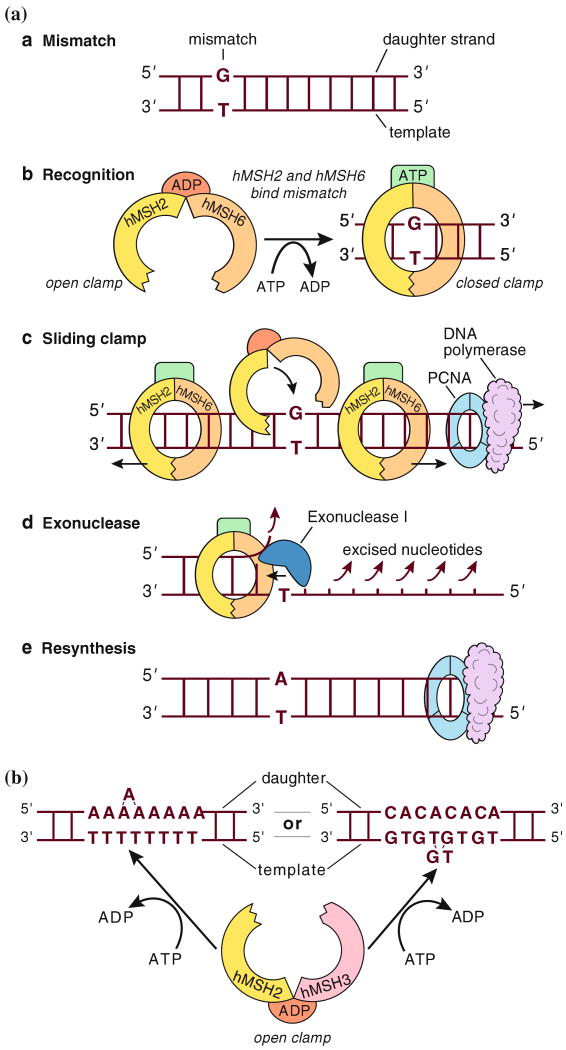
    1. Gradia S, Subramanian D, Wilson T, et al. hMSH2-hMSH6 forms a hydrolysis-independent sliding clamp on mismatched DNA. Mol Cell. 1999;3(2):255–261. - PubMed
    1. Kadyrov FA, Dzantiev L, Constantin N, Modrich P. Endonucleolytic function of MutLalpha in human mismatch repair. Cell. 2006;126(2):297–308. - PubMed
    1. Heinen CD, Schmutte C, Fishel R. DNA repair and tumorigenesis: lessons from hereditary cancer syndromes. Cancer Biol Ther. 2002;1(5):477–485. - PubMed

MeSH terms