. 2007 Aug;7(7):738-45.
doi: 10.1016/j.modgep.2007.06.003. Epub 2007 Jun 15.

Mrf4 (myf6) is dynamically expressed in differentiated zebrafish skeletal muscle

Affiliations

Mrf4 (myf6) is dynamically expressed in differentiated zebrafish skeletal muscle

Yaniv Hinits et al. Gene Expr Patterns. 2007 Aug.

Abstract

Mrf4 (Myf6) is a member of the basic helix-loop-helix (bHLH) myogenic regulatory transcription factor (MRF) family, which also contains Myod, Myf5 and myogenin. Mrf4 is implicated in commitment of amniote cells to skeletal myogenesis and is also abundantly expressed in many adult muscle fibres. The specific role of Mrf4 is unclear both because mrf4 null mice are viable, suggesting redundancy with other MRFs, and because of genetic interactions at the complex mrf4/myf5 locus. We report the cloning and expression of an mrf4 gene from zebrafish, Danio rerio, which shows conservation of linkage to myf5. Mrf4 mRNA accumulates in a subset of terminally differentiated muscle fibres in parallel with myosin protein in the trunk and fin. Although most, possibly all, trunk muscle expresses mrf4, the level of mRNA is dynamically regulated. No expression is detected in muscle precursor cell populations prior to myosin accumulation. Moreover, mrf4 expression is not detected in head muscles, at least at early stages. As fish mature, mrf4 expression is pronounced in the region of slow muscle fibres.


Figures
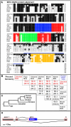
Figure 1
Figure 1. Family relationship, sequence and structural comparison of Mrf4
A. Clustal alignment of the conceptual translation product of the zebrafish mrf4 gene (Genebank accession No. AY335193) with representatives of other major vertebrate groups (human, mouse, chicken, Xenopus laevis and Fugu rubripes). The blue line above the sequence encoded by the third exon indicates the region missing in the alternatively spliced mRNA, which encodes the residues indicated. B. Sequence pair distances of representative Mrf4 proteins (top) and bHLH domains (bottom) using the Clustal method with PAM 250 residue weight table. C. An evolutionary tree showing the phylogenetic distance among predicted mrf4 proteins. D. Exon structure of mrf4, showing the DANA retrotransposon insertion (blue) and accompanying alternative splice. Canonical exons are shaded. The termination of the 3′ untranslated region in exons 3 and alt. 3 is unknown. Distances are approximately to scale.
Figure 2
Figure 2. Mrf4 mRNA is expressed in terminally differentiated skeletal muscle
In situ mRNA hybridisation for mrf4 (purple, A-N,P,Q,R,S), myoD (red, B,H), myogenin (myog red, D; purple, O) actin (actc red, A,C) prdm1 (red, F) and wholemount immunohistochemistry for MyHC (brown, G,J,S) in zebrafish embryos, viewed in dorsal flatmount (A-H), wholemount (K,L,N centre,O,Q,S lateral view; I,N right,P left dorsal view; N left ventral view) or transverse cryosection (J,M,R). Anterior is to the top (A-I,K,S), or left (L, N-Q) and dorsal to top (M,R) A. Mrf4 is first detected adaxially in each somite at 5-6s, after actin (right panel). The horizontal line indicates the newest somite border (A-H). B-D. At 10-12s, mrf4 mRNA is restricted to somitic adaxial cells, whereas myoD (B) and actin (C) expression are strong in adaxial cells of presomitic mesoderm (arrows) and myod (B) and myog (D) in the lateral fast muscle precursors (arrowheads). Myogenin mRNA (D) shows that mrf4 is not expressed in fast precursors at this stage. Dual immunohistochemical detection of two mRNAs in a single embryo reveals expression of mrf4 exclusively in the adaxial location of slow muscle obscuring the red myod or actin label. Note that myogenin expression is not detected in presomitic adaxial cells (Weinberg et al., 1996) due to short incubation with substrate. E-H. Mrf4 is down regulated in rostral somites before 14s (E). Mrf4 and prdm1 mRNAs are co-expressed in differentiated adaxial cells whereas prdm1 alone is expressed in presomitic adaxial cells (F). Mrf4 transcripts co-localise with MyHC protein in adaxial cells of the newly-forming somite (G, asterisks). Mrf4 expression is strongest in the six most recently formed somites, obscuring the MyHC signal (G, arrowheads; box shown magnified at right). In more mature somites, declining mrf4 is detected in a similar location (G, bracketed region shown in inset, green arrowheads), whereas MyHC-expressing slow fibres are increasing (G inset, arrows). By 18s, mrf4 is expressed most strongly in six nascent somites (H). I. At 22s, mrf4 is up-regulated once more in rostral somites. J. By 25s, mrf4 mRNA is present throughout the somite and at high level in slow fibres after their migration (arrowheads). Re-staining of the same section shows that slow MyHC (F59) overlaps the high mrf4 expressing cells (arrowheads, right panel). K. At 26hpf, mrf4 is broadly expressed in muscle. L,M. Mrf4 expression at 48hpf appears stronger in the superficial slow muscle cells. N,O. Mrf4 and myogenin are expressed in dorsal and ventral muscle masses of pectoral fin at 48hpf (arrows). High magnifications of the fin show that mrf4 and myogenin expression in dorsal and ventral muscle masses. Myogenin is detected in numerous ventral head muscle anlage (O left), but mrf4 mRNA shows no such pattern (N left). P. Mrf4 mRNA persists in pectoral fin at 72hpf. Q,R. Mrf4 is weakly expressed at 5d and is more readily detected superficial slow cells. S. Embryos injected with myf5 and myoD antisense morpholinos have no MyHC and lack mrf4 expression at 12s.
Figure 3
Figure 3. Mrf4 mRNA is expressed late in muscle terminal differentiation
In situ mRNA hybridisation for mrf4 (blue, A,C-K), eng2a (B) and mylz2 (K) and wholemount immunohistochemistry for MyHC (brown, B,F) in zebrafish embryos viewed in wholemount (E,G,H,K lateral view, dorsal to right) or flatmount (A-D,J, dorsal view), anterior to top, or transverse section (F,I; dorsal to top). A. Mrf4 expression is missing in gli2aty119/ty119 embryos and reduced in heterozygotes gli2aty119/+ compared to wild type siblings. B. At 15s, shha mutant embryos lack eng2a-expressing MPs, but have residual MyHC-expressing SSF precursors (black arrows), whereas sibling embryos show eng2a expression in MPs anterior to the three youngest somites (between red arrows) and MyHC in SSF precursors (black arrows). C. shha mutants show no mrf4 MP expression prior to 16s, in contrast to their siblings. D. Similarly, prdm1 mutants show faint mrf4 expression in adaxial cells but lack transient strong expression. E. Mrf4 is widely expressed at 24 hpf in wild type. Anteriorly, mrf4 expression fills the bulk of the somite chevron. Note transient dorsoventral midline expression in the most caudal newly-formed somites (arrowhead). shha mutant lacks MP mrf4 staining in caudal somites (arrowhead), but retains strong expression in more anterior U-shaped somites (arrow). F. Rostral section of a 21s shha embryo, identified by U-shape somites and lack of caudal MP mrf4 expression, showing that MyHC closely co-localises with mrf4 mRNA in differentiated SSF precursor fibres migrating towards the lateral surface of the somite. Eosin counterstain. G,H. At 24-25s, both prdm1 (G) and gli2a (H) mutants and sibling show mrf4 expression in fast muscle cells in more rostral somites, whereas only the sibling shows caudal slow expression (arrowheads). I. Gli2a mutant with no slow muscle (MPs or SSFs) expresses mrf4 in fast muscle cells. J. Fgf8 morphants show no change in mrf4 expression at 15s. K. Mylz2 (fast myosin light chain 2) co-localizes with mrf4 in fast fibres but not in slow fibres (arrowhead) in fgf8 mutants and siblings. In fgf8 mutants, mrf4 mRNA is reduced in parallel with differentiated fast muscle (insets), but is unchanged in slow.

Similar articles

Cited by

References

    1. Baxendale S, Davison C, Muxworthy C, Wolff C, Ingham PW, Roy S. The B-cell maturation factor Blimp-1 specifies vertebrate slow-twitch muscle fiber identity in response to Hedgehog signaling. Nat Genet. 2004;36:88–93. - PubMed
    1. Bergstrom DA, Tapscott SJ. Molecular distinction between specification and differentiation in the myogenic basic helix-loop-helix transcription factor family. Mol Cell Biol. 2001;21:2404–12. - PMC - PubMed
    1. Blagden CS, Currie PD, Ingham PW, Hughes SM. Notochord induction of zebrafish slow muscle mediated by Sonic Hedgehog. Genes Dev. 1997;11:2163–2175. - PMC - PubMed
    1. Bober E, Lyons GE, Braun T, Cossu G, Buckingham M, Arnold HH. The muscle regulatory gene, Myf-6, has a biphasic pattern of expression during early mouse development. J Cell Biol. 1991;113:1255–1265. - PMC - PubMed
    1. Braun T, Arnold HH. Inactivation of Myf-6 and Myf-5 genes in mice leads to alterations in skeletal muscle development. EMBO J. 1995;14:1176–1186. - PMC - PubMed

Publication types

MeSH terms

LinkOut - more resources