. 2007 Nov 1;110(9):3398-406.
doi: 10.1182/blood-2007-06-094748. Epub 2007 Jul 17.

The Laminin 511/521-binding site on the Lutheran blood group glycoprotein is located at the flexible junction of Ig domains 2 and 3

Affiliations

The Laminin 511/521-binding site on the Lutheran blood group glycoprotein is located at the flexible junction of Ig domains 2 and 3

Tosti J Mankelow et al. Blood. .

Abstract

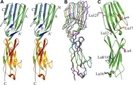
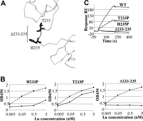
The Lutheran blood group glycoprotein, first discovered on erythrocytes, is widely expressed in human tissues. It is a ligand for the alpha5 subunit of Laminin 511/521, an extracellular matrix protein. This interaction may contribute to vaso-occlusive events that are an important cause of morbidity in sickle cell disease. Using x-ray crystallography, small-angle x-ray scattering, and site-directed mutagenesis, we show that the extracellular region of Lutheran forms an extended structure with a distinctive bend between the second and third immunoglobulin-like domains. The linker between domains 2 and 3 appears to be flexible and is a critical determinant in maintaining an overall conformation for Lutheran that is capable of binding to Laminin. Mutagenesis studies indicate that Asp312 of Lutheran and the surrounding cluster of negatively charged residues in this linker region form the Laminin-binding site. Unusually, receptor binding is therefore not a function of the domains expected to be furthermost from the plasma membrane. These studies imply that structural flexibility of Lutheran may be essential for its interaction with Laminin and present a novel opportunity for the development of therapeutics for sickle cell disease.


Figures

Figure 1
Figure 1
Structure of Lu gp D1D2. (A) Graphic representation in stereo showing the Lu gp structure. D1 is shown at the top and D2 at the bottom. The strands in each domain are labeled, as are the amino (N) and carboxy termini (C). (B) Overlay of Cα traces from the 3 independent copies of D1D2 in the 2 crystal structures. Orientation is as in panel A. (C) Location of residues that are altered in the known inherited forms of Lu gp that can be attributed to D1D2. Mutated residues are shown in stick form, and are colored as follows: Lu a/b is shown in red; Lu4, purple; Lu5, yellow; Lu8/14, blue; Lu12, pink; Lu16, gray; and Lu17, orange.
Figure 2
Figure 2
SAXS analysis of Lu gp D1D2D3. (A) Experimental scattering data for Lu gp D1D2D3 plotted in black as a function of q and compared with a theoretic fit generated with SASREF. (B) Shapes obtained using a bead model using the program GASBOR from 20 independent simulations, each differently colored. The average shape is shown as orange wire-frame density in (C) representative rigid-body fit of the D1D2 crystal structure and D3 model to the scattering data, overlaid on average density obtained as in panel B. D1 is shown in blue; D2, green; and D3, red. (D) Shapes obtained from the scattering curve for Lu gp (D1-D5) as described in panel B. The average shape is shown as orange wire-frame density in (E) overlay of the D1D2D3 model on the envelope derived for Lu gp (D1-D5). D4 and D5 are expected to occupy the additional density at the base of the figure, proximal to the cell membrane.
Figure 3
Figure 3
Mutation of acidic residues of Lu gp reduces Ln511/521 binding. (A) Representative ELISA titrations of 5, 0.5, 0.05, and 0.005 nM Lu gp binding to 5 nM Ln511/521 for the E309A, D310A, D312A, and D315A (solid line) mutations in comparison with native Lu gp (dashed line). Standard deviation for each point is less than 0.1. ELISA results of all other mutations can be seen in Figure S3. (B) The level of binding to Ln511/521 of mutant Lu gp compared with the native protein at 0.05 nM measured in an ELISA. Proteins were assayed in duplicate and the results shown are the mean of 2 separate ELISA plates and are expressed as the percentage of the absorption seen from wells containing native Lu gpFc (OD450 = 0.95). (C) Ln511/521 binding assessed by surface plasmon resonance using a Biacore X. Shown is the mean (± SEM) change in response units (RU) over the course of a 100-μL injection of 10 nM Ln511/521 for 2 assays per protein. In both panels B-C, Ln511/521 binding to Muc18 acted as a negative control. (D) Overlaid sensorgrams for a representative sample of the different proteins showing the association and dissociation curves of Ln511/521 binding to Lu gpFc. Vertical arrows indicate the beginning and end points of the Ln511/521 injection.
Figure 4
Figure 4
Location of charged amino acid residues involved in interactions with Ln511/521 on the surface of Lu gp. (A) Lu gp domains D1D2 (dark gray) and D3 (light gray) are shown in an orientation consistent with the SAXS envelope. Indicated are the positions of acidic residues that have been mutated to alanine. The residues are color coded according to their effect on Ln511/521 binding: blue indicates no effect; yellow, slight effect; orange, marked effect; and red, severe effect. (B) Lu D1D2D3 in the same orientation, shown as an electrostatic surface.
Figure 5
Figure 5
Ln511/521 binding to D2-D3 linker mutants. (A) Three proteins, T233P, H235P, and Δ233-235 (darker shade), containing mutations within the domain 2 to 3 hinge were made. (B) Representative ELISA titrations of hinge region mutants to Ln511/521 in comparison with binding to the native protein. Standard deviations were less than 0.1 OD units for any point. (C) The sensorgrams of representative Biacore assays with these mutants are shown.

Similar articles

Cited by

References

    1. Parsons SF, Mallinson G, Holmes CH, et al. The Lutheran blood group glycoprotein, another member of the immunoglobulin superfamily, is widely expressed in human tissues and is developmentally regulated in human liver. Proc Natl Acad Sci U S A. 1995;92:5496–5500. - PMC - PubMed
    1. Parsons SF, Mallinson G, Judson PA, et al. Evidence that the Lub blood group antigen is located on red cell membrane glycoproteins of 85 and 78 kd. Transfusion. 1987;27:61–63. - PubMed
    1. Campbell IG, Foulkes WD, Senger G, et al. Molecular cloning of the B-CAM cell surface glycoprotein of epithelial cancers: a novel member of the immunoglobulin superfamily. Cancer Res. 1994;54:5761–5765. - PubMed
    1. Rahuel C, Le Van KC, Mattei MG, Cartron JP, Colin Y. A unique gene encodes spliceoforms of the B-cell adhesion molecule cell surface glycoprotein of epithelial cancer and of the Lutheran blood group glycoprotein. Blood. 1996;88:1865–1872. - PubMed
    1. El Nemer W, Colin Y, Bauvy C, et al. Isoforms of the Lutheran/basal cell adhesion molecule glycoprotein are differentially delivered in polarized epithelial cells: mapping of the basolateral sorting signal to a cytoplasmic di-leucine motif. J Biol Chem. 1999;274:31903–31908. - PubMed

Publication types

MeSH terms