

HER4 D-box sequences regulate mitotic progression and degradation of the nuclear HER4 cleavage product s80HER4
- PMID: 17638867
- PMCID: PMC2917069
- DOI: 10.1158/0008-5472.CAN-06-4145
HER4 D-box sequences regulate mitotic progression and degradation of the nuclear HER4 cleavage product s80HER4
Abstract
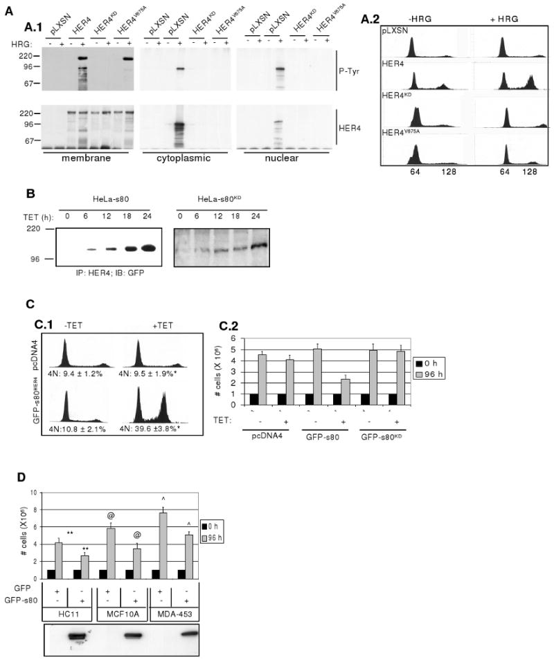
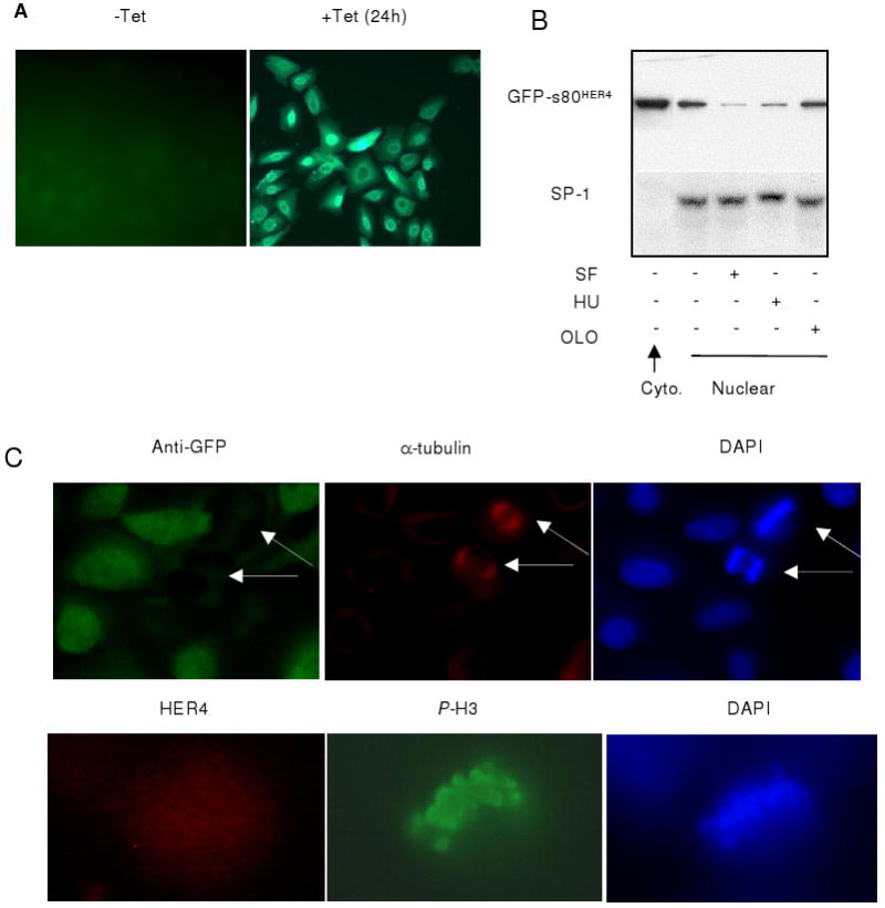
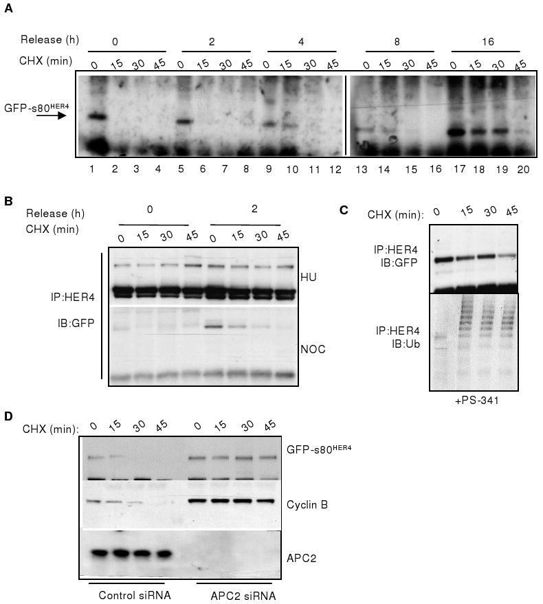
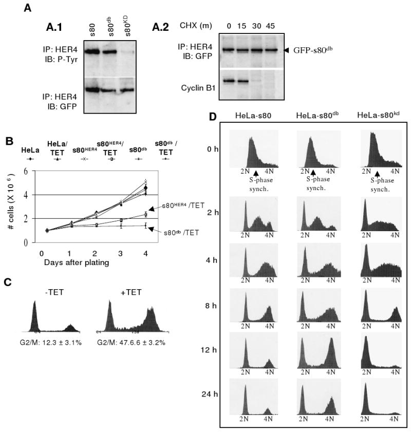
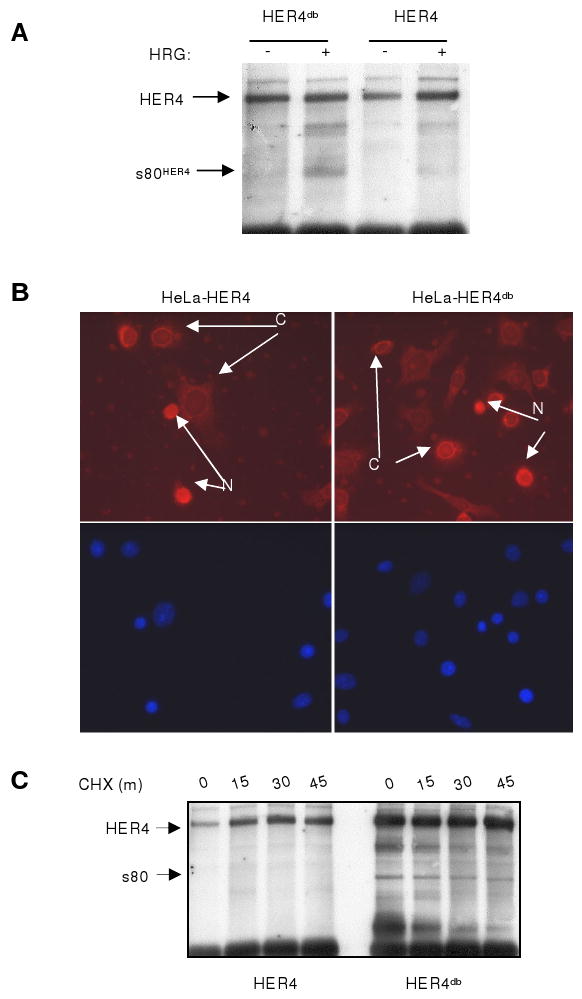
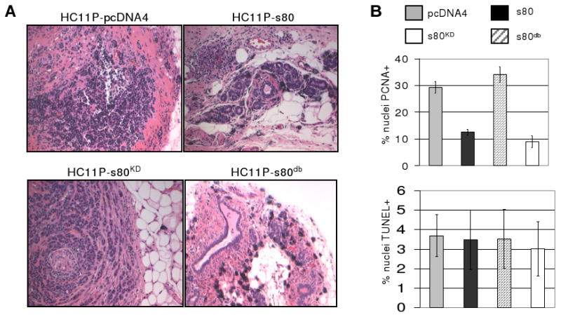
Heregulin-mediated activation of HER4 initiates receptor cleavage (releasing an 80-kDa HER4 intracellular domain, s80(HER4), containing nuclear localization sequences) and results in G(2)-M delay by unknown signaling mechanisms. We report herein that s80(HER4) contains a functional cyclin B-like sequence known as a D-box, which targets proteins for degradation by anaphase-promoting complex (APC)/cyclosome, a multisubunit ubiquitin ligase. s80(HER4) ubiquitination and proteasomal degradation occurred during mitosis but not during S phase. Inhibition of an APC subunit (APC2) using short interfering RNA knockdown impaired s80(HER4) degradation. Mutation of the s80(HER4) D-box sequence stabilized s80(HER4) during mitosis, and s80(HER4)-dependent growth inhibition via G(2)-M delay was significantly greater with the D-box mutant. Polyomavirus middle T antigen-transformed HC11 cells expressing s80(HER4) resulted in smaller, less proliferative, more differentiated tumors in vivo than those expressing kinase-dead s80(HER4) or the empty vector. Cells expressing s80(HER4) with a disrupted D-box did not form tumors, instead forming differentiated ductal structures. These results suggest that cell cycle-dependent degradation of s80(HER4) limits its growth-inhibitory action, and stabilization of s80(HER4) enhances tumor suppression, thus providing a link between HER4-mediated growth inhibition and cell cycle control.
Figures
Similar articles
-
The E3 ubiquitin ligase WWP1 selectively targets HER4 and its proteolytically derived signaling isoforms for degradation.Mol Cell Biol. 2009 Feb;29(3):892-906. doi: 10.1128/MCB.00595-08. Epub 2008 Dec 1. Mol Cell Biol. 2009. PMID: 19047365 Free PMC article.
-
The HER4 cytoplasmic domain, but not its C terminus, inhibits mammary cell proliferation.Mol Endocrinol. 2007 Aug;21(8):1861-76. doi: 10.1210/me.2006-0101. Epub 2007 May 15. Mol Endocrinol. 2007. PMID: 17505063 Free PMC article.
-
The intracellular domain of ErbB4 induces differentiation of mammary epithelial cells.Mol Biol Cell. 2006 Sep;17(9):4118-29. doi: 10.1091/mbc.e06-02-0101. Epub 2006 Jul 12. Mol Biol Cell. 2006. PMID: 16837552 Free PMC article.
-
Impressionist portraits of mitotic exit: APC/C, K11-linked ubiquitin chains and Cezanne.Cell Cycle. 2019 Mar-Apr;18(6-7):652-660. doi: 10.1080/15384101.2019.1593646. Epub 2019 Mar 28. Cell Cycle. 2019. PMID: 30874463 Free PMC article. Review.
-
Processive ubiquitin chain formation by the anaphase-promoting complex.Semin Cell Dev Biol. 2011 Aug;22(6):544-50. doi: 10.1016/j.semcdb.2011.03.009. Epub 2011 Apr 6. Semin Cell Dev Biol. 2011. PMID: 21477659 Free PMC article. Review.
Cited by
-
The Yin and Yang of ERBB4: Tumor Suppressor and Oncoprotein.Pharmacol Rev. 2022 Jan;74(1):18-47. doi: 10.1124/pharmrev.121.000381. Pharmacol Rev. 2022. PMID: 34987087 Free PMC article. Review.
-
The E3 ubiquitin ligase WWP1 selectively targets HER4 and its proteolytically derived signaling isoforms for degradation.Mol Cell Biol. 2009 Feb;29(3):892-906. doi: 10.1128/MCB.00595-08. Epub 2008 Dec 1. Mol Cell Biol. 2009. PMID: 19047365 Free PMC article.
-
ErbB4 splice variants Cyt1 and Cyt2 differ by 16 amino acids and exert opposing effects on the mammary epithelium in vivo.Mol Cell Biol. 2009 Sep;29(18):4935-48. doi: 10.1128/MCB.01705-08. Epub 2009 Jul 13. Mol Cell Biol. 2009. PMID: 19596786 Free PMC article.
-
ErbB4/HER4: role in mammary gland development, differentiation and growth inhibition.J Mammary Gland Biol Neoplasia. 2008 Jun;13(2):235-46. doi: 10.1007/s10911-008-9080-x. Epub 2008 Apr 25. J Mammary Gland Biol Neoplasia. 2008. PMID: 18437540 Free PMC article. Review.
-
HER4 intracellular domain (4ICD) activity in the developing mammary gland and breast cancer.J Mammary Gland Biol Neoplasia. 2008 Jun;13(2):247-58. doi: 10.1007/s10911-008-9076-6. Epub 2008 May 13. J Mammary Gland Biol Neoplasia. 2008. PMID: 18473151 Free PMC article. Review.
References
-
- Harper JW, Burton JL, Solomon MJ. The anaphase-promoting complex: it's not just for mitosis any more. Genes Dev. 2002;16:2179–2206. - PubMed
-
- Peters JM. The anaphase-promoting complex: proteolysis in mitosis and beyond. Mol Cell. 2002;9:931–943. - PubMed
-
- Glotzer M, Murray AW, Kirschner MW. Cyclin is degraded by the ubiquitin pathway. Nature. 1991;349:132–138. - PubMed
-
- Visintin R, Prinz S, Amon A. CDC20 and CDH1: a family of substrate-specific activators of APC-dependent proteolysis. Science. 1997;278:460–463. - PubMed
-
- Zachariae W, Nasmyth K. Whose end is destruction: cell division and the anaphase-promoting complex. Genes Dev. 1999;13:2039–2058. - PubMed
Publication types
MeSH terms
Substances
Grants and funding
LinkOut - more resources
Full Text Sources
Molecular Biology Databases
Miscellaneous
