. 2007 Jul 15;21(14):1767-78.
doi: 10.1101/gad.1565107.

LEDGF/p75 functions downstream from preintegration complex formation to effect gene-specific HIV-1 integration

Affiliations

LEDGF/p75 functions downstream from preintegration complex formation to effect gene-specific HIV-1 integration

Ming-Chieh Shun et al. Genes Dev. .

Abstract

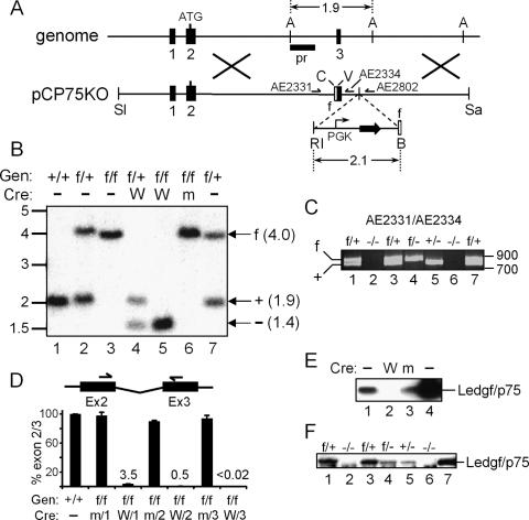
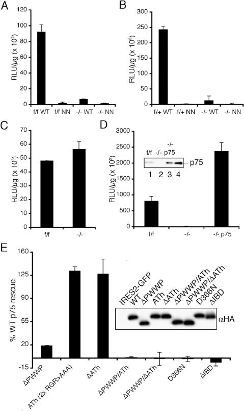
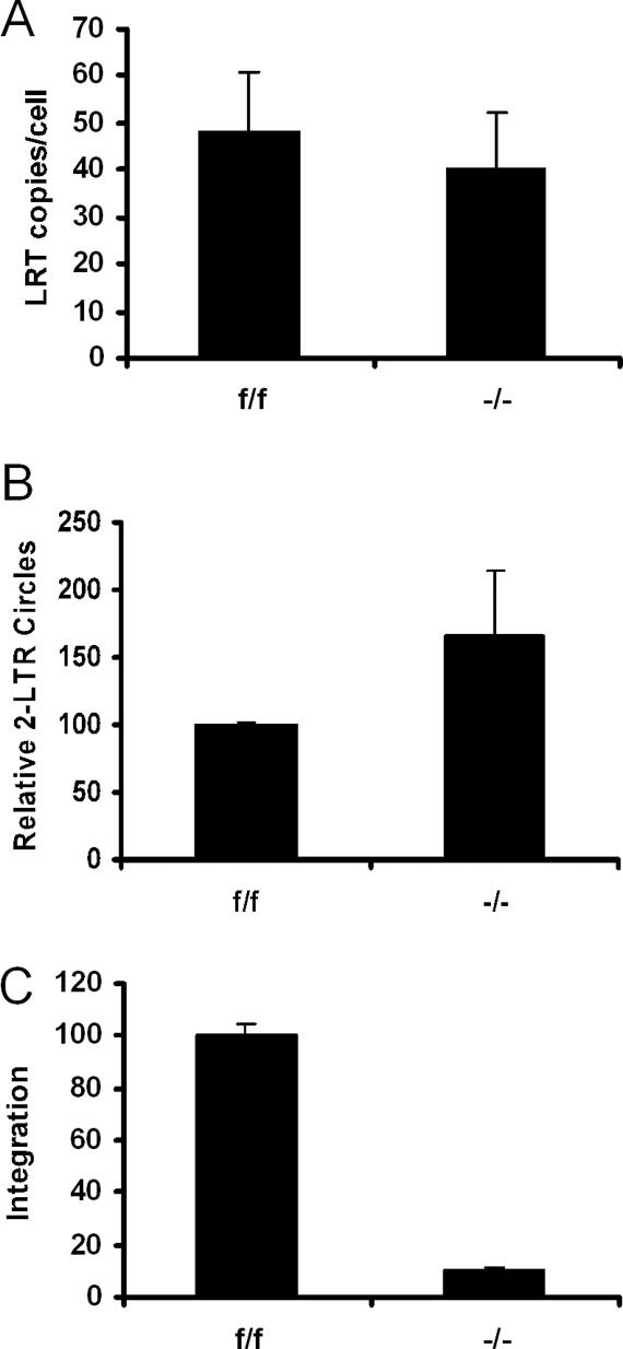
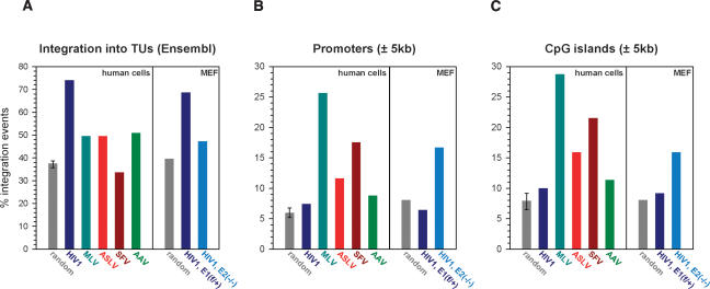
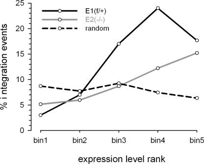
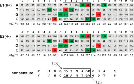
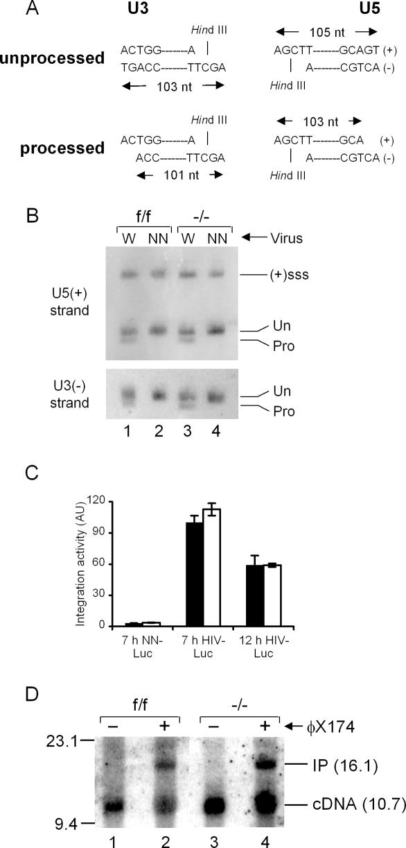
LEDGF/p75 directly interacts with lentiviral integrase proteins and can modulate their enzymatic activities and chromosomal association. A novel genetic knockout model was established that allowed us for the first time to analyze HIV-1 integration in the absence of LEDGF/p75 protein. Supporting a crucial role for the cofactor in viral replication, HIV-1 vector integration and reporter gene expression were significantly reduced in LEDGF-null cells. Yet, integrase processed the viral cDNA termini normally and maintained its local target DNA sequence preference during integration. Preintegration complexes extracted from knockout cells moreover supported normal levels of DNA strand transfer activity in vitro. In contrast, HIV-1 lost its strong bias toward integrating into transcription units, displaying instead increased affinity for promoter regions and CpG islands. Our results reveal LEDGF/p75 as a critical targeting factor, commandeering lentiviruses from promoter- and/or CpG island-proximal pathways that are favored by other members of Retroviridae. Akin to yeast retrotransposons, disrupting the lentiviral targeting mechanism significantly perturbs overall integration.


Figures

Figure 1.
Figure 1.
Creation and characterization of Psip1/Ledgf knockout cells. (A) The pCP75KO targeting vector harbors ∼7.5 kb of chromosome 4 (Psip1 exons 1–3); the 2.1-kb EcoRI (RI)–BamHI (B) fragment confers resistance to neomycin (horizontal arrows). (A) AflIII; (SI) SalI; (C) ClaI; (V) EcoRV; (Sa) SacII; (pr) Southern blotting probe; (half-head arrows) PCR primers; (empty boxes) flox (f) sites. Cre deletes ∼2.6 kb, yielding an ∼1.4-kb AflIII fragment. (B) Southern analysis of AflIII-digested DNA. (Lanes 13) Samples from wild-type, f/+, or f/f MEFs. (Lanes 46) DNA prepared after three rounds of transduction with wild-type (W) or mutant (m) Cre expression vector. (Lane 7) Founder B5a ES cell DNA. Migration positions (in kilobases) of size standards are indicated at the left. (Gen) Genotype. (C) PCR analysis of indicated MEF cell DNAs. AE2331/AE2334 amplify 802-bp and 745-bp f and wild-type allele products, respectively. Recombination removes AE2334-specific DNA, yielding a novel 324-bp AE2331/AE2802 product (not shown). (Lane 7) B5a ES cell DNA; standard migrations are to the right. (D) mRNA levels in f/f and +/+ MEFs (set to 100%) following one, two, or three rounds of exposure to W or m Cre. Standard errors from duplicate qRT–PCR assays are shown. (E) Western analysis of 1.3 μg of HeLa (lane 1) or f/f cell nuclear proteins after three rounds of exposure to W (lane 2) or m (lane 3) Cre. (Lane 4) Five nanograms of purified LEDGF/p75 protein. (F) Western analysis of nuclear proteins (5 μg) from indicated primary MEF cultures. (Lane 7) One nanogram of purified protein.
Figure 2.
Figure 2.
LEDGF/p75 requirements for HIV-1 infection. (A) Normalized Luc values in f/f and −/− cell lysates following infection with wild-type (WT) or NN mutant virus. (B) Same as in A, except the f/+ and −/− cell pair was analyzed. (C) Normalized MLV-Luc infectivities. (D) LEDGF/p75 expression rescues HIV-1 infection in −/− cells. NN-Luc activities were subtracted from the presented values. (Inset) Western analysis of 5 μg of f/f (lane 1), −/− (lane 2), and −/− p75 (lane 3) nuclear extract. (Lane 4) One nanogram of purified protein. (E) LEDGF/p75 effector functions important for HIV-1 infection. NN-Luc-corrected values expressed as percent wild-type LEDGF/p75 activity. (Inset) Anti-HA immunoblot of 160 ng of whole-cell extract. The C-terminal HA tag reduced wild-type LEDGF/p75 function approximately twofold, but did not affect the fractional activity of any mutant protein (data not shown). (AD) Error bars derived from duplicate Luc assays of duplicate infections. (E) Averages from a minimum of two independent experiments (duplicate Luc assays of quadruplicate infections).
Figure 3.
Figure 3.
HIV-1 integration is severely reduced in Ledgf knockout cells. (A) LRT products normalized on a per cell basis. (B) 2-LTR circle levels normalized to f/f cell content (set to 100%). (C) Integration in −/− cells relative to f/f cells (set to 100%). Standard errors from duplicate qPCR assays are shown.
Figure 4.
Figure 4.
Ledgf channels HIV-1 to integrate into TUs away from promoters and CpG islands. Frequencies of integration within Ensembl TUs (A), ±5 kb of RefSeq transcription start sites (B), and ±5 kb of CpG islands (C) are shown. Random integration sites (n = 857) in the mouse genome were generated employing a computer simulation of the procedures used to clone and map the sites from infected cells. For the human genome, three random sets of sites were generated (Supplementary Table S1); the resulting average depicts the highest and lowest frequencies observed among the data sets. GenBank accession numbers for the integration sites in E1f/+ and E2−/− cells are ER870925–ER871372 and ER871373–ER872153, respectively.
Figure 5.
Figure 5.
Ledgf directs HIV-1 integration to a subset of highly expressed genes. Ensembl TUs ranked based on expression levels in E1f/+ and E2−/− MEFs were divided into five equal bins of increasing expression: bin1 < bin2 < bin3 < bin4 < bin5; TUs that hosted HIV-1 or simulated random integration sites in each bin were counted. The expression profile of E2−/− cells was used to generate the random plot (dashed line), which was almost indistinguishable from the plot generated using E1f/+ cell data (not shown).
Figure 6.
Figure 6.
The local HIV-1 integration site consensus sequence is preserved in Ledgf-null cells. Tabulated is the percent expected nucleotide frequency at each position. The position 0 nucleotide was joined to the processed U3 end of the LTR. Nucleotide sequences for positions 0–12 were experimentally determined by sequencing; those for positions −8 to −1 were assumed from genomic sequences upstream of mapped integration sites. Positions 0–4, which become duplicated following integration and gap repair, are boxed. Statistical significance, expressed as −log10(P), was calculated for the difference between observed and expected nucleotide frequency at each position. Frequencies <70% or >130% at positions with P < 0.001 are shown in bold; those that were used to derive the consensus are highlighted in green (>130%) or red (<70%). The arrowheads indicate the nucleotides within the target consensus sequence that become joined to the U3 and U5 ends of proviral DNA. (D) A, G, or T; (V) A, C, or G; (B) G, C, or T; (H) A, C, or T.
Figure 7.
Figure 7.
IN 3′ processing and DNA strand transfer activities. (A) 3′ Processing schematic. HindIII digestion yields the depicted end-specific fragments. IN processing converts the 103-nt U3 strand to a 101-base product; the product of U5 end processing is 103 nt. (B) Strand-specific indirect end labeling of unintegrated wild-type (W) and NN-SIN-Luc DNA isolated from the indicated cells. (Un) Migration positions of unprocessed DNA strands; (Pro) 3′ processing products. Gel mobilities were confirmed via side-by-side comparison to an M13 sequencing ladder. [(+)sss] Plus-strand strong-stop DNA. (C) In vitro integration activities of wild-type and NN-Luc PICs isolated from f/f (filled bars) or −/− (empty bars) cell extracts at the indicated times post-infection. Activities plotted relative to 7-h HIV-Luc f/f PICs (set to 100%); 7-h samples, average ± standard deviation (n = 10); NN-Luc and 12-h HIV-Luc error bars, standard error of duplicate qPCR assays. (AU) Arbitrary units. (D) Integration activities of nuclear PICs extracted at 7 h post-infection. Target DNA was omitted from the reactions in lanes 1 and 3. (Lane 2) The f/f cell complexes converted 46% of the 10.7-kb cDNA substrate into the 16.1-kb integration product (IP). (Lane 4) Knockout cell PICs displayed 43% activity.

Similar articles

Cited by

References

    1. Ariumi Y., Turelli P., Masutani M., Trono D., Turelli P., Masutani M., Trono D., Masutani M., Trono D., Trono D. DNA damage sensors ATM, ATR, DNA-PKcs, and PARP-1 are dispensable for human immunodeficiency virus type 1 integration. J. Virol. 2005;79:2973–2978. - PMC - PubMed
    1. Berry C., Hannenhalli S., Leipzig J., Bushman F.D., Hannenhalli S., Leipzig J., Bushman F.D., Leipzig J., Bushman F.D., Bushman F.D. Selection of target sites for mobile DNA integration in the human genome. PLoS Comput. Biol. 2006;2:e157. doi: 10.1371/journal.pcbi.0020157. - DOI - PMC - PubMed
    1. Brown P.O., Bowerman B., Varmus H.E., Bishop J.M., Bowerman B., Varmus H.E., Bishop J.M., Varmus H.E., Bishop J.M., Bishop J.M. Correct integration of retroviral DNA in vitro. Cell. 1987;49:347–356. - PubMed
    1. Bushman F., Lewinski M., Ciuffi A., Barr S., Leipzig J., Hannenhalli S., Hoffmann C., Lewinski M., Ciuffi A., Barr S., Leipzig J., Hannenhalli S., Hoffmann C., Ciuffi A., Barr S., Leipzig J., Hannenhalli S., Hoffmann C., Barr S., Leipzig J., Hannenhalli S., Hoffmann C., Leipzig J., Hannenhalli S., Hoffmann C., Hannenhalli S., Hoffmann C., Hoffmann C. Genome-wide analysis of retroviral DNA integration. Nat. Rev. Microbiol. 2005;3:848–858. - PubMed
    1. Busschots K., Vercammen J., Emiliani S., Benarous R., Engelborghs Y., Christ F., Debyser Z., Vercammen J., Emiliani S., Benarous R., Engelborghs Y., Christ F., Debyser Z., Emiliani S., Benarous R., Engelborghs Y., Christ F., Debyser Z., Benarous R., Engelborghs Y., Christ F., Debyser Z., Engelborghs Y., Christ F., Debyser Z., Christ F., Debyser Z., Debyser Z. The interaction of LEDGF/p75 with integrase is lentivirus-specific and promotes DNA binding. J. Biol. Chem. 2005;280:17841–17847. - PubMed

Publication types

MeSH terms