. 2007 Jul 15;21(14):1817-31.
doi: 10.1101/gad.1541807.

TGF-beta signaling-mediated morphogenesis: modulation of cell adhesion via cadherin endocytosis

Affiliations

TGF-beta signaling-mediated morphogenesis: modulation of cell adhesion via cadherin endocytosis

Souichi Ogata et al. Genes Dev. .

Abstract

The molecular mechanisms governing the cell behaviors underlying morphogenesis remain a major focus of research in both developmental biology and cancer biology. TGF-beta ligands control cell fate specification via Smad-mediated signaling. However, their ability to guide cellular morphogenesis in a variety of biological contexts is poorly understood. We report on the discovery of a novel TGF-beta signaling-mediated cellular morphogenesis occurring during vertebrate gastrulation. Activin/nodal members of the TGF-beta superfamily induce the expression of two genes regulating cell adhesion during gastrulation: Fibronectin Leucine-rich Repeat Transmembrane 3 (FLRT3), a type I transmembrane protein containing extracellular leucine-rich repeats, and the small GTPase Rnd1. FLRT3 and Rnd1 interact physically and modulate cell adhesion during embryogenesis by controlling cell surface levels of cadherin through a dynamin-dependent endocytosis pathway. Our model suggests that cell adhesion can be dynamically regulated by sequestering cadherin through internalization, and subsequent redeploying internalized cadherin to the cell surface as needed. As numerous studies have linked aberrant expression of small GTPases, adhesion molecules such as cadherins, and TGF-beta signaling to oncogenesis and metastasis, it is tempting to speculate that this FLRT3/Rnd1/cadherin pathway might also control cell behavior and morphogenesis in adult tissue homeostasis.


Figures
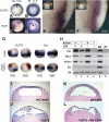
Figure 1.
Figure 1.
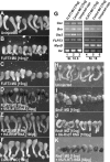
FLRT3 is a direct target of activin and regulates cell adhesion. (AF) In situ hybridization of FLRT3 (A,B,E) and Rnd1 (C,D,F) in Xenopus gastrula, vegetal view (A–D), and cross-section bisecting Spemann’s organizer (E–F). Arrowheads mark the dorsal lip of the blastopore. (G) Overexpression of Cer-S together with β-galactosiade mRNA at the ventral marginal zone (VMZ) or dorsal marginal zone (DMZ) blocks FLRT3 and brachyury (Bra) expression in the gastrula-stage embryos. Arrowheads indicate the dorsal blastopore lips and brown corresponds to β-galactosidase expression. (H) RT–PCR analysis of animal cap explants. (CHX) Cycloheximide; (H4) histone H4; (WE) whole embryos. (I,J) FLRT3-expressing cells (0.3 ng per embryo) detach from the blastocoel roof and fall into the cavity. (K,L) Coexpression of a dominant-negative form of FGF receptor (2 μg of DN-FGFR mRNA) does not affect the cell detachment property conferred by FLRT3.
Figure 2.
Figure 2.
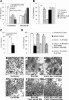
FLRT3 interacts directly with Rnd1 and affects cell adhesion. (A) Overexpression of FLRT3 promotes cell detachment in animal caps. (B) Coinjection of FLRT3 and Rnd1 synergistically increases the percentage of the detached cells. (C–E) Injection of Rnd1 MO (10 ng per embryo) in the equatorial region of four-cell stage embryos disrupts gastrulation movement. (E) Coinjection of HA-xRnd1 (1 ng) RNA rescues this phenotype. (F,G) Cross-section of a stage 9 embryo showing that coinjection of Rnd1 MO partially rescues the loss of cell adhesion conferred by the overexpression of FLRT3. (H) A constitutively active form of xRhoA (xRhoA-V14, 3 pg per embryo) rescues the cell detachment phenotype. (I) Rnd1 directly interacts with FLRT3 in vitro. (Lane 3) FLRT3-GST pulldown assay shows that only Rnd1 interacts with FLRT3. (J) Deletion of Lys580 and Lys581 (FLRT3CP-KK) significantly weakens interaction with 35S-labeled Rnd1 (arrow). Each experiment was repeated three times and reproducible results were obtained.
Figure 3.
Figure 3.
Rnd1 and FLRT1/3 loss-of-function phenotypes. In situ hybridization of Bra (A–F), gsc (G–L), and Xnot (M–R) in Xenopus mid-gastrula-stage embryos. A–R are dorsal external views, and A′–R′ are cross-sections bisecting through Spemann’s organizer. Note the lack of notochord extension in the mid-line of Rnd1-injected (12 ng) and FLRT1/3 MO-injected (30 ng each) embryos.
Figure 4.
Figure 4.
Loss of FLRT3 blocks activin-mediated animal cap elongation. (A–F) Animal cap explants isolated from embryos injected with FLRT3 MO fail to extend properly (B,C) after activin treatment (5 ng/mL), while animal caps both from uninjected (A) and control MO-injected (F) embryos elongate. The FLRT3 MO effect was rescued by coinjection of FLRT3 rescue mRNA lacking the MO annealing sequences (D) or by coinjection of HA-Rnd1 mRNA (E). (G) RT–PCR analysis of RNAs isolated from activin-treated animal caps that have been injected with control or FLRT3 MOs. RNAs are from animal cap explants equivalent to stage 10.5 and stage 18 embryos. (H–K) Rnd1 MO also blocks activin-mediated animal cap elongation. (J) Coinjection of HA-Rnd1 mRNA rescues the Rnd1 MO phenotype. (K) V5-FLRT3 mRNA was unable to rescue.
Figure 5.
Figure 5.
FLRT3 and Rnd1 affect cadherin-mediated cell adhesion. (A) FLRT3- and Rnd1-expressing cells interact weakly with the cadherin substrate. Percentages of cells remaining attached to glass slides before and after shaking were scored. (B) Overexpression of C-cadherin rescues the FLRT3-mediated cell detachment on the cadherin substrate. (C) Anti-cadherin antibody inhibits animal cap cell interaction with cadherin substrate. (D) Inhibition of FLRT3 and Rnd1 promotes cadherin-mediated cell adhesion. (E–J) Loss of FLRT1/3 or Rnd1 in dorsal mesodermal cells promotes cadherin-mediated cell adhesion and cell–cell interaction. (F,G) Rnd1 MO-injected (12 ng per embryo) or FLRT1/3 MO-injected (30 ng each per embryo) cells spread and coalesced together on cadherin-coated slides. (E,H) Control MO-injected or uninjected cells did not adhere to cadherin-coated slide.
Figure 6.
Figure 6.
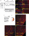
Expression of FLRT3 and Rnd1 promotes cadherin internalization. (A) C-cadherin expression in FLRT3-expressing cells. Western blot analysis was performed using extracts obtained from uninjected and FLRT3 mRNA-injected animal cap cells. (B) Expression of C-cadherin in animal cap explants before and after trypsin treatment. An arrow indicates native C-cadherin protein, a closed arrowhead indicates processed C-cadherin, and an open arrowhead indicates β-tubulin. The full-length integrin-β1 protein is indicated with a single asterisk (*), while the processed integrin-β1 is indicated with double asterisks (**). (C–H) Immunofluorescent staining of C-cadherin in activin-treated or untreated animal cap explants. C-cadherin (in red) is predominantly localized at the cell membrane, while the explants treated with activin show an increase of cytoplasmic cadherin (shown in C,D). (E,G) Inhibition of FLRT3 or Rnd1 expression blocks the internalization of C-cadherin even after an activin treatment. (F,H) Overexpression of FLRT3 or Rnd1 reverts the localization of C-cadherin to the cytoplasm. DAPI staining is in blue. (I) Expression of C-cadherin in deep cells of the dorsal marginal zone before and after a trypsin treatment. An arrow indicates native C-cadherin protein, and a closed arrowhead indicates processed C-cadherin. (J) Schematic diagram of a mid-gastrula-stage embryo. A boxed area in red represents the magnified regions in K–M. (K–M) Subcellular localization of C-cadherin in the organizer of a wild-type, a Rnd1 MO-injected (10 ng per embryo), and a FLRT1/3 MO-injected (30 ng each per embryo) gastrula-stage embryo (stage 11). (K) In the wild-type embryo, C-cadherin staining is predominantly on the cell surface of peripheral cells, but not in the internal cells. The boundary between the peripheral layer and the internal layer is indicated with a dotted line, and the tip of the cleft is marked with an asterisk (*). Arrowheads indicate interstitial spaces between cells.
Figure 7.
Figure 7.
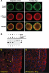
C-cadherin internalization is mediated by endocytosis. (A) Colocalization of C-cadherin and Rab5. mRNAs encoding GFP-tagged C-cadherin (1 ng per embryo) and mCherry-tagged Rab5 (1 ng per embryo) were injected animally into two-cell stage embryos with or without FLRT3 mRNA (0.6 ng per embryo). Animal cap cells were dissociated at the early gastrula stage and subjected to confocal immunofluorescent microscopy. (B) Animal cap cells overexpressing FLRT3 promote cadherin internalization and dominant-negative dynamin blocks the internalization. An arrow represents native C-cadherin protein, and a closed arrowhead represents processed C-cadherin. (C) Subcellular localization of C-cadherin in control uninjected embryo. Arrowheads indicate interstitial spaces between cells. (D) Overexpression of dominant-negative dynamin in the dorsal marginal region blocks cadherin internalization.

References

    1. Adams C.L., Nelson W.J., Smith S.J., Nelson W.J., Smith S.J., Smith S.J. Quantitative analysis of cadherin-catenin-actin reorganization during development of cell-cell adhesion. J. Cell Biol. 1996;135:1899–1911. - PMC - PubMed
    1. Asashima M., Nakano H., Shimada K., Kinoshita K., Ishii K., Shibai H., Ueno N., Nakano H., Shimada K., Kinoshita K., Ishii K., Shibai H., Ueno N., Shimada K., Kinoshita K., Ishii K., Shibai H., Ueno N., Kinoshita K., Ishii K., Shibai H., Ueno N., Ishii K., Shibai H., Ueno N., Shibai H., Ueno N., Ueno N. Mesodermal induction in early amphibian embryos by activin A (erythroid differentiation factor) Rouxs Arch. Dev. Biol. 1990;198:330–335. - PubMed
    1. Blitz I.L., Shimmi O., Wunnenberg-Stapleton K., O’Connor M.B., Cho K.W., Shimmi O., Wunnenberg-Stapleton K., O’Connor M.B., Cho K.W., Wunnenberg-Stapleton K., O’Connor M.B., Cho K.W., O’Connor M.B., Cho K.W., Cho K.W. Is chordin a long-range- or short-range-acting factor? Roles for BMP1-related metalloproteases in chordin and BMP4 autofeedback loop regulation. Dev. Biol. 2000;223:120–138. - PubMed
    1. Bottcher R.T., Pollet N., Delius H., Niehrs C., Pollet N., Delius H., Niehrs C., Delius H., Niehrs C., Niehrs C. The transmembrane protein XFLRT3 forms a complex with FGF receptors and promotes FGF signalling. Nat. Cell Biol. 2004;6:38–44. - PubMed
    1. Braga V.M., Machesky L.M., Hall A., Hotchin N.A., Machesky L.M., Hall A., Hotchin N.A., Hall A., Hotchin N.A., Hotchin N.A. The small GTPases Rho and Rac are required for the establishment of cadherin-dependent cell–cell contacts. J. Cell Biol. 1997;137:1421–1431. - PMC - PubMed

Publication types

MeSH terms