. 2007 Apr;216(2-3):107-16.
doi: 10.1007/s00232-007-9040-1. Epub 2007 Jul 17.

Palytoxin acts on Na(+),K (+)-ATPase but not nongastric H(+),K (+)-ATPase

Affiliations

Palytoxin acts on Na(+),K (+)-ATPase but not nongastric H(+),K (+)-ATPase

Saida Guennoun-Lehmann et al. J Membr Biol. 2007 Apr.

Abstract

Palytoxin (PTX) opens a pathway for ions to pass through Na,K-ATPase. We investigate here whether PTX also acts on nongastric H,K-ATPases. The following combinations of cRNA were expressed in Xenopus laevis oocytes: Bufo marinus bladder H,K-ATPase alpha(2)- and Na,K-ATPase beta(2)-subunits; Bufo Na,K-ATPase alpha(1)- and Na,K-ATPase beta(2)-subunits; and Bufo Na,K-ATPase beta(2)-subunit alone. The response to PTX was measured after blocking endogenous Xenopus Na,K-ATPase with 10 microM ouabain. Functional expression was confirmed by measuring (86)Rb uptake. PTX (5 nM: ) produced a large increase of membrane conductance in oocytes expressing Bufo Na,K-ATPase, but no significant increase occurred in oocytes expressing Bufo H,K-ATPase or in those injected with Bufo beta(2)-subunit alone. Expression of the following combinations of cDNA was investigated in HeLa cells: rat colonic H,K-ATPase alpha(1)-subunit and Na,K-ATPase beta(1)-subunit; rat Na,K-ATPase alpha(2)-subunit and Na,K-ATPase beta(2)-subunit; and rat Na,K-ATPase beta(1)- or Na,K-ATPase beta(2)-subunit alone. Measurement of increases in (86)Rb uptake confirmed that both rat Na,K and H,K pumps were functional in HeLa cells expressing rat colonic HKalpha(1)/NKbeta(1) and NKalpha(2)/NKbeta(2). Whole-cell patch-clamp measurements in HeLa cells expressing rat colonic HKalpha(1)/NKbeta(1) exposed to 100 nM PTX showed no significant increase of membrane current, and there was no membrane conductance increase in HeLa cells transfected with rat NKbeta(1)- or rat NKbeta(2)-subunit alone. However, in HeLa cells expressing rat NKalpha(2)/NKbeta(2), outward current was observed after pump activation by 20 mM K(+) and a large membrane conductance increase occurred after 100 nM PTX. We conclude that nongastric H,K-ATPases are not sensitive to PTX when expressed in these cells, whereas PTX does act on Na,K-ATPase.


Figures
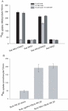
Figure 1
Figure 1
(A) Measurements of 86Rb uptake by HeLa cells. The four groups of 3 bars represent data obtained from HeLa cells transiently transfected with rat NKα1/NKβ1 cDNAs encoding for rat Na,K-ATPase (group 1 from the left), NKβ1 cDNA encoding for rat Na,K-ATPase β1-subunit (group 2), HKα2/NKβ2 cDNAs encoding for rat ngH,K-ATPase (group 3), and NKβ2 cDNA encoding for rat Na,K-ATPase β2-subunit (group 4). Assays for 86Rb uptake (nMol/mg protein/10 min) were performed on each category of cells exposed to: a) 10 μM ouabain (black bars), b) 10 mM ouabain (light grey), and c) 10 μM ouabain plus 10 mM SCH-28080 (grey). The mean ± SEM is shown for 6 assays under each condition. HeLa cells expressing rat Na,K-ATPase or colonic ngH,K-ATPase exhibited increases of 86Rb uptake of 8.8 ± 0.2 fold and 6.9 ± 0.3 fold respectively over the 86Rb uptake background observed with cells transfected with rat Na,K-ATPase β1 or β2 subunits alone. (B) Measurements of 86Rb uptake by Xenopus oocytes. The mean ± SEM of86Rb uptake (pMol/oocyte/12 minutes, 8-10 oocytes in each group) was measured in oocytes microinjected with cDNA encoding for Bufo Na,K-ATPase β2-subunit alone (β2)(left), cDNAs encoding for Bufo bladder H,K-ATPase and β2 (middle), and cDNA encoding for Bufo Na,K-ATPase and β2 (right). Oocytes expressing Bufo bladder H,K-ATPase and bufo Na,K-ATPase exhibited an increase of 86Rb uptake of 4.09 ± 1.04 fold and 4.35 ± 0.57 fold respectively, over that measured in oocytes microinjected with β2-subunit alone. Oocytes were Na+ loaded prior to the uptake measurements. The solutions for uptake measurements in both A and B contained 10 μM ouabain to inhibit endogenous Na,K-pumps, and 10 μM bumetanide to block 86Rb uptake mediated by the Na-K-2Cl co-transporter.
Figure 2
Figure 2
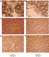
Effect of PTX on confluent HeLa cells morphology. 1 μM PTX was applied for 90 minutes to 35 mm Petri dishes containing 1 ml of culture medium previously grown to confluence of HeLa cells transfected with rat NKα1/NKβ1 cDNAs encoding for rat Na,K-ATPase (Fig. 2A, 2B), rat ngHKα2/NKβ2 cDNAs encoding rat colonic ngH,K-ATPase (Fig. 2C, 2D), and NKβ1 cDNA encoding rat Na,K-ATPase β1-subunit (Fig. 2E, 2F). Large clusters of cells expressing Na,K-ATPase (Fig. 2A and 2B) have swollen and detached from their neighboring cells and the substrate. They eventually become small, round and are found freely floating in the medium. Cells expressing rat Na,K-ATPase (Fig. 2B) show cytoplasmic granulations, whereas no granulations are seen within cells expressing ngH,K-ATPase (Fig. 2D) or β1 subunit alone (Fig. 2F). Fig. 2A, 2C, and 2E were photographed with phase contrast illumination at 250X magnification. Fig. 2B, 2D, and 2F were photographed with phase contrast illumination at 400X magnification. Petri dishes were treated with 20 μM ouabain for 30 minutes prior to PTX application. Four such experiments all showed similar morphological changes in cells expressing rat Na,K-ATPase and no similar changes were observed in cells expressing the rat colonic ngH,K-ATPase or those transfected with rat Na,K-ATPase β1-subunit cDNA alone.
Figure 3
Figure 3
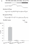
Conductance changes produced by PTX in Xenopus oocytes. A, Representative traces of currents recorded using the two microelectrode voltage clamp technique in oocytes expressing Bufo Na,K-ATPase (top), Bufo bladder H,K-ATPase (middle), and Bufo Na,K-ATPase β2-subunit alone (bottom). After clamping the oocyte at -50 mV in K+-free solution (flow rate of 1 ml/min), solution containing 10 mM K+ was applied for 30-60 s. Oocytes expressing Na,K-ATPase and exposed to 10 mM K+ produced a small rapid increase in outward current (∼0.2 μA) while oocytes expressing H,K-ATPase or those injected by β2-subunit alone did not produce a response. PTX (5 nM) generated a large inward current that continued to increase for several min after PTX removal in oocytes expressing Bufo Na,K-ATPase. No similar inward current was produced in oocytes expressing Bufo bladder ngH,K-ATPase or those injected with β2-subunit alone. B. Palytoxin-induced conductance, Gm (μS) measured from oocytes injected with cRNA coding for Bufo Na,K-ATPase β2-subunit (left column), Bufo Na,K-ATPase/β2 (middle column), and Bufo bladder ngH,K-ATPase β2 (right column). A large increase of membrane conductance occured in oocytes expressing Bufo Na,K-ATPase after exposure to 5 nM PTX (middle column). No significant membrane conductance changes were produced by 5 nM PTX in oocytes expressing ngH,K-ATPase or oocytes injected with β2-subunit cRNA alone. The oocytes were exposed to 10 μM ouabain prior to electrophysiological measurements. Values are means ± SEM of 8-10 measurements.
Figure 4
Figure 4
Measurements of conductance changes produced by PTX in HeLa cells. A. Typical current traces obtained in whole-cell patch clamp experiments (-40 mV holding potential) in HeLa cells expressing rat Na,K-ATPase (top trace), ngH,K-ATPase (middle), or rat Na,K-ATPase β1-subunit cDNA alone (bottom). Cells expressing Na,K-ATPase produced an outward current presumably mediated by the Na,K pump whereas cells expressing ngH,K-ATPase or rat Na,K-ATPase β1-subunit alone did not show an immediate response to K+ (middle and lower traces respectively). PTX application produced a large inward current in cells expressing rat Na,K-ATPase while no significant intward current after 3 min application of PTX to cells expressing rat ngH,K-ATPase or those transfected with rat Na,K-ATPase β1-subunit cDNA. B. Mean values of PTX-induced membrane conductance changes (Gm). Vertical bars indicate the mean values ± SEM of conductance changes measured from cells expressing rat Na,K-ATPase (left column), rat ngH,K-ATPase (middle column), and cells transfected with rat Na,K-ATPase β1-subunit (right column). A large conductance was produced by PTX application on cells expressing rat Na,K-ATPase but no significant membrane conductance increase occurred after 3 min application of 100 nM PTX to cells expressing rat ngH,K-ATPase or transfected with rat Na,K-ATPase β-subunit.
Figure 5
Figure 5
Views of rat Na,K-ATPase (left) and rat ngH,K-ATPase (right) models based on E2 (PDB ID: 1WPG) crystal structures of SERCA. Transmembrane segments M1 are shown in red. The actuator domains are shown in blue and N-termini in green. The boxes indicate the M1-2 loops, and the arrows indicate N-termini. The remaining parts of the models are quite similar and are shown in grey.

Similar articles

Cited by

References

    1. Artigas P, Gadsby DC. Na+/K+-pump ligands modulate gating of palytoxin-induced ion channels, Proc. Natl. Acad. Sci. USA. 2003;100:501–505. - PMC - PubMed
    1. Artigas P, Gadsby DC. Large diameter of palytoxin-induced Na/K pump channels and modulation of palytoxin interaction by Na/K pump ligands. J. Gen. Physiol. 2004 Apr;123(4):357–76. - PMC - PubMed
    1. Burnay M, Crambert G, Geering K, Horisberger J-D. Bufo marinus bladder H-K-ATPase carries out electroneutral ion transport. Am. J. Physiol. 2001;281:F869–F874. - PubMed
    1. Caplan MJ. Ion pumps in epithelial cells: sorting, stabilization, and polarity. Am J Physiol. 1997;272:G1304–G1313. - PubMed
    1. Cantley LG, Resh MD, Guidoti G. Vanadate Inhibits the red cell Na,K-ATPase from the cytoplasmic side. Nature. 1978;272:552–554. - PubMed

Publication types

MeSH terms

LinkOut - more resources