

Polyetheretherketone (PEEK) cage filled with cancellous allograft in anterior cervical discectomy and fusion
- PMID: 17639386
- PMCID: PMC2551716
- DOI: 10.1007/s00264-007-0378-x
Polyetheretherketone (PEEK) cage filled with cancellous allograft in anterior cervical discectomy and fusion
Abstract
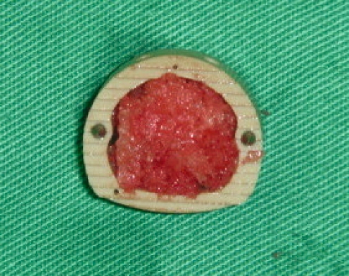
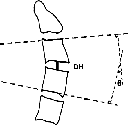
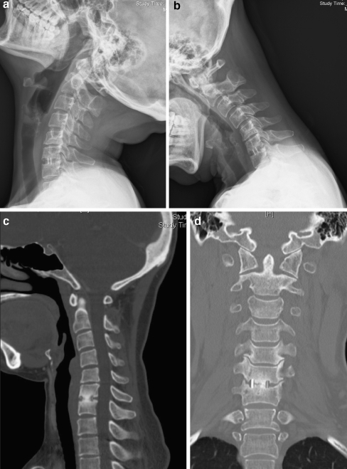
From July 2004 to June 2005, 19 patients with 25 discs underwent anterior cervical discectomy and interbody fusion (ACDF) in which polyetheretherketone (PEEK) cages were filled with freeze-dried cancellous allograft bone. This kind of bone graft was made from femoral condyle that was harvested during total knee arthroplasty. Patient age at surgery was 52.9 (28-68) years. All patients were followed up at least 1 year. We measured the height of the disc and segmental sagittal angulation by pre-operative and post-operative radiographs. CT scan of the cervical spine at 1 year was used to evaluate fusion rates. Odom's criteria were used to assess the clinical outcome. All interbody disc spaces achieved successful union at 1-year follow-up. The use of a PEEK cage was found to increase the height of the disc immediately after surgery (5.0 mm pre-operatively, 7.3 mm immediately post-operatively). The final disc height was 6.2 mm, and the collapse of the disc height was 1.1 mm. The segmental lordosis also increased after surgery (2.0 degrees pre-operatively, 6.6 degrees immediately post-operatively), but the mean loss of lordosis correction was 3.3 degrees at final follow-up. Seventy-four percent of patients (14/19) exhibited excellent/good clinical outcomes. Analysis of the results indicated the cancellous allograft bone-filled PEEK cage used in ACDF is a good choice for patients with cervical disc disease, and avoids the complications of harvesting iliac autograft.
De juillet 2004 à juin 2005, 19 patients ont bénéficié de 25 discectomies cervicales suivies d’arthrodèses intercorporéales (ACDF) avec l’utilisation de cages de type PEEK utilisant des allogreffes cryoconservées. Le matériel osseux a été obtenu à partir de condyles fémoraux récupérés lors d’une prothèse totale du genou. Matériel et méthode : la moyenne d’âge lors de la chirurgie a été de 52.9 ans (28 à 68). Tous les patients ont été suivis au moins un an. Nous avons mesuré radiologiquement la hauteur du disque et les angulations sagittales pré et post-opératoires. Afin d’évaluer la bonne fusion nous avons utilisé un scanner cervical un an après l’intervention. Les critères d’Odomi ont été appréciés de façon à évaluer le résultat clinique. Les disques arthrodésés sont considérés comme consolidés après un an post-opératoire. L’utilisation de cages de type PEEK semble améliorer la conservation de la hauteur du disque immédiatement après l’intervention (5.0 mm pré op, 7.3 mm post-op). La hauteur finale du disque étant de 6.2 mm et le collapsus du disque de 1.1 mm. La lordose segmentaire augmente également après l’intervention chirurgicale (2° en pré op et 6.6° immédiatement en post-opératoire) la perte de lordose est de 3.3° au dernier suivi. 74% des patients (14/19) ont un excellent résultat clinique. L’analyse de ces résultats nous indique que l’utilisation d’allogreffes cryoconservées associées à une cage de type PEEK dans l’arthrodèse cervicale permet d’obtenir une bonne fusion en évitant les complications secondaires à la prise de greffes iliaque.
Figures
Comment in
-
Polyetheretherketone (PEEK) cage filled with bone morphogenic protein and demineralised bone matrix in anterior cervical discectomy and fusion.Int Orthop. 2008 Oct;32(5):717. doi: 10.1007/s00264-007-0450-6. Epub 2007 Aug 28. Int Orthop. 2008. PMID: 17724590 Free PMC article. No abstract available.
References
MeSH terms
Substances
LinkOut - more resources
Full Text Sources
Medical
