. 2007 Aug;65(4):1106-21.
doi: 10.1111/j.1365-2958.2007.05851.x. Epub 2007 Jul 19.

The essential peptidoglycan glycosyltransferase MurG forms a complex with proteins involved in lateral envelope growth as well as with proteins involved in cell division in Escherichia coli

Affiliations

The essential peptidoglycan glycosyltransferase MurG forms a complex with proteins involved in lateral envelope growth as well as with proteins involved in cell division in Escherichia coli

Tamimount Mohammadi et al. Mol Microbiol. 2007 Aug.

Abstract

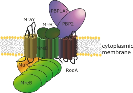
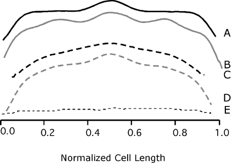
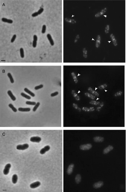
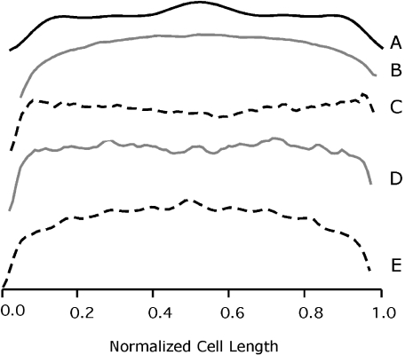
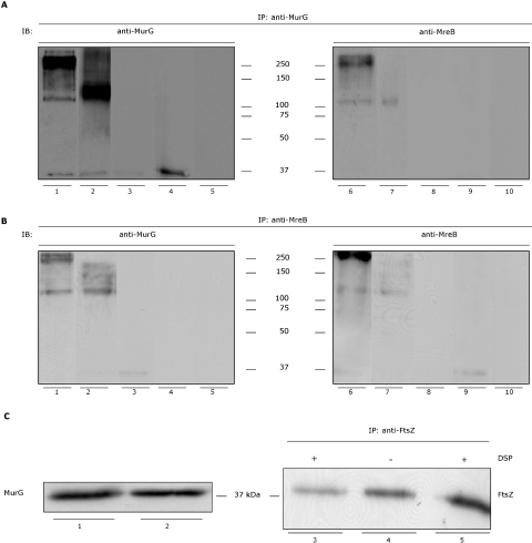
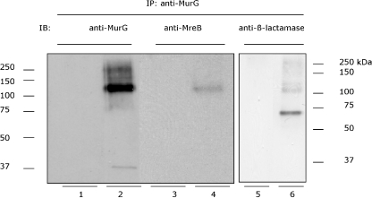
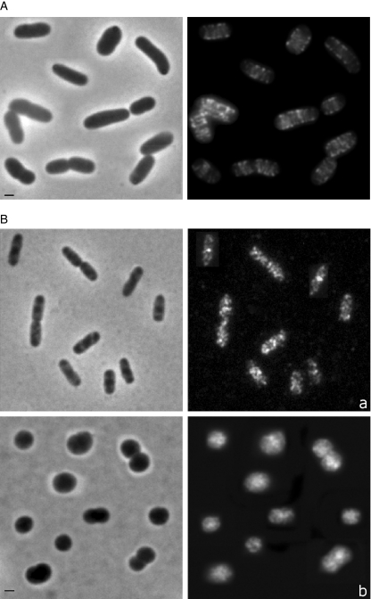
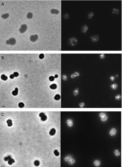
In Escherichia coli many enzymes including MurG are directly involved in the synthesis and assembly of peptidoglycan. MurG is an essential glycosyltransferase catalysing the last intracellular step of peptidoglycan synthesis. To elucidate its role during elongation and division events, localization of MurG using immunofluorescence microscopy was performed. MurG exhibited a random distribution in the cell envelope with a relatively higher intensity at the division site. This mid-cell localization was dependent on the presence of a mature divisome. Its localization in the lateral cell wall appeared to require the presence of MreCD. This could be indicative of a potential interaction between MurG and other proteins. Investigating this by immunoprecipitation revealed the association of MurG with MreB and MraY in the same protein complex. In view of this, the loss of rod shape of DeltamreBCD strain could be ascribed to the loss of MurG membrane localization. Consequently, this could prevent the localized supply of the lipid II precursor to the peptidoglycan synthesizing machinery involved in cell elongation. It is postulated that the involvement of MurG in the peptidoglycan synthesis concurs with two complexes, one implicated in cell elongation and the other in division. A model representing the first complex is proposed.


Figures

Fig. 8
Fig. 8
Schematic representation of the possible constituents of the putative cell elongation complex in E. coli. MraY, MurG and MreB interact with the MreCD. The complex of MreBCD, PBP2 and RodA was published by Kruse et al. (2005). As MreC was shown to interact with three of the four high-molecular-weight class A PBPs of B. subtilis (Van den Ent et al., 2006), it was postulated that PBP1A could be part of the complex (see Discussion). For simplicity only one MurG molecule is illustrated. However, the possibility of formation of dimmers that might be part of the complex is not excluded.
Fig. 2
Fig. 2
Fluorescence intensity profiles of LMC500 cells grown at 28°C in TY (A and B) or in GB1 (C and D) and immunolabelled with anti-MurG. The average normalized cell length is given on the x-axis. The average normalized fluorescence intensity (AU) is reflected on the y-axis. Fluorescence intensity was determined in cells (n ≥ 500) that have no visible constriction (A and C) and in constricting cells (B and D). To indicate the background fluorescence (unspecific binding of secondary antibody), the immunolabelling procedure was carried out with LMC500 cells where the incubation with the first antibody (anti-MurG) was omitted (E). Profiles were measured using Object-image as described in Experimental procedures.
Fig. 1
Fig. 1
Localization of MurG in wild-type E. coli LMC500 (A) and in the MurG(Ts) strain GS58 (B and C). Phase-contrast images are given on the left and fluorescence images on the right. LMC500 cells were grown to steady state at 28°C in GB1 (A), fixed, permeabilized and immunolabelled with anti-MurG. GS58 cells were first grown to mid-exponential phase at 28°C in TY medium (B) shifted to 42°C and allowed to grow for 2 MDs (C). Thereafter they were fixed, permeabilized and subjected to MurG IFM. The arrows point to the band at mid-cell. All panels have the same exposure time. Scale bar equals 1 μm.
Fig. 3
Fig. 3
Fluorescence intensity profiles of LMC509 (FtsZ(Ts), A and B), LMC510 (PBP3(Ts), C), LMC531 (FtsQ(Ts), D) and LMC500 (wild-type, E) cells. After growth at 28°C in 1/2 GB1 LMC509 cells were shifted to 42°C for 2 MDs, and immunolabelled with anti-MurG (A, 28°C) and (B, 42°C). LMC510 and LMC531 cells were grown at 28°C in GB1 to steady state and shifted to 42°C for 2 MDs, and immunolabelled with anti-MurG. LMC500 cells were grown at 28°C in GB1 after which aztreonam was added and growth was continued for 2 MDs before the cells were immunolabelled with anti-MurG (E). Similar patterns shown in A were also obtained for the other temperature-sensitive strains when grown at 28°C in GB1. The average normalized cell length is given on the x-axis. The y-axis represents the average normalized fluorescence intensity.
Fig. 4
Fig. 4
Immunoblotting (IB) analysis of the protein complex with anti-MurG and anti-MreB. IP was performed on Triton X-100 solubilized membranes prepared from wild-type E. coli cells (LMC500), the MraY–β-lactamase–His fusion protein expression strain BW25113ΔmraY/pMAKmraYec, the ΔmreBCD mutant PA340-678 cells, the MurG(Ts) strain GS58, and cross-linked with DSP. The detection was carried out with anti-MurG (A, lanes 1–5, and B, lanes 1–5) or anti-MreB (A, lanes 6–10, and B, lanes 6–10). The protein complex is visible among a smeared background in LMC500 (A, lanes 1 and 6). The dominant bands of this complex have a molecular weight of about 120 kDa and approximately 250 kDa. In the strain BW25113ΔmraY/pMAKmraYec (A, lane 2) the 120 kDa band is more pronounced. The band at the position of 37 kDa is free MurG. No protein complex was yielded in the control where the IP procedure was carried out without the membrane fractions (A, lanes 5 and 10). The same complexes (of 250 kDa and 120 kDa) are also seen when the immunoblot was probed with anti-MreB (A, lanes 6 and 7 respectively). In the strain ΔmreBCD PA340-678 the protein complex is absent (A, lanes 3 and 8) and only free MurG is visible at the level of about 37 kDa when the detection is carried out with anti-MurG (A, lane 3). The complexes of 250 kDa and 120 kDa were undetectable in the MurG(Ts) strain GS58 (A, lanes 4 and 9). The protein band of about 37 kDa is free MurG. Performing the IP with anti-MreB resulted as well in a protein complex of approximately 250 kDa and 120 kDa in LMC500 when probing with anti-MurG or anti-MreB (B, lanes 1 and 6 respectively). The 120 kDa band was more pronounced in the strain BW25113ΔmraY/pMAKmraYec (B, lanes 2 and 7). The complex was not visible in the ΔmreBCD PA340-678 cells (B, lanes 3 and 8). In this strain only a MurG band at the position of 37 kDa was detected when probing with anti-MurG (B, lane 3). In the MurG(Ts) the protein complex was absent (B, lanes 4 and 9). A faint MreB band is visible when anti-MreB was used for detection (B, lane 9). To assess background binding of the secondary antibodies the incubation with the primary antibodies was omitted from the procedure. As depicted in Fig. S5 (see Supplementary materials) no protein bands are detected. One tenth of the membrane fraction used for the IP experiments was applied (without cross-linking) on SDS-PAGE and probed with anti-MurG (C, lanes 1 and 2). In LMC500 and BW25113ΔmraY/pMAKmraYec the intensity of the band (corresponding to MurG) is comparable (lanes 1 and 2 respectively). The interaction between MurG and MreB is specific. To show this the IP procedure with the membrane fraction was performed with anti-FtsZ and the immunoblot was probed with anti-FtsZ. Only a band (∼40 kDa) at the position of FtsZ is detectable in the presence (lane 3) and absence (lane 4) of DSP under non-reducing (lanes 3 and 4) or reducing (lane 5) conditions. The molecular weight of MurG, MreB and MraY–β-lactamase–His is 37.8, 36.9 and 71 kDa respectively. The data are representative of at least three separate experiments. Although some samples were not run in the same gel, the IB procedure (i.e. electrophoresis, blotting and ECL detection conditions) was the same for all of them.
Fig. 5
Fig. 5
Immunoblotting (IB) analysis of the protein complex with anti-MurG, anti-MreB and anti-β-lactamase under non-reducing conditions. The membranes extracted from BW25113ΔmraY/pMAKmraYec strain (MraY–β-lactamase–His expressing strain) were used for the IP (with cross-linking) with anti-MurG. The blot was then probed with anti-MurG (lanes 1 and 2), anti-MreB (lanes 3 and 4) or anti β-lactamase (lanes 5 and 6). In parallel the same IP was performed without the membrane fraction (lanes 1, 3 and 5). A cross-linked product with a molecular weight of about 120 kDa is visible in all samples except in control samples (lanes 1, 3 and 5). The faint band in lane 2 is MurG, which is not completely cross-linked in the protein complex. The band in lane 6 with the molecular weight of approximately 70 kDa corresponds to the MraY–β-lactamase–His protein that is not completely cross-linked in the protein complex.
Fig. 6
Fig. 6
Localization of MreB in the MurG(Ts) strain GS58 grown at the restrictive temperature (42°C) in TY for 2 MDs (A). The MreB helical structure was maintained in the absence of the functional full-length MurG protein. (See for MurG localization in these cells Fig. 1C.) MurG localization in the absence of MreB helix (B). IFM was performed with PA340 (wild type) and PA340-678 (ΔmreBCD) cells grown at 28°C in TY to mid-exponential phase. MurG localizes as multiple foci in the cell envelope and at mid-cell in the wild-type strain (a) and is evenly distributed in the cytoplasm with no clear mid-cell localization in the ΔmreBCD strain, although a packed localization is visible (b). Phase contrast (left) and fluorescence images (right) are shown. The image contains a collection of representative cell. Scale bar equals 1 μm.
Fig. 7
Fig. 7
MurG and MreB localization are independent of the spherical cell morphology (A and B). LMC500 cells were grown for 2 MDs in the presence of mecillinam (inhibitor of PBP2), at 28°C in GB1. Cells were immunolabelled with anti-MreB (A) or anti-MurG (B). The MreB helical structure is preserved and the multi-foci localization pattern of MurG is observed. Arrangement of MurG is dependent on the presence of MreCD (C). IFM was performed with PA340-678pMEW1 strain (mreBCD deletion strain with the pMEW1 plasmid that expresses MreC and MreD constitutively) grown at 28°C in TY to mid-exponential phase. In these spherical cells MurG localized normally as multiple foci in the cell envelope and at mid-cell. Phase contrast (left) and fluorescence images (right) are shown. Scale bar equals 1 μm.

Similar articles

Cited by

References

    1. Aaron M, Charbon G, Lam H, Schwarz H, Vollmer W, Jacobs-Wagner C. The tubulin homologue FtsZ contributes to cell elongation by guiding cell wall precursor synthesis in Caulobacter crescentus. Mol Microbiol. 2007;64:938–952. - PubMed
    1. Aarsman ME, Piette A, Fraipont C, Vinkenvleugel TM, Nguyen-Distèche M, den Blaauwen T. Maturation of the Escherichia coli divisome occurs in two steps. Mol Microbiol. 2005;55:1631–1645. - PubMed
    1. Addinall SG, Cao C, Lutkenhaus J. Temperature shift experiments with an FtsZ84(Ts) strain reveal rapid dynamics of FtsZ localization and indicate that the Z Ring is required throughout septation and cannot reoccupy division sites once constriction has initiated. J Bacteriol. 1997;179:4277–4284. - PMC - PubMed
    1. Auger G, van Heijenoort J, Mengin-Lecreulx D, Blanot D. A MurG assay which utilises a synthetic analogue of lipid I. FEMS Microbiol Lett. 2003;219:115–119. - PubMed
    1. Bertsche U, Kast T, Wolf B, Fraipont C, Aarsman ME, Kannenberg K, et al. Interaction between two murein (peptidoglycan) synthases, PBP3 and PBP1B, in Escherichia coli. Mol Microbiol. 2006;61:675–690. - PubMed

Publication types

MeSH terms

Substances

LinkOut - more resources