. 2007;9(4):R69.
doi: 10.1186/ar2267.

Human meniscus cells express hypoxia inducible factor-1alpha and increased SOX9 in response to low oxygen tension in cell aggregate culture

Affiliations

Human meniscus cells express hypoxia inducible factor-1alpha and increased SOX9 in response to low oxygen tension in cell aggregate culture

Adetola B Adesida et al. Arthritis Res Ther. 2007.

Abstract

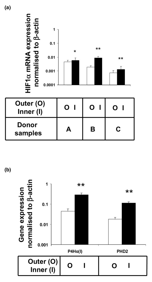
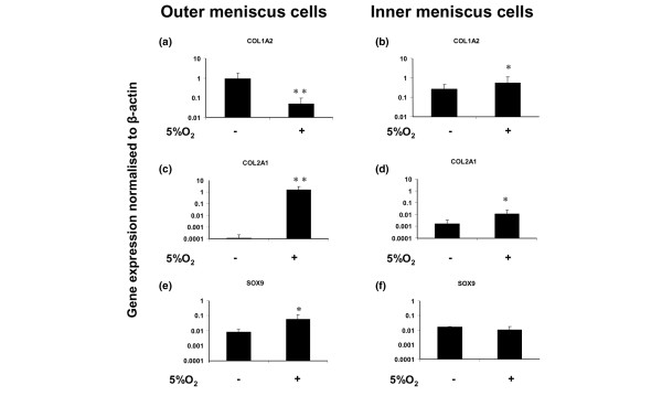
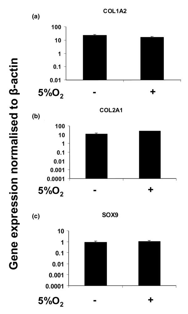
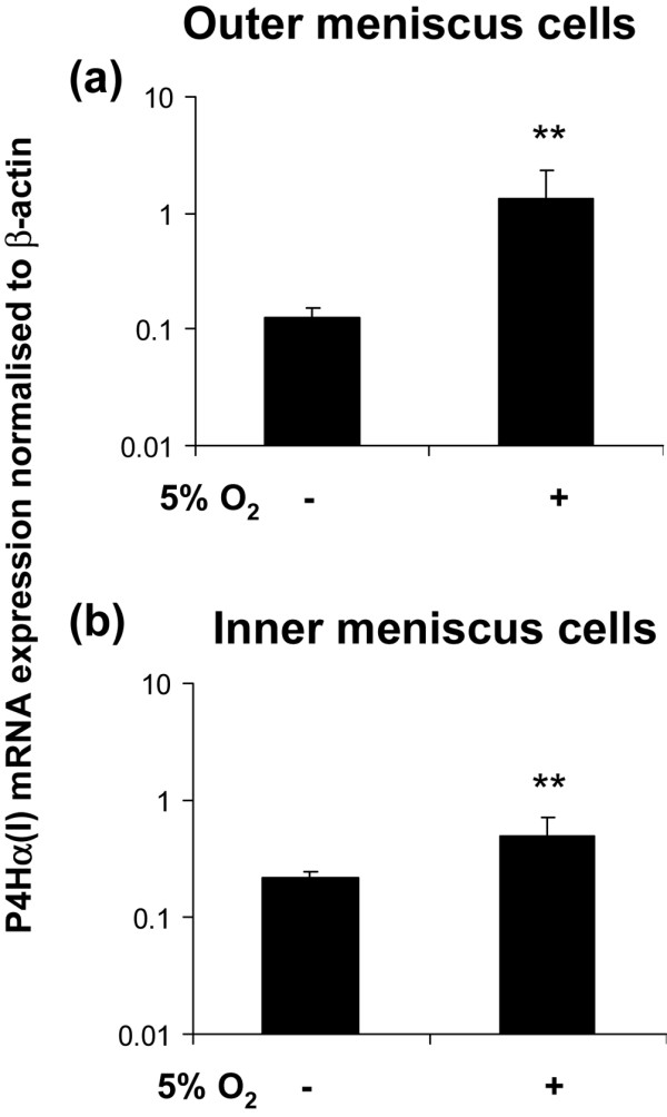
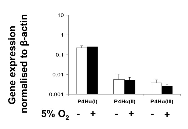
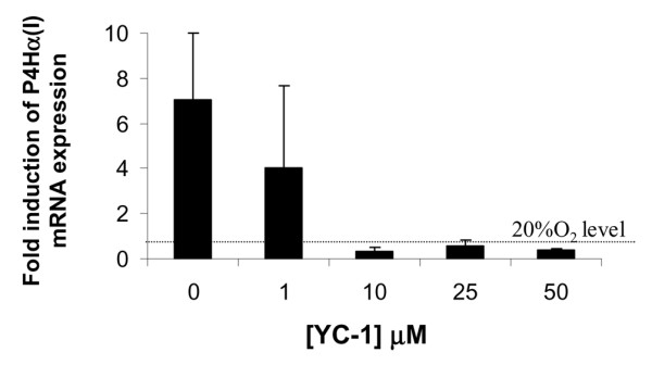
In previous work we demonstrated that the matrix-forming phenotype of cultured human cells from whole meniscus was enhanced by hypoxia (5% oxygen). Because the meniscus contains an inner region that is devoid of vasculature and an outer vascular region, here we investigate, by gene expression analysis, the separate responses of cells isolated from the inner and outer meniscus to lowered oxygen, and compared it with the response of articular chondrocytes. In aggregate culture of outer meniscus cells, hypoxia (5% oxygen) increased the expression of type II collagen and SOX9 (Sry-related HMG box-9), and decreased the expression of type I collagen. In contrast, with inner meniscus cells, there was no increase in SOX9, but type II collagen and type I collagen increased. The articular chondrocytes exhibited little response to 5% oxygen in aggregate culture, with no significant differences in the expression of these matrix genes and SOX9. In both aggregate cultures of outer and inner meniscus cells, but not in chondrocytes, there was increased expression of collagen prolyl 4-hydroxylase (P4H)alpha(I) in response to 5% oxygen, and this hypoxia-induced expression of P4H alpha(I) was blocked in monolayer cultures of meniscus cells by the hypoxia-inducible factor (HIF)-1alpha inhibitor (YC-1). In fresh tissue from the outer and inner meniscus, the levels of expression of the HIF-1alpha gene and downstream target genes (namely, those encoding P4H alpha(I) and HIF prolyl 4-hydroxylase) were significantly higher in the inner meniscus than in the outer meniscus. Thus, this study revealed that inner meniscus cells were less responsive to 5% oxygen tension than were outer meniscus cells, and they were both more sensitive than articular chondrocytes from a similar joint. These results suggest that the vasculature and greater oxygen tension in the outer meniscus may help to suppress cartilage-like matrix formation.


Figures

Figure 1
Figure 1
Gene expression of HIF-1α and target genes in the outer and inner meniscus. (a) Hypoxia inducible factor (HIF)-1α gene expression in the outer (O; white bars) and inner (I; black bars) region of meniscus tissue from three donors (n = 3). (b) Prolyl 4-hydroxylase (P4H)α(I) and HIF prolyl-hydroxylase (PHD)2 gene expression in the outer (O; white bars) and inner (I; black bars) region of meniscus tissue (n = 3). Values are expressed as mean ± standard deviation. *P < 0.05, **P < 0.01 (by Student's unpaired t-test) in inner versus outer region of the meniscus matched against the same donor.
Figure 2
Figure 2
Collagen types I and II, and SOX9 gene expression in human meniscus cells. (a, b) Collagen type I alpha 2 (COL1A2) gene expression levels of cell aggregate cultures of (panel a) outer and (panel b) inner meniscus cells (n = 6) in 5% oxygen versus 20% oxygen. (c, d) Collagen type II alpha 1 (COL2A1) gene expression in aggregate cultures of (panel c) outer and (panel d) inner meniscus cells (n = 6) in 5% oxygen versus 20% oxygen. (e, f) SOX9 (Sry-related HMG box-9) gene expression levels of aggregate cultures of (panel e) outer and (panel f) inner meniscus cells (n = 6) in 5% oxygen versus 20% oxygen. *P < 0.05, **P < 0.01 (by Student's unpaired t-test).
Figure 3
Figure 3
Collagen types I and II, and SOX9 gene expression in human articular chondrocytes. (a) Collagen type I alpha 2 (COL1A2) gene expression levels of aggregate cultures of articular chondrocytes (n = 3) in 5% oxygen versus 20% oxygen. (b) Collagen type II alpha 1 (COL2A1) gene expression in aggregate cultures of articular chondrocytes (n = 3) in 5% oxygen versus 20% oxygen. (c) SOX9 (Sry-related HMG box-9) gene expression levels of cell aggregate cultures of articular chondrocytes (n = 3) in 5% oxygen versus 20% oxygen. Student's unpaired t-test. Data are expressed as mean ± standard deviation (n = 3).
Figure 4
Figure 4
Collagen P4Hα(I) gene expression in meniscus cell aggregates. Prolyl 4-hydroxylase (P4H)α(I) gene expression in (a) outer meniscus cell aggregates (n = 4) and (b) inner meniscus cell aggregates (n = 4) in 5% oxygen versus 20% oxygen. Data are expressed as mean ± standard deviation (n = 3). **P < 0.01 (by Student's unpaired t-test).
Figure 5
Figure 5
Collagen P4H isoenzyme gene expression in articular chondrocytes aggregates. Prolyl 4-hydroxylase (P4H)α isoenzyme gene expression in cell aggregates of articular chondrocytes in 20% oxygen (white bars) and 5% oxygen (black bars). Data are expressed as mean ± standard deviation (n = 3).
Figure 6
Figure 6
YC-1 mediated inhibition of hypoxic induction of P4H in meniscus cell aggregates. Ratio of hypoxia (5% oxygen) induced prolyl 4-hydroxylase (P4H)α(I) gene expression (post-normalization to β-actin) to P4Hα(I) gene expression (post-normalization to β-actin) in 20% oxygen in monolayer culture of meniscus cells in the presence and absence of the hypoxia inducible factor-1α inhibitor YC-1. Data are expressed as mean ± standard deviation (n = 3).

Similar articles

Cited by

References

    1. Ahmed AM. The load-bearing role of the knee meniscus. In: Mow VC, Arnoczky SP, Jackson DW, editor. Knee Meniscus: Basic and Clinical Foundations. New York: Raven Press, Ltd; 1992. pp. 59–73.
    1. Levy IM, Torzilli PA, Fisch ID. The contribution of the menisci to the stabilty of the knee. In: Mow VC, Arnoczky SP, Jackson DW, editor. Knee Meniscus: Basic and Clinical Foundations. New York: Raven Press, Ltd; 1992. pp. 107–115.
    1. Fairbank T. Knee joint changes after menisectomy. J Bone Joint Surg. 1948;30B:664–670. - PubMed
    1. Aagaard H, Verdonk R. Function of the normal meniscus and consequences of meniscal resection. Scand J Med Sci Sports. 1999;9:134–140. - PubMed
    1. Wyland DJ, Guilak F, Elliott DM, Setton LA, Vail TP. Chondropathy after meniscal tear or partial meniscectomy in a canine model. J Orthop Res. 2002;20:996–1002. doi: 10.1016/S0736-0266(02)00022-0. - DOI - PubMed

Publication types

MeSH terms