

Evolution of Class I cytokine receptors
- PMID: 17640376
- PMCID: PMC1963337
- DOI: 10.1186/1471-2148-7-120
Evolution of Class I cytokine receptors
Abstract
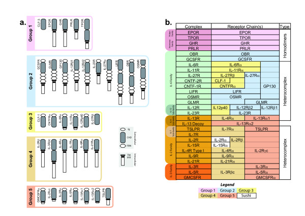
Background: The Class I cytokine receptors have a wide range of actions, including a major role in the development and function of immune and blood cells. However, the evolution of the genes encoding them remains poorly understood. To address this we have used bioinformatics to analyze the Class I receptor repertoire in sea squirt (Ciona intestinalis) and zebrafish (Danio rerio).
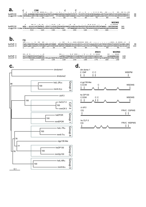
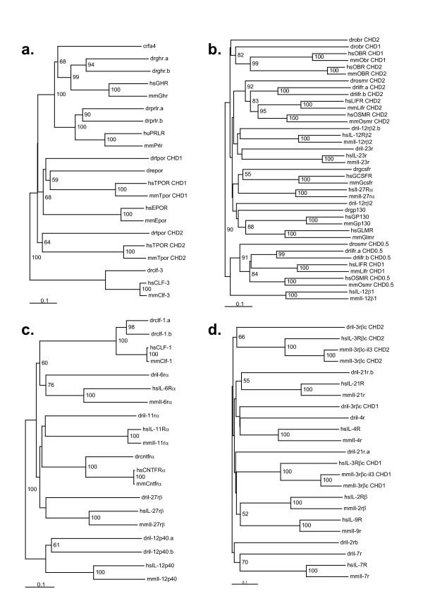
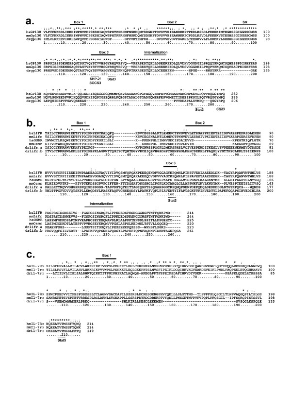
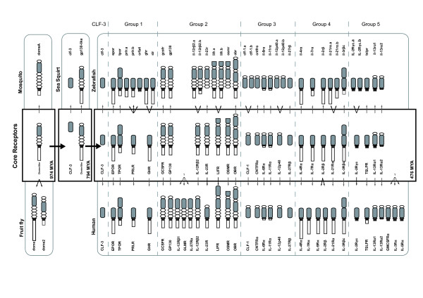
Results: Only two Class I receptors were identified in sea squirt, one with homology to the archetypal GP130 receptor, and the other with high conservation with the divergent orphan receptor CLF-3. In contrast, 36 Class I cytokine receptors were present in zebrafish, including representative members for each of the five structural groups found in mammals. This allowed the identification of 27 core receptors belonging to the last common ancestor of teleosts and mammals.
Conclusion: This study suggests that the majority of diversification of this receptor family occurred after the divergence of urochordates and vertebrates approximately 794 million years ago (MYA), but before the divergence of ray-finned from lobe-finned fishes around 476 MYA. Since then, only relatively limited lineage-specific diversification within the different Class I receptor structural groups has occurred.
Figures
Similar articles
-
The repertoire of trace amine G-protein-coupled receptors: large expansion in zebrafish.Mol Phylogenet Evol. 2005 May;35(2):470-82. doi: 10.1016/j.ympev.2004.12.003. Epub 2005 Jan 25. Mol Phylogenet Evol. 2005. PMID: 15804416
-
Two divergent leptin paralogues in zebrafish (Danio rerio) that originate early in teleostean evolution.J Endocrinol. 2009 Jun;201(3):329-39. doi: 10.1677/JOE-09-0034. Epub 2009 Mar 17. J Endocrinol. 2009. PMID: 19293295
-
Evolution of the Class 2 cytokines and receptors, and discovery of new friends and relatives.Pharmacol Ther. 2005 Jun;106(3):299-346. doi: 10.1016/j.pharmthera.2004.12.002. Epub 2005 Apr 14. Pharmacol Ther. 2005. PMID: 15922016 Review.
-
The evolution of tachykinin/tachykinin receptor (TAC/TACR) in vertebrates and molecular identification of the TAC3/TACR3 system in zebrafish (Danio rerio).Mol Cell Endocrinol. 2012 Sep 25;361(1-2):202-12. doi: 10.1016/j.mce.2012.04.007. Epub 2012 May 2. Mol Cell Endocrinol. 2012. PMID: 22580006
-
Phylogeny and evolution of class-I helical cytokines.J Endocrinol. 2006 Apr;189(1):1-25. doi: 10.1677/joe.1.06591. J Endocrinol. 2006. PMID: 16614377 Review.
Cited by
-
The Function of Fish Cytokines.Biology (Basel). 2016 May 24;5(2):23. doi: 10.3390/biology5020023. Biology (Basel). 2016. PMID: 27231948 Free PMC article. Review.
-
Hematopoietic Cytokine Gene Duplication in Zebrafish Erythroid and Myeloid Lineages.Front Cell Dev Biol. 2018 Dec 20;6:174. doi: 10.3389/fcell.2018.00174. eCollection 2018. Front Cell Dev Biol. 2018. PMID: 30619854 Free PMC article. Review.
-
Immunobiology of the TAM receptors.Nat Rev Immunol. 2008 May;8(5):327-36. doi: 10.1038/nri2303. Nat Rev Immunol. 2008. PMID: 18421305 Free PMC article. Review.
-
Hepatocyte Specific gp130 Signalling Underlies APAP Induced Liver Injury.Int J Mol Sci. 2022 Jun 25;23(13):7089. doi: 10.3390/ijms23137089. Int J Mol Sci. 2022. PMID: 35806094 Free PMC article.
-
Zebrafish Granulocyte Colony-Stimulating Factor Receptor Maintains Neutrophil Number and Function throughout the Life Span.Infect Immun. 2019 Jan 24;87(2):e00793-18. doi: 10.1128/IAI.00793-18. Print 2019 Feb. Infect Immun. 2019. PMID: 30455199 Free PMC article.
References
-
- Taga T. Gp130, a shared signal transducing receptor component for hematopoietic and neuropoietic cytokines. J Neurochem. 1996;67:1–10. - PubMed
Publication types
MeSH terms
Substances
Associated data
- Actions
LinkOut - more resources
Full Text Sources
Molecular Biology Databases
