Review
. 2007 Jul 19:2:32.
doi: 10.1186/1750-1172-2-32.

Ehlers-Danlos syndrome type IV

Affiliations
Review

Ehlers-Danlos syndrome type IV

Dominique P Germain. Orphanet J Rare Dis. .

Abstract

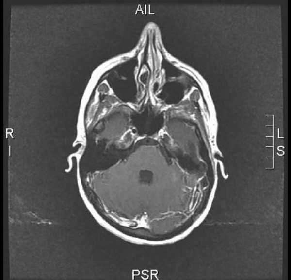
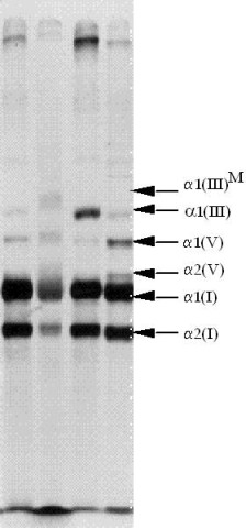
Ehlers-Danlos syndrome type IV, the vascular type of Ehlers-Danlos syndromes (EDS), is an inherited connective tissue disorder defined by characteristic facial features (acrogeria) in most patients, translucent skin with highly visible subcutaneous vessels on the trunk and lower back, easy bruising, and severe arterial, digestive and uterine complications, which are rarely, if at all, observed in the other forms of EDS. The estimated prevalence for all EDS varies between 1/10,000 and 1/25,000, EDS type IV representing approximately 5 to 10% of cases. The vascular complications may affect all anatomical areas, with a tendency toward arteries of large and medium diameter. Dissections of the vertebral arteries and the carotids in their extra- and intra-cranial segments (carotid-cavernous fistulae) are typical. There is a high risk of recurrent colonic perforations. Pregnancy increases the likelihood of a uterine or vascular rupture. EDS type IV is inherited as an autosomal dominant trait that is caused by mutations in the COL3A1 gene coding for type III procollagen. Diagnosis is based on clinical signs, non-invasive imaging, and the identification of a mutation of the COL3A1 gene. In childhood, coagulation disorders and Silverman's syndrome are the main differential diagnoses; in adulthood, the differential diagnosis includes other Ehlers-Danlos syndromes, Marfan syndrome and Loeys-Dietz syndrome. Prenatal diagnosis can be considered in families where the mutation is known. Choriocentesis or amniocentesis, however, may entail risk for the pregnant woman. In the absence of specific treatment for EDS type IV, medical intervention should be focused on symptomatic treatment and prophylactic measures. Arterial, digestive or uterine complications require immediate hospitalisation, observation in an intensive care unit. Invasive imaging techniques are contraindicated. Conservative approach is usually recommended when caring for a vascular complication in a patient suffering from EDS type IV. Surgery may, however, be required urgently to treat potentially fatal complications.


Figures

Figure 1
Figure 1
Extensive bruising of the legs in a child affected with Ehlers-Danlos syndrome type IV.
Figure 2
Figure 2
Angio-MRI (coronal section) of the supra-aortic vessels after injection of 20 cc of gadolinium in a woman aged 24 with EDS type IV, revealing a dissecting haematoma of the left internal carotid (black arrow), a bilateral dissection of the vertebral arteries in their V1 and V2 segments (white arrows) and a dissection of the middle and distal third of the right subclavian artery (head of arrow).
Figure 3
Figure 3
Cerebral angio-MRI after injection of 20 cc of gadolinium in a woman aged 24 with EDS type IV displaying a dissecting haematoma of the left internal carotid.
Figure 4
Figure 4
Surgical laparotomy scar following sigmoid colon perforation in a young patient presenting with vascular Ehlers-Danlos syndrome.
Figure 5
Figure 5
Sodium dodecyl sulfate-PolyAcrylamide Gel Electrophoresis (SDS-PAGE) analysis of collagens secretion in a patient affected with vascular EDS and a control. The band corresponding to type III collagen is indicated by α1(III). Columns 1 and 3 correspond to the supernatant culture medium of the cutaneous fibroblasts. Colums 2 and 4 correspond to cell extracts. Secretion of type III collagen in the medium is reduced in the patient (column 1) as compared to the control (column 3). Conversely, there is intracellular retention of abnormal collagen in the cell extracts of the patient (column 2) as compared to the control. In addition, the mutant collagen III has a higher molecular weight due to additional post-translational modification α1(III)M.
Figure 6
Figure 6
Detection of a heterozygote missense mutation (G514V) in the COL3A1 gene in a 47-year old female patient affected with Ehlers-Danlos syndrome type IV. A G to A substitution was found at nucleotide position 2042 starting from the initiation codon (ATG) of the COL3A1 gene. This nucleotide substitution alters the codon (GGT) for glycine to the codon (GTT) for valine at position 514 of the α-chain of collagen type III protein. Two electrophoregrams of the same patient are shown. (Figure courtesy of Prof. X. Jeunemaitre)

Similar articles

Cited by

References

    1. Pepin M, Schwarze U, Superti-Furga A, Byers PH. Clinical and genetic features of Ehlers-Danlos syndrome type IV, the vascular type. N Engl J Med. 2000;342:673–680. doi: 10.1056/NEJM200003093421001. - DOI - PubMed
    1. Byers PH. Ehlers-Danlos syndrome type IV: a genetic disorder in many guises. J Invest Dermatol. 1995;105:311–313. doi: 10.1111/1523-1747.ep12319926. - DOI - PubMed
    1. Beighton P, De Paepe A, Steinmann B, Tsipouras P, Wenstrup RJ. Ehlers-Danlos syndromes: revised nosology, Villefranche, 1997. Am J Med Genet. 1998;77:31–37. doi: 10.1002/(SICI)1096-8628(19980428)77:1<31::AID-AJMG8>3.0.CO;2-O. - DOI - PubMed
    1. Barabas AP. Vascular complications in the Ehlers-Danlos syndrome, with special reference to the "arterial type" or Sack's syndrome. J Cardiovasc Surg (Torino) 1972;13:160–167. - PubMed
    1. Germain DP, Herrera-Guzman Y. Vascular Ehlers-Danlos syndrome. Ann Genet. 2004;47:1–9. - PubMed

MeSH terms