. 2007 Aug 15;308(2):621-31.
doi: 10.1016/j.ydbio.2007.06.002. Epub 2007 Jun 12.

The function and regulation of Ultrabithorax in the legs of Drosophila melanogaster

Affiliations

The function and regulation of Ultrabithorax in the legs of Drosophila melanogaster

Gregory K Davis et al. Dev Biol. .

Abstract

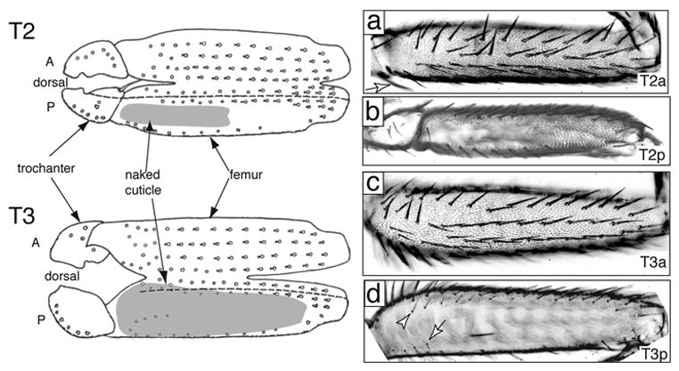
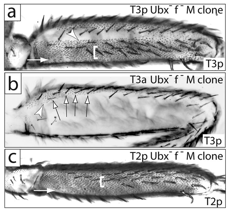
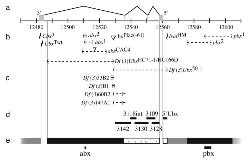
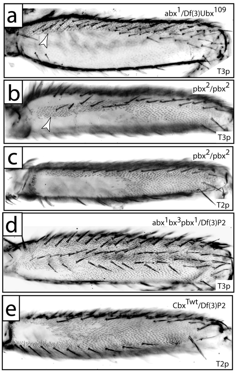
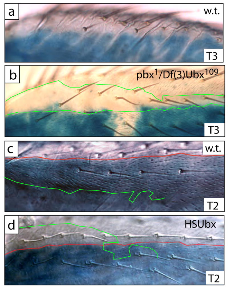
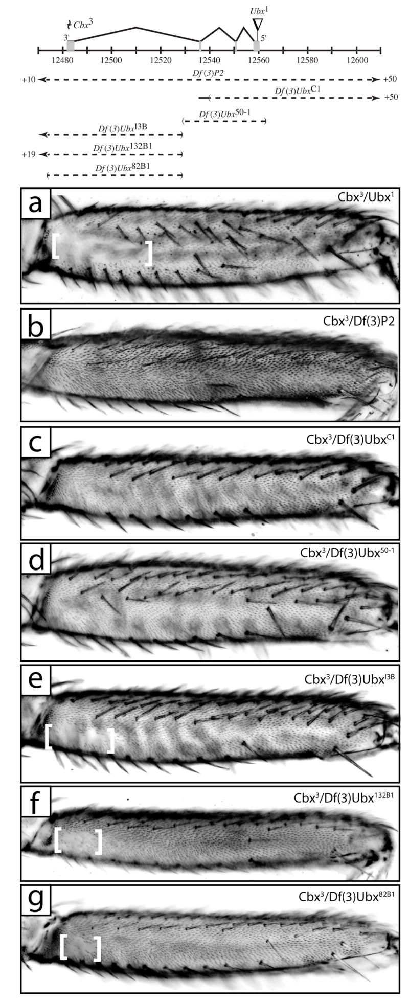
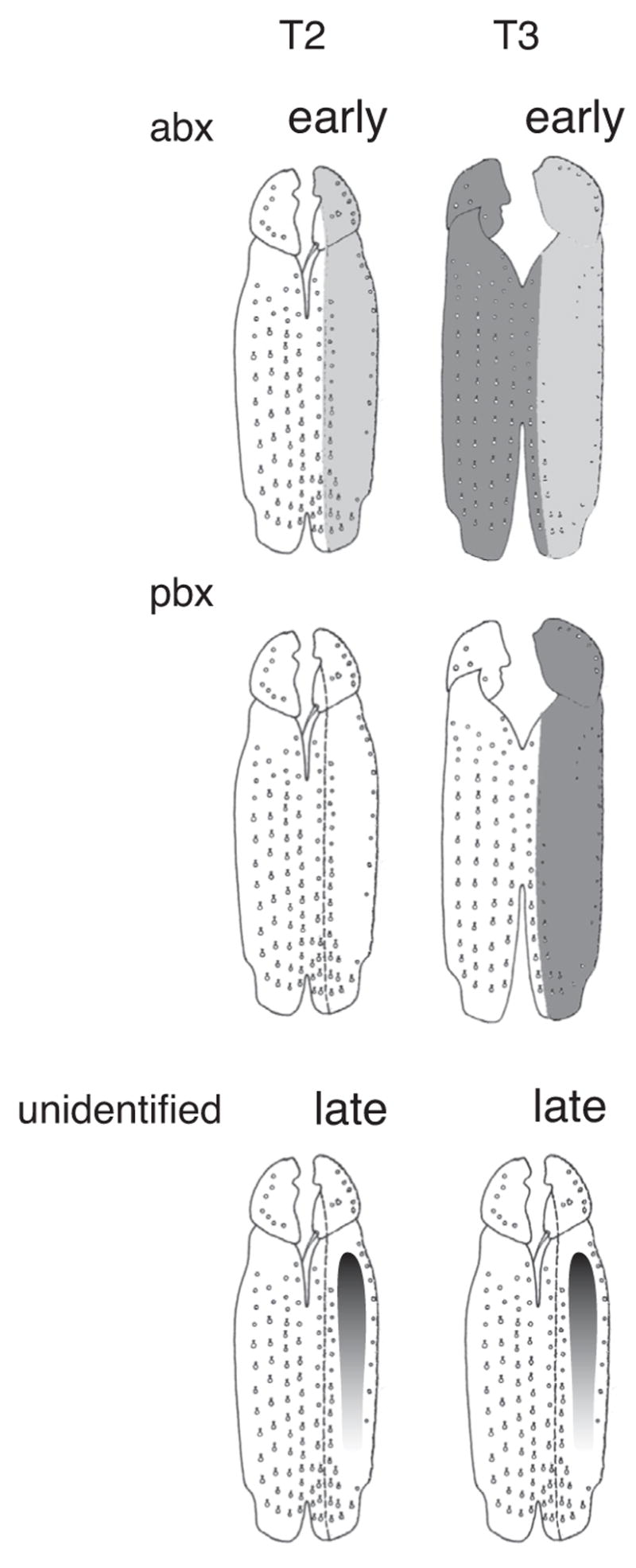
Alterations in Hox gene expression patterns have been implicated in both large and small-scale morphological evolution. An improved understanding of these changes requires a detailed understanding of Hox gene cis-regulatory function and evolution. cis-regulatory evolution of the Hox gene Ultrabithorax (Ubx) has been shown to contribute to evolution of trichome patterns on the posterior second femur (T2p) of Drosophila species. As a step toward determining how this function of Ubx has evolved, we performed a series of experiments to clarify the role of Ubx in patterning femurs and to identify the cis-regulatory regions of Ubx that drive expression in T2p. We first performed clonal analysis to further define Ubx function in patterning bristle and trichome patterns in the legs. We found that low levels of Ubx expression are sufficient to repress an eighth bristle row on the posterior second and third femurs, whereas higher levels of expression are required to promote the development and migration of other bristles on the third femur and to repress trichomes. We then tested the hypothesis that the evolutionary difference in T2p trichome patterns due to Ubx was caused by a change in the global cis-regulation of Ubx expression. We found no evidence to support this view, suggesting that the evolved difference in Ubx function reflects evolution of a leg-specific enhancer. We then searched for the regulatory regions of the Ubx locus that drive expression in the second and third femur by assaying all existing regulatory mutations of the Ubx locus and new deficiencies in the large intron of Ubx that we generated by P-element-induced male recombination. We found that two enhancer regions previously known to regulate Ubx expression in the legs, abx and pbx, are required for Ubx expression in the third femur, but that they do not contribute to pupal expression of Ubx in the second femur. This analysis allowed us to rule out at least 100 kb of DNA in and around the Ubx locus as containing a T2p-specific enhancer. We then surveyed an additional approximately 30 kb using enhancer constructs. None of these enhancer constructs produced an expression pattern similar to Ubx expression in T2p. Thus, after surveying over 95% of the Ubx locus, we have not been able to localize a T2p-specific enhancer. While the enhancer could reside within the small regions we have not surveyed, it is also possible that the enhancer is structurally complex and/or acts only within its native genomic context.


Figures

Fig. 1
Fig. 1
Wildtype mesothoracic (T2) and metathoracic (T3) femurs. A diagrammatic representation of the legs is shown to the left, with the legs split open along the ventral boundary of the anterior/posterior compartment (modified from Steiner 1976). Anterior and posterior compartments are labelled with A and P and are separated by a dashed line. Regions of naked cuticle are shaded gray. The photographs are oriented in the same way as the diagram: in (a) and (c) ventral is up and in (b) and (d) dorsal is up. (A) Anterior T2 femur has five rows of bristles and is completely covered in trichomes. (B) Posterior T2 has one row of bristles dorsally and one ventral row that is incomplete distally. A proximal lateral patch of naked cuticle varies in size between different strains. The remainder of the posterior surface is covered with trichomes. (C) Anterior T3 has five rows of bristles and is covered in trichomes over all but a dorsal proximal region (see D). (D) Posterior T3 has one row of bristles ventrally, several fine bristles in the proximal region (arrow), and a dorsal row of small bristles. Most of the surface is naked, and a small distal region normally produces trichomes. The dorsal anterior-posterior compartment boundary lies approximately along the dorsal bristle row (arrowhead) and does not reflect an obvious morphological discontinuity. The dorsal boundary between trichomes and naked cuticle extends into the proximal anterior dorsal region.
Fig. 2
Fig. 2
Ubx+ represses most of the trichomes and one lateral bristle row on posterior T2 and T3. (a) Posterior of a T3 femur carrying a Minute+ Ubx clone that fills the posterior compartment. A small patch of naked cuticle is found proximal-ventrally (arrow). Trichomes differentiate throughout the rest of the posterior compartment. Naked cuticle in the anterior compartment (dorsally) is unaffected. The fine bristles normally found in the proximal lateral region are repressed, and the dorsal bristles are apparently transformed into more robust bristles. Finally, an ectopic, eighth row of bristles differentiates laterally (white bracket). All Minute+ Ubx clones found on the posterior T3 exhibited a sharp dorsal boundary between derepressed trichomes and naked cuticle (arrowhead). (b) A view of the posterior and dorsal anterior of a femur with a Minute+ Ubx clone that fills the anterior compartment. Trichomes differentiate in the normally naked cuticle region of the anterior dorsal femur (arrowhead). The small bristles in the dorsal posterior compartment are unaffected by the clone and are forked+, indicating that they originated in the posterior compartment (arrows). (c) T2 posterior femur carrying a Minute+ Ubx clone that fills the posterior compartment. Trichomes differentiate throughout most of the posterior femur except for a small patch of cuticle proximal-ventrally (arrow). An ectopic, eighth row of bristles is indicated with a white bracket. Clones in (a) and (c) are marked with forked bristles both dorsal and ventral to the small ventral naked patches.
Fig. 3
Fig. 3
Map of the Ubx locus. (a) The four exons are illustrated above a scale showing genomic positions in kilobases (Drosophila melanogaster Genome Release 4.3). The centromere is to the left. (b) The most informative alleles used in this study are shown beside symbols indicating the type of lesion. Deletions are indicated by parentheses separated by a dashed line, insertions by inverted triangles (not to scale), inversion breakpoints by horizontal lines bisected by a wavy line. Uncertainty in the location of breakpoints is indicated by the range of the solid horizontal lines. (c) Four new deficiencies generated in this study by P-element induced male recombination using the bxPlac(−61) insertion are shown. (d) The positions of the six enhancer constructs are indicated with horizontal bars. (e) Regions scanned with deficiencies are indicated by black bars. Regions scanned by inversion are indicated by grey bars. Regions scanned with enhancer constructs are indicated with stippled boxes. The two minimal regions identified by this scan, the previously identified abx and pbx regions, are indicated with solid lines at the bottom of the figure. Regions of the Ubx gene not scanned by any technique are bounded by light lines extending from the molecular map vertically to the bottom of the figure.
Fig. 4
Fig. 4
The posterior femurs of Ubx regulatory mutants. (a) T3p from a fly carrying abx1/Df(3)Ubx109 displays ectopic trichomes in the dorsal anterior compartment (arrowhead, compare with Fig. 2c). (b) T3p of a pbx2 homozygote displays a stripe of ectopic trichomes proximally (arrowhead) and distally. Naked cuticle is observed dorsally and ventrally to the ectopic proximal trichomes. (c) The distribution of naked cuticle on T2p of a pbx2 homozygote is approximately equal to the ventral naked cuticle on T3p (b). (d) T3p of a fly carrying abx1bx3pbx1/Df(3)P2 displays a trichome pattern that is approximately a composite of the abx and pbx patterns, with a naked patch of cuticle only on the posterior proximal surface. (e) T2p from a fly carrying CbxTwt/Df(3)P2 displays a wild type distribution of trichomes.
Fig. 5
Fig. 5
The boundary of naked cuticle and trichomes does not obey the compartment boundary in T2 or T3, and the proximal dorsal anterior femur of both T2 and T3 is competent to repress trichomes when high levels of Ubx are expressed in these cells. (a) In proximal dorsal regions of wild-type T3, naked cuticle is found in both posterior and anterior compartments. Blue en-lacZ staining marks the posterior compartment. (b) In a pbx1 hemizygote naked cuticle is still found in the anterior compartment, but cells in the posterior compartment differentiate trichomes. In this preparation, en-lacZ staining failed in a small patch of cells, revealing the faint trichomes. In most specimens this boundary of en-lacZ staining is complete and approximately straight, and the ectopic trichomes are only found within the region of en-lacZ staining. The boundaries of naked cuticle and trichomes are marked with green lines. (c) In dorsal proximal regions of wild-type T2 legs trichomes differentiate in both the anterior and posterior compartment. The boundary of en-lacZ staining is marked with a red line, and the boundary of trichomes is marked with a green line. (d) When Ubx is expressed ectopically at high levels at 24H APF, cells in this dorsal region now differentiate naked cuticle in both the posterior and anterior compartments. The boundary of en-lacZ staining is marked with a red line, and the border of naked cuticle is marked with a green line.
Fig. 6
Fig. 6
Cbx3 causes ectopic T2p and T3a trichomes in a transvection dependent manner. A map of the Ubx locus, as in Fig. 3, is shown at the top illustrating Cbx3 and Ubx1 lesions above and positions of deficiencies used in transvection mapping below. (a) T2p from a fly with the genotype Cbx3/Ubx1 displays a normal sized patch of naked cuticle. (b) T2p from Cbx3/Df(3)P2 flies exhibits only a small patch of proximal-ventral naked cuticle similar to that observed in Ubx clones (Fig. 2a). (c,d) T2p from flies with the genotype Cbx3/Df(3)UbxC1 (c) and Cbx3/Df(3)Ubx50-1 (d). (e,f,g) T2p from flies with the genotype Cbx3/Df(3)UbxI3B (e), Cbx3/Df(3)Ubx132B1 (f), and Cbx3/Df(3)Ubx82B1 (g) show a patch of proximal naked cuticle (between white brackets) indicative of trichome repression due to Ubx expression.
Fig. 7
Fig. 7
Domains of Ubx regulatory function in the femurs of Drosophila melanogaster. Weak expression is illustrated as light gray shading, strong expression as dark gray shading. The abx region contains elements promoting early embryonic expression in T2p and T3p and later high levels of expression in T3a. The pbx region contains elements required for T3p throughout development. Unidentified regions contain elements driving a proximal-distal expression gradient in T2p and T3p femurs.

Similar articles

Cited by

References

    1. Ashburner M. Drosophila: A Laboratory Handbook. Cold Spring Harbor Laboratory Press; Cold Spring Harbor: 1989.
    1. Averof M, Patel NH. Crustacean appendage evolution associated with changes in Hox gene expression. Nature. 1997;388:682–686. - PubMed
    1. Barolo S, Carver LA, Posakony JW. GFP and beta-galactosidase transformation vectors for promoter/enhancer analysis in Drosophila. Biotechniques 29, 726, 728. 2000;730:732. - PubMed
    1. Bender W, Akam M, Karch F, Beachy PA, Peifer M, Spierer P, Lewis EB, Hogness DS. Molecular genetics of the bithorax complex in Drosophila melanogaster. Science. 1983;221:23–29. - PubMed
    1. Bienz M, Saari G, Tremml G, Müller J, Züst B, Lawrence PA. Differential regulation of Ultrabithorax in two germ layers of Drosophila. Cell. 1988;53:567–576. - PubMed

Publication types

MeSH terms

LinkOut - more resources