. 2007;40(16):3570-9.
doi: 10.1016/j.jbiomech.2007.06.001. Epub 2007 Jul 20.

Inter-joint coupling effects on muscle contributions to endpoint force and acceleration in a musculoskeletal model of the cat hindlimb

Affiliations

Inter-joint coupling effects on muscle contributions to endpoint force and acceleration in a musculoskeletal model of the cat hindlimb

Keith W van Antwerp et al. J Biomech. 2007.

Abstract

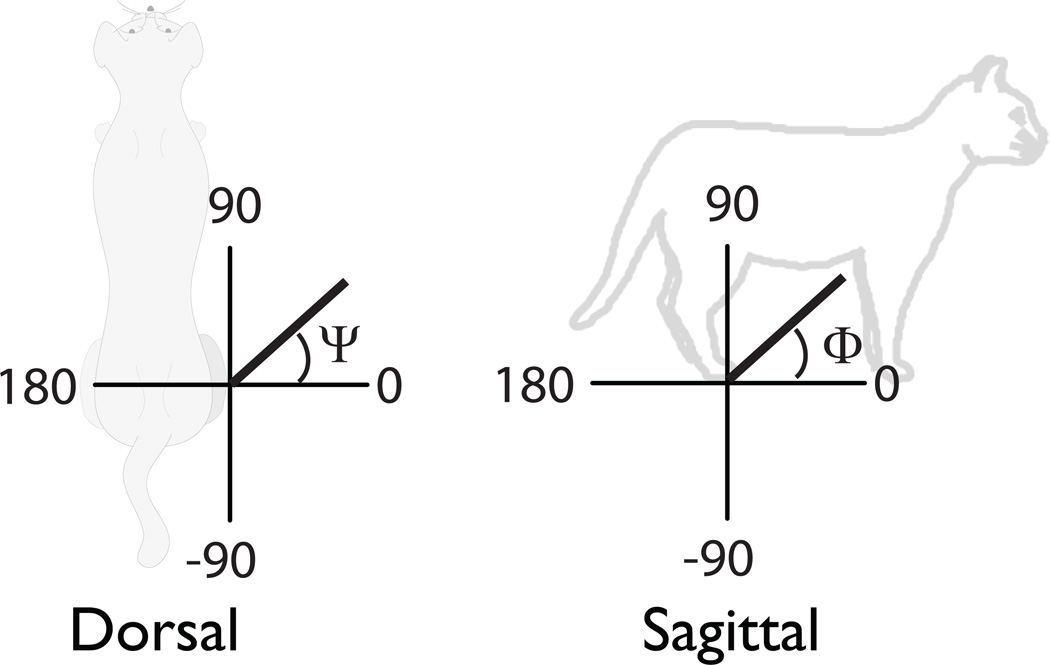
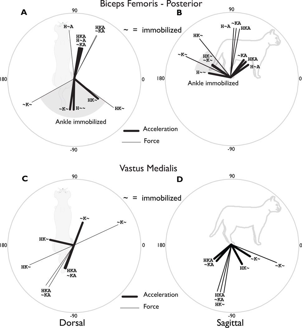
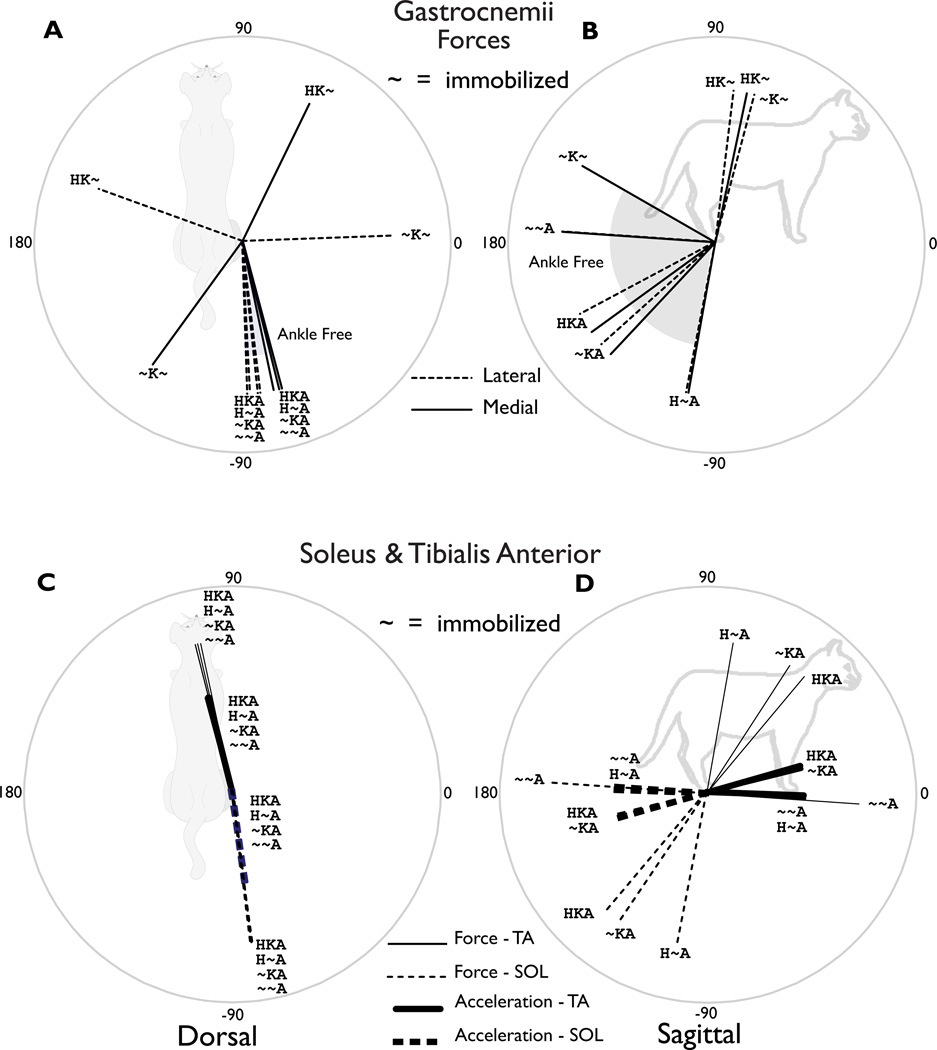
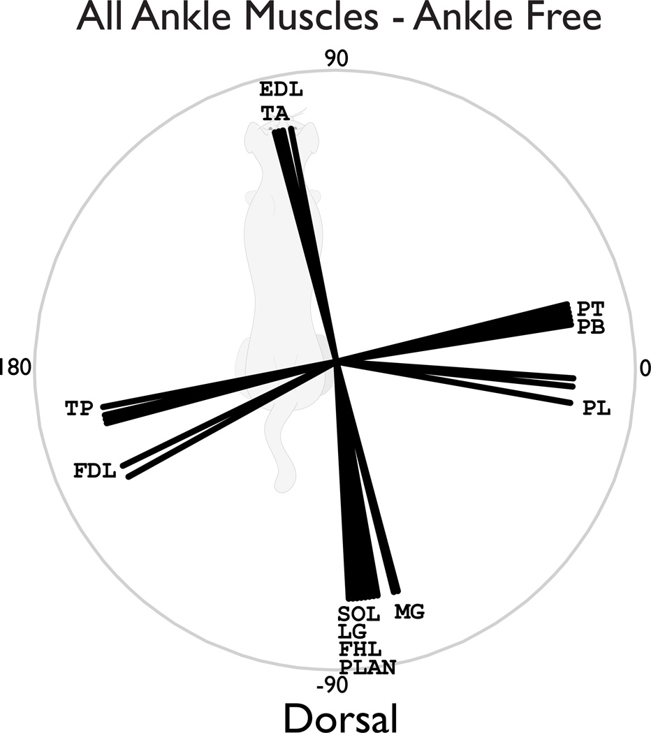
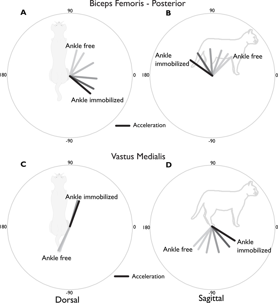
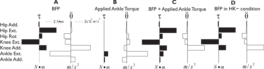
The biomechanical principles underlying the organization of muscle activation patterns during standing balance are poorly understood. The goal of this study was to understand the influence of biomechanical inter-joint coupling on endpoint forces and accelerations induced by the activation of individual muscles during postural tasks. We calculated induced endpoint forces and accelerations of 31 muscles in a 7 degree-of-freedom, three-dimensional model of the cat hindlimb. To test the effects of inter-joint coupling, we systematically immobilized the joints (excluded kinematic degrees of freedom) and evaluated how the endpoint force and acceleration directions changed for each muscle in 7 different conditions. We hypothesized that altered inter-joint coupling due to joint immobilization of remote joints would substantially change the induced directions of endpoint force and acceleration of individual muscles. Our results show that for most muscles crossing the knee or the hip, joint immobilization altered the endpoint force or acceleration direction by more than 90 degrees in the dorsal and sagittal planes. Induced endpoint forces were typically consistent with behaviorally observed forces only when the ankle was immobilized. We then activated a proximal muscle simultaneous with an ankle torque of varying magnitude, which demonstrated that the resulting endpoint force or acceleration direction is modulated by the magnitude of the ankle torque. We argue that this simple manipulation can lend insight into the functional effects of co-activating muscles. We conclude that inter-joint coupling may be an essential biomechanical principle underlying the coordination of proximal and distal muscles to produce functional endpoint actions during motor tasks.


Figures

Figure 1
Figure 1. Coordinate system for computing endpoint force and acceleration direction
(A) dorsal plane angles (Ψ). (B) sagittal plane angles (Φ). These coordinates were used in the reporting of single muscle-induced endpoint accelerations and forces in Tables 3 and 4.
Figure 2
Figure 2. Induced endpoint force and acceleration directions for a representative hamstring and quadriceps muscle
(A) Dorsal plane endpoint force and acceleration directions induced by BFP. (B) Sagittal plane endpoint force and acceleration directions induced by BFP. The anatomical description of BFP (“hip extensor”/ “knee flexor”) suggests that a posterior and upward force would be generated. Forces and accelerations in these directions were found in all conditions where the ankle was immobilized (gray shaded area). However, when the ankle was free, the forces and accelerations were in the anterior direction. Even when the ankle was immobilized, the medial-lateral direction could vary by more than 90°, depending upon the immobilization of the hip joint (gray shaded area). (C) Dorsal plane directions induced by VM. Only when the hip and ankle were both immobilized did the VM produce a forward-directed endpoint force. It also produced substantial medial/lateral forces and accelerations due to the knee ad/abduction moment arms depending on the mechanical status of the hip joint. (D) Sagittal plane directions induced by VM. The VM force and accelerations were always directed downward, supporting its known role as an anti-gravity muscle. VM and BFP acted as near antagonists in the sagittal plane under all conditions. VI also exhibited this behavior, but not VL. Closer examinations of the moment arms revealed that VI, VM and BFP have similar ratios between the moment arms about the two joints (knee flexion/extension and knee aB/Adduction), whereas VL deviates drastically.
Figure 3
Figure 3. Induced endpoint force and acceleration directions for bi- and mono-articular muscles crossing the ankle
(A) Dorsal plane directions induced by MG and LG. When the ankle was free, both the MG and LG generated a posterior and slightly lateral force. These directions were similar whether the hip and knee joints were free or immobilized. However, the aBduction/aDduction action when the ankle was immobilized depended on whether the hip was immobilized. (B) Sagittal plane directions induced by MG and LG. The conditions where the ankle was free exhibited substantial variations in the sagittal plane, but always provided antigravity support and propulsive directed forces. When the ankle was immobilized, the MG actions switched from posterior to anterior. (C) Dorsal plane directions induced by the TA and SOL. The SOL generated posterior and slightly lateral force and accelerations, while the TA generated anterior and slightly medial forces and accelerations. Force and acceleration directions were consistent across all conditions. (D) Sagittal plane directions induced by TA and SOL were relatively consistent across all conditions, with the primary components in the posterior and anterior directions, respectively (thick lines). However, the sagittal plane forces rotated by almost 90° depending on the immobilization of the hip and knee joints (thin lines).
Figure 4
Figure 4. Induced endpoint force directions in the dorsal plane for all ankle muscles under all of the ankle-free conditions
The EDL and TA consistently induced anterior force directions, while the SOL, MG, LG, FHL, and PLAN consistently induced posterior force directions. The ankle evertors: PL, PT, and PB induced laterally directed endpoint forces, while TP and FDL induced medial force directions. These force direction were similar regardless of whether the knee and/or hip were immobilized.
Figure 5
Figure 5. Endpoint acceleration direction induced by hamstrings and quadriceps muscles simultaneous with varying ankle torque
BFP and VM were maximally activated simultaneously with ankle flexion/extension and ad/abduction torques that canceled 0 to 100% of their induced acceleration at the ankle, respectively. (A) Dorsal plane directions induced by activation of BFP simultaneous with varying ankle torque magnitudes. (B) Sagittal plane directions induced by activation of BFP simultaneous with varying ankle torque magnitudes. All other joints were free. (C) Dorsal plane directions induced by VM and varying ankle torque. (D) Sagittal plane directions induced by VM and varying ankle torque. The hip was immobilized. In both muscles, increasing the ankle torque rotated the endpoint acceleration within the plane subtended by the ankle immobilized and ankle free conditions shown in Figure 2. Total ankle torque levels required to functionally immobilize the joint were relatively low, never greater than 20% of the muscle torques produced by VM or BFP. These results demonstrate how multi-muscle coordination or even the contributions of passive joint torques can dramatically alter the endpoint action of the limb when muscles are activated.
Figure 6
Figure 6. Joint torques and their corresponding induced accelerations in all 7 degrees of freedom for four conditions
(A) BFP in the all joints free (HKA) condition. BFP induces only relatively small accelerations at the proximal joints, and large accelerations at the ankle. (B) Torque applied at the ankle. The magnitude of the ankle torques exactly cancel the induced ankle accelerations of BFP. The ankle torques also induce small accelerations at the proximal joints. (C) Combined activation of BFP and ankle torque. (D) BFP in the ankle immobilized (HK~) condition. The net accelerations in C and D are identical, but only the former reveals the necessary applied ankle torque and the accelerations that it induces.

Similar articles

Cited by

References

    1. Abelew TA, Huyghues-Despointes CMJI, Nichols TR. Three dimensional knee torques produced by the quadriceps and hamstrings muscles in the cat. 1996
    1. Anderson FC, Goldberg SR, Pandy MG, Delp SL. Contributions of muscle forces and toe-off kinematics to peak knee flexion during the swing phase of normal gait: an induced position analysis. Journal of Biomechanics. 2004;37(5):731–737. - PubMed
    1. Bernstein N. The Coordination and Regulation of Movements. New York: Pergamon Press; 1967.
    1. Biewener AA, Konieczynski DD, Baudinette RV. In vivo muscle force-length behavior during steady-speed hopping in tammar wallabies. J Exp Biol. 1998;201(Pt 11):1681–1694. - PubMed
    1. Bonasera SJ, Nichols TR. Mechanical actions of heterogenic reflexes among ankle stabilizers and their interactions with plantarflexors of the cat hindlimb. J Neurophysiol. 1996;75(5):2050–2070. - PubMed

Publication types