. 2007 Sep 7;372(1):50-66.
doi: 10.1016/j.jmb.2007.03.046. Epub 2007 Mar 24.

Structural analysis and dynamics of retinal chromophore in dark and meta I states of rhodopsin from 2H NMR of aligned membranes

Affiliations

Structural analysis and dynamics of retinal chromophore in dark and meta I states of rhodopsin from 2H NMR of aligned membranes

Andrey V Struts et al. J Mol Biol. .

Abstract

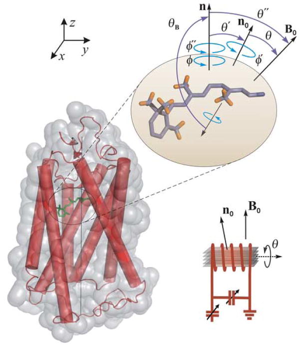
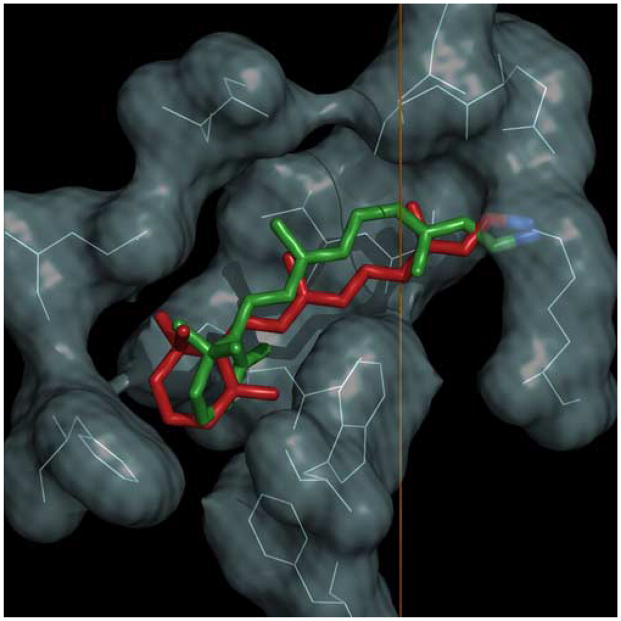
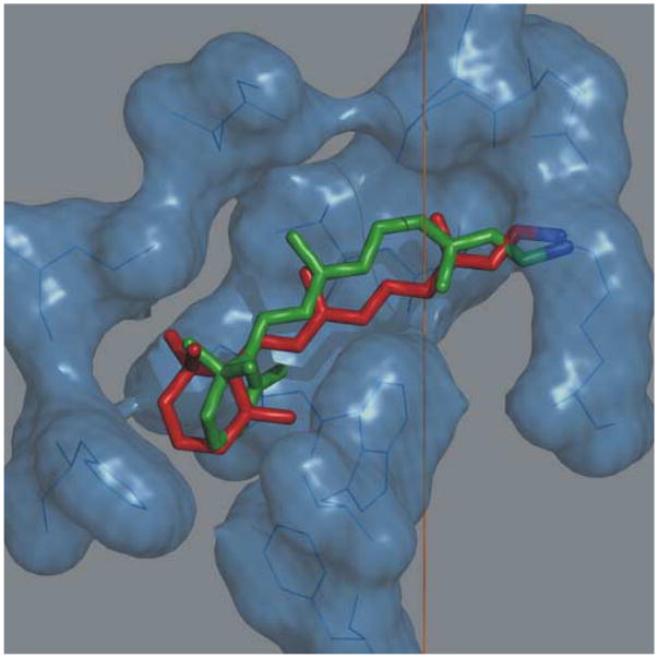
Rhodopsin is a prototype for G protein-coupled receptors (GPCRs) that are implicated in many biological responses in humans. A site-directed (2)H NMR approach was used for structural analysis of retinal within its binding cavity in the dark and pre-activated meta I states. Retinal was labeled with (2)H at the C5, C9, or C13 methyl groups by total synthesis, and was used to regenerate the opsin apoprotein. Solid-state (2)H NMR spectra were acquired for aligned membranes in the low-temperature lipid gel phase versus the tilt angle to the magnetic field. Data reduction assumed a static uniaxial distribution, and gave the retinylidene methyl bond orientations plus the alignment disorder (mosaic spread). The dark-state (2)H NMR structure of 11-cis-retinal shows torsional twisting of the polyene chain and the beta-ionone ring. The ligand undergoes restricted motion, as evinced by order parameters of approximately 0.9 for the spinning C-C(2)H(3) groups, with off-axial fluctuations of approximately 15 degrees . Retinal is accommodated within the rhodopsin binding pocket with a negative pre-twist about the C11=C12 double bond that explains its rapid photochemistry and the trajectory of 11-cis to trans isomerization. In the cryo-trapped meta I state, the (2)H NMR structure shows a reduction of the polyene strain, while torsional twisting of the beta-ionone ring is maintained. Distortion of the retinal conformation is interpreted through substituent control of receptor activation. Steric hindrance between trans retinal and Trp265 can trigger formation of the subsequent activated meta II state. Our results are pertinent to quantum and molecular mechanics simulations of ligands bound to GPCRs, and illustrate how (2)H NMR can be applied to study their biological mechanisms of action.


Figures

Figure 1
Figure 1
Powder-type 2H NMR spectra for 2H-labeled retinal in the dark state are indicative of rotating methyl groups with large order parameters. (a)–(c) Experimental 2H NMR spectra for 11-Z-[9-C2H3]-retinylidene rhodopsin, i.e. having 11-cis-retinal deuterated at the C9 methyl group, in gel-phase POPC membranes (1:50 molar ratio) at pH 7 and T = −30 °C (blue), −60 °C (magenta), and −150 °C (green), respectively. (d) Theoretical 2H NMR spectrum for randomly oriented C–C2H3 groups undergoing fast three-fold rotation on the 2H NMR time scale (< (3χQ/8)−1 ≈ 10 μs). Probability density p(ξ±) is plotted against reduced frequency ξ± for the two I=1 spectral branches, which is expressed in units of (3/4)χQeff = 41.75 kHz for rotating methyl groups. (e),(f) Representative 2H NMR spectra for dark-state 11-Z-[5-C2H3]-retinylidene rhodopsin and 11-Z-[13-C2H3]-retinylidene rhodopsin, i.e. with 11-cis-retinal deuterated at the C5 methyl (yellow) or C13 methyl group (red), respectively, in POPC membranes (1:50) at pH 7 and T = −100 °C. Theoretical powder-type 2H NMR spectra for C–C2H3 groups undergoing axial rotation (continuous color lines) are superimposed on the experimental spectra, with residuals below.
Figure 2
Figure 2
Structure and orientation of retinal ligand of rhodopsin in membranes are investigated by solid-state 2H NMR spectroscopy. Geometry of the tilt experiments is shown for aligned bilayers. (Top) 11-cis-retinylidene chromophore of rhodopsin in the dark state. For a given methyl group, θB is angle of C–C2H3 bond axis to the local membrane normal n, with static rotational symmetry given by the azimuthal angle ϕ. Alignment disorder is described by angle θ′ of n relative to the average membrane normal n0, and is likewise uniaxially distributed as characterized by ϕ′. Next, θ is the tilt angle from n0 to the main magnetic field B0, about which there is cylindrical symmetry. Lastly, θ″ and ϕ″ are the angles for overall transformation from n to B0. (Bottom left) Membrane-bound rhodopsin including N-retinylidene cofactor within its binding cavity. The van der Waals surface of rhodopsin is depicted in light grey, where the seven transmembrane helices are indicated by rods; the N-terminus is at top (extracellular side) and C-terminus at bottom (cytoplasmic side). (Bottom right) Schematic depiction of stack of aligned membranes containing rhodopsin within the radiofrequency coil of the NMR spectrometer, showing geometry relative to the magnetic field.
Figure 3
Figure 3
Tilt series of 2H NMR spectra for 11-cis-retinal in the dark state depends on methyl bond orientations and mosaic spread of aligned membranes. (a)–(c) 2H NMR spectra for 11-Z-[5-C2H3]-retinylidene rhodopsin (blue), 11-Z-[9-C2H3]-retinylidene rhodopsin (magenta), and 11-Z-[13-C2H3]-retinylidene rhodopsin (green), respectively. Experimental 2H NMR spectra are shown as a function of tilt angle θ of the average normal of membrane stack n0 to magnetic field B0 for rhodopsin/POPC bilayers (1:50) at pH 7 and T = −150°C. Theoretical lineshapes (continuous color lines) assume a static uniaxial distribution of rhodopsin molecules, with rotating C–C2H3 groups of retinylidene cofactor.
Figure 4
Figure 4
Angular sequence of 2H NMR spectra for trans-retinal in meta I shows differences versus dark state. (a)–(c) Tilt series of 2H NMR spectra for 11-E-[5-C2H3]-retinylidene rhodopsin, 11-E-[9-C2H3]-retinylidene rhodopsin, and 11-E-[13-C2H3]-retinylidene rhodopsin. 2H NMR spectra are plotted at different values of tilt angle θ for meta I/POPC (1:50) membranes at pH 7 and T = −100 °C. Calculated lineshapes (continuous lines) assume a static uniaxial distribution of rhodopsin molecules with rotating methyl groups.
Figure 5
Figure 5
Error surfaces for 2H NMR spectra of 11-cis-retinal in the dark state give accurate methyl bond orientations. (a)–(c) Root mean square deviation (RMSD) of calculated versus experimental 2H NMR spectra (normalized; cf. Figure 3) for rhodopsin with retinal deuterated at C5, C9, or C13 methyl groups, respectively. (d)–(f) Cross-sections through hypersurfaces at left showing well-defined minima in bond orientation and mosaic spread of aligned membranes. Fitting parameters at T = −150 °C are: C5 (θB =70±3°, σ =18±3°, ΔνQpowder=39.0kHz , FWHM = 3.8 kHz); C9 (θB =52±3°, σ =21±3°, ΔνQpowder=39.0kHz, FWHM = 5.0 kHz); and C13 (θB =68±2°, σ =20±2°, ΔνQpowder=37.5-40.0kHz, FWHM = 3.2 kHz). Global fitting parameters at T = −60 °C (spectra not shown) are: C5 (θB=70±5°, σ=18±4°, ΔνQpowder=39.0kHz, FWHM = 3.8 kHz); C9 (θB =52±5°, σ =21±4°, ΔνQpowder=39.0kHz, FWHM = 5.0 kHz); and C13 (θB =68±4°, σ =20±4°, ΔνQpowder=37.5-40.0kHz, FWHM = 3.2 kHz).
Figure 6
Figure 6
Error analysis for 2H NMR spectra of trans-retinal in meta I indicate differences in methyl bond orientations from dark state. (a)–(c) RMSD error hypersurfaces for meta I (cf. Figure 4) with retinal 2H-labeled at C5, C9, or C13 methyl groups, respectively. (d)–(f) Cross-sections through error hypersurfaces displaying minima in bond orientation θB and mosaic spread σ. Fitting parameters for meta I at T = −100 °C are: C5 (θB=72±4°, σ=24±3°, ΔνQpowder=37.0-40.0kHz, FWHM = 3.2 kHz); C9 (θB =53± 3°, σ =25± 3°, ΔνQpowder=38.0kHz, FWHM = 4.4 kHz); and C13 (θB =59±3°, σ =22±3°, ΔνQpowder=35.0-40.0kHz, FWHM = 3.2 kHz).
Figure 7
Figure 7
Solid-state 2H NMR yields structure and orientation of 11-cis-retinal in the dark state of rhodopsin. The retinal conformation is described by three planes of unsaturation (designated A, B, C). (a) Simple three-plane model with torsional twisting only about C6–C7 and C12–C13 bonds. (b) Extended three-plane model with additional pre-twisting about C11=C12 double bond. Extracellular side of rhodopsin is at top and the cytoplasmic side is at bottom (cf. text).
Figure 8
Figure 8
Orientation and structure of trans-retinal in meta I are illuminated by solid-state 2H NMR spectroscopy. Three planes of unsaturation (A, B, C) are depicted; the structures are mirror images related by a vertical reflection plane. Extracellular side of rhodopsin points up and the cytoplasmic side is down (cf. text).
Figure 9
Figure 9
Solid-state NMR provides structure of retinal cofactor in the dark and pre-activated meta I states of rhodopsin. Vertical direction corresponds to membrane normal; the extracellular side is up and cytoplasmic side is down. (a) Comparison of NMR structure (green) of 11-cis-retinal in the dark state versus retinal structure (red) from X-ray crystallography (PDB accession code 1U19). Inset: NMR structure calculated with twisting of polyene about C12–C13 bond only (simple three-plane model; red) compared to structure with twisting about both C11=C12 and C12–C13 bonds (extended three-plane model; green); view angle is with horizontal rotation of β-ionone ring towards observer. (b) NMR structure for 11-cis-retinal in the dark state (green) compared to NMR structure of trans-retinal in the meta I state (red). (Figure produced with PyMOL.)
Cover
Cover
Solid-state 2H NMR structure of retinal ligand of G protein-coupled receptor rhodopsin in the dark (green) and pre-activated metarhodopsin I (red) states. [Four alternate cover figures are shown]
Cover
Cover
Solid-state 2H NMR structure of retinal ligand of G protein-coupled receptor rhodopsin in the dark (green) and pre-activated metarhodopsin I (red) states. [Four alternate cover figures are shown]
Cover
Cover
Solid-state 2H NMR structure of retinal ligand of G protein-coupled receptor rhodopsin in the dark (green) and pre-activated metarhodopsin I (red) states. [Four alternate cover figures are shown]
Cover
Cover
Solid-state 2H NMR structure of retinal ligand of G protein-coupled receptor rhodopsin in the dark (green) and pre-activated metarhodopsin I (red) states. [Four alternate cover figures are shown]

Similar articles

Cited by

References

    1. Pierce KL, Premont RT, Lefkowitz RJ. Seven-transmembrane receptors. Nat Rev Mol Cell Biol. 2002;3:639–650. - PubMed
    1. Fanelli F, De Benedetti PG. Computational modeling approaches to structure-function analysis of G protein-coupled receptors. Chem Rev. 2005;105:3297–3351. - PubMed
    1. Sakmar TP, Menon ST, Marin EP, Awad ES. Rhodopsin: Insights from recent structural studies. Annu Rev Biophys Biomol Struct. 2002;31:443–484. - PubMed
    1. Hubbell WL, Altenbach C, Hubbell CM, Khorana HG. Rhodopsin structure, dynamics, and activation: a perspective from crystallography, site-directed spin labeling, sulfhydryl reactivity, and disulfide cross-linking. Adv Prot Chem. 2003;63:243–290. - PubMed
    1. Lüdeke S, Beck R, Yan ECY, Sakmar TP, Siebert F, Vogel R. The role of Glu181 in the photoactivation of rhodopsin. J Mol Biol. 2005;353:345–356. - PubMed

Publication types