. 2007 Aug;92(8):1013-20.
doi: 10.3324/haematol.11407. Epub 2007 Jul 20.

Functional characterization of novel telomerase RNA (TERC) mutations in patients with diverse clinical and pathological presentations

Affiliations

Functional characterization of novel telomerase RNA (TERC) mutations in patients with diverse clinical and pathological presentations

Anna Marrone et al. Haematologica. 2007 Aug.

Abstract

Background and objectives: Functional characterization of heterozygous TERC (telomerase RNA component) and TERT (telomerase reverse transcriptase) mutations found in autosomal dominant dyskeratosis congenita (DC) and aplastic anemia (AA) shows that telomerase function is defective and that this is associated with short telomeres. This leads to reduced cell longevity with maximal impact on tissues with high proliferate potential. The aim of this study was to establish the role of TERC in the pathophysiology of uncharacterized patients with AA with some features of DC.

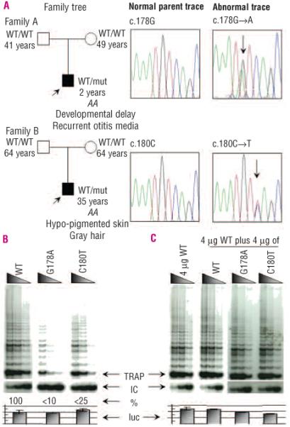
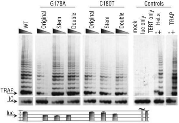
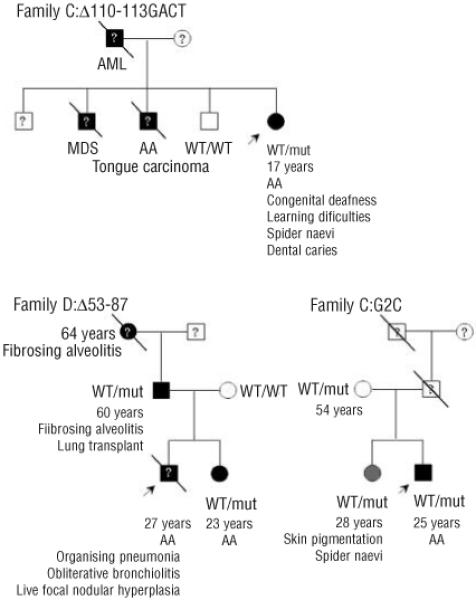
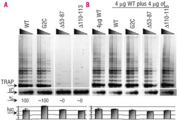
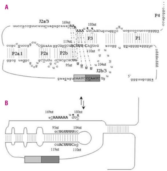
Design and methods: The TERC gene was screened for mutations by denaturing high performance liquid chromatography. To determine the functional significance of TERC mutations telomerase activity was assessed in an in vitro (TRAP) assay and telomere length of patients' samples was determined using Southern blot analysis. RESULTS This study led to the identification of four novel TERC mutations (G178A, C180T, D52-86 and G2C) and a recurrent TERC mutation (D110-113GACT).

Interpretation and conclusions: Two of the de novo TERC mutations (G178A and C180T) found uniquely produce a clinical phenotype in the first generation, differing from previously published cases in which individuals in the first generation are usually asymptomatic. Curiously these mutations are located near the triple-helix domain of TERC. We also observed that the recurrent D110-113GACT can present with AA, myelodysplasia or leukemia. The D52-86 is associated with varied phenotypes including pulmonary disease (pulmonary fibrosis) as the first presentation. In summary, this study reports the functional characterization of several novel TERC mutations associated with varied hematologic and extra-hematologic presentations.


Figures

Figure 1
Figure 1
TERC mutation identification and analysis for Families A and B. A. Sequence traces depicting the normal and abnormal heterozygous TERC traces of G178A and C180T for families A and B, respectively. Age (years), TERC allele status (WT: as published TERC sequence; mut: nucleotide substitution as indicated in the sequence trace) and relevant family clinical history are indicated in the family trees in which the index cases (black squares) are indicated by an arrow and the parents appear asymptomatic (white squares or circles). B. Reconstituted TRAP assays showed that G178A and C180T reduce in vitro telomerase activity to less than 10% and 25% of WT control levels, respectively. C. In mixing experiments, activity did not drop below 50% of WT activity, suggesting no dominant negative effect for these two de novo heterozygous TERC mutations. The arrows denote the start of the TRAP ladder, the corresponding internal control (IC), the amount of activity in relationship to the WT TRAP control (%) and the levels of luciferase (luc) to control for the corresponding TRAP experiment. Serial dilutions of each transfected lysate were assayed as described in the methods section.
Figure 2
Figure 2
Telomere lengths of investigated families compared to those of normal controls. A. Normal subjects are indicated by small white circles. The different families are represented as follows: G178A affected and normal parents, black and white squares; C180T affected and normal parents, black and white triangles; 110-113delGACT affected, black circle; 53-87del affected and normal parent, black and white diamonds; G2C affected and asymptomatic sister and mother, gray and white circles respectively. The line of best fit through this normal range is shown as a black line which corresponds to the equation Y=17.821 – 0.0407X. Deviation from the best-fit-line is highlighted as a dark gray box for 68%, a lighter gray box for 90% and the palest grey box for 95%. Therefore the index cases lie on or below the 90% deviation range, which represents the 5th percentile when compared to the panel of normal controls. B. The Δtel values from normal subjects from panel A (n=112) are represented on a linear graph and compared to the telomere lengths from previously reported patients with TERC mutations (n=38; black lines) and the patients from this report (n=7). The patients in this report have similar Δtel values to those of other patients with TERC mutations. Other labels are as in panel A.
Figure 3
Figure 3
Reconstituted telomerase activity of disrupted and restored pseudoknot helices. Original represents the TERC mutation found in the index case, Stem represents the mutation formed on the corresponding stem opposite the original mutation and Double represents the TERC RNA when both the original and stem mutation are present together in the same molecule, reconstituting the stem but with the mutated primary sequence. Other labels are as in Figure 1.
Figure 4
Figure 4
Clinical history and inheritance of TERC mutations in families C, D and E. The arrow denotes the index case and a line through the person represented indicates that the person has died. Black squares represent an AA/DC phenotype while gray circles represent borderline phenotypes. White squares and circles indicate asymptomatic people. ? denotes that a sample was not available for TERC mutation screening. Other labels are as in Figure 1.
Figure 5
Figure 5
Reconstituted telomerase activity of other TERC mutations studied in vitro. A. Reconstituted TRAP assays show that 53-87del and 110-113delGACT mutations reduce in vitro telomerase activity to near 0% of that of WT controls while the G2C mutation appears to have near normal activity. B. In mixing experiments, there was no observed drop below 50% WT activity, suggesting no dominant negative effect for these three heterozygous TERC mutations. Other labels are as in Figure 1.
Figure 6
Figure 6
TERC pseudoknot structure and proposed molecular switch model. A. The secondary structure of the TERC pseudoknot has been elucidated by functional analysis. Every tenth base of the 451bp molecule is underlined and <80% vertebrate nucleotide conservation is denoted by upper case letters. The RNA template contains partial repeats of the telomere repeat known as the alignment region (lighter gray box). The larger dark grey box contains the templating region which is utilized to ensure correct base-pairing and correct translocation and re-alignment for the next round of template synthesis. Critical bases involved in the triple helix structure are highlighted in bold. Thermodynamics and mutational analysis suggest that nucleotides 171-172AA form a Hoogsteen base-pair with 97-98UG, which are helically-bound by conventional Watson-Crick base-pairing to 116-117CA. Nucleotides 100-102UUU then form a Hoogsteen base-pair with 174-176AAA, which are helically-bound by conventional Watson-Crick base-pairing to 113-115UUU. Nucleotides 99U and 173A can then pair up between these two triple helices to bridge the physical gap. The Watson-Crick base-pairing is denoted by gray lines while the dashed black lines represent the Hoogsteen basepairing. The de novo mutations described in this paper are in italics and are highlighted with *. B. While the two novel de novo mutations are not directly involved in the loop formed during the proposed molecular switch, the complementary nucleotides G110 and C112 are critical to the correct formation of the new TERC RNA structure.

Comment in

Similar articles

Cited by

References

    1. Heiss NS, Knight SW, Vulliamy TJ, Klauck SM, Wiemann S, Mason PJ, et al. X-linked dyskeratosis congenita is caused by mutations in a highly conserved gene with putative nucleolar functions. Nat Genet. 1998;19:32–8. - PubMed
    1. Vulliamy T, Marrone A, Goldman F, Dearlove A, Bessler M, Mason PJ, et al. The RNA component of telomerase is mutated in autosomal dominant dyskeratosis congenita. Nature. 2001;413:432–5. - PubMed
    1. Armanios M, Chen JL, Chang YP, Brodsky RA, Hawkins A, Griffin CA, et al. Haploinsufficiency of telomerase reverse transcriptase leads to anticipation in autosomal dominant dyskeratosis congenita. Proc Natl Acad Sci USA. 2005;102:15960–4. - PMC - PubMed
    1. Chen JL, Greider CW. Telomerase RNA structure and function: implications for dyskeratosis congenita. Trends Biochem Sci. 2004;29:183–92. - PubMed
    1. Marrone A, Dokal I. Dyskeratosis congenita: a disorder of telomerase deficiency and its relationship to other diseases. Expert Rev Derm. 2006;1:463–79.

Publication types

MeSH terms