. 2007 Jul 24;104(30):12571-6.
doi: 10.1073/pnas.0701737104. Epub 2007 Jul 17.

Crystal structure of the chromophore binding domain of an unusual bacteriophytochrome, RpBphP3, reveals residues that modulate photoconversion

Affiliations

Crystal structure of the chromophore binding domain of an unusual bacteriophytochrome, RpBphP3, reveals residues that modulate photoconversion

Xiaojing Yang et al. Proc Natl Acad Sci U S A. .

Abstract

Bacteriophytochromes RpBphP2 and RpBphP3 from the photosynthetic bacterium Rhodopseudomonas palustris work in tandem to modulate synthesis of the light-harvesting complex LH4 in response to light. Although RpBphP2 and RpBphP3 share the same domain structure with 52% sequence identity, they demonstrate distinct photoconversion behaviors. RpBphP2 exhibits the "classical" phytochrome behavior of reversible photoconversion between red (Pr) and far-red (Pfr) light-absorbing states, whereas RpBphP3 exhibits novel photoconversion between Pr and a near-red (Pnr) light-absorbing states. We have determined the crystal structure at 2.2-A resolution of the chromophore binding domains of RpBphP3, covalently bound with chromophore biliverdin IXalpha. By combining structural and sequence analyses with site-directed mutagenesis, we identify key residues that directly modulate the photochemical properties of RpBphP3 and RpBphP2. Remarkably, we identify a region spanning residues 207-212 in RpBphP3, in which a single mutation, L207Y, causes this unusual bacteriophytochrome to revert to the classical phenotype that undergoes reversible photoconversion between the Pr and Pfr states. The reverse mutation, Y193L, in the corresponding region in RpBphP2 significantly diminishes the formation of the Pfr state. We propose that residues 207-212 and the spatially adjacent conserved residues, Asp-216 and Tyr-272, interact with the chromophore and form part of the interface between the chromophore binding domains and the PHY domain that modulates photoconversion.


Conflict of interest statement

The authors declare no conflict of interest.

Figures

Fig. 1.
Fig. 1.
Crystal structure of RpBphP3-CBD. (a) Domain structure of the full-length Bph RpBphP3. (b) Ribbon diagram of the two RpBphP3-CBD molecules in the asymmetric unit. The PAS domain is in yellow, and the GAF domain is in green. (c) Superposition of the crystal structures of RpBphP3-CBD (PDB ID code 2OOL, in yellow and green) and DrBphP-CBD (PDB ID code 2O9C, in light blue). (d) The FoFc omit map in the region of chromophore. The chromophore is shown as 2(S),3(E)-PΦB covalently linked to Cys-28.
Fig. 2.
Fig. 2.
The chromophore binding pocket. (a) Residues in the chromophore (cyan) pocket examined by site-directed mutagenesis. Lys-183, Ser-297, and His-299 form the 15Za pocket; Leu-207, Phe-210, and Phe-212 form part of the 15Ea pocket; and Tyr-272 and Asp-216 are colored in pink. Potential hydrogen bonds are shown in red dashed lines, and the corresponding distances are given in the text. (b) The chromophore cavity surrounding ring D. (c) Sequence alignment of representative Bphs and Phy-like photoreceptors in the regions of the 15Za and 15Ea pockets in the GAF domain (residues colored as in a). The consensus sequence motif, PASDIP, is highlighted in green. The sequences are as follows: RpBphP3 (R. palustris CGA009 PhyB2), RpBphP2 (R. palustris CGA009 PhyB1), DrBphP (D. radiodurans R1 BphP), AtBphP1/Agp1 (Agrobacterium tumefaciens BphP1), PsBphP (Pseudomonas syringae DC3000 BphP), XaBphP (Xanthomonas axonopodis BphP), XcBphP (Xanthomonas campestris ATCC 33913 BphP), RcPPH (R. centenum PPH), TtPPD (Thermochromatium tepidum Ppd), AtBphP2/Agp2 (A. tumefaciens BphP2), RpBphP5 (R. palustris CGA009 PhyB5), PaBphP (Pseudomonas aeruginosa PA01 BphP), Cph1 (Synechocystis PCC6803 Cph1), and Cph2 (Synechocystis PCC6803 Cph2).
Fig. 3.
Fig. 3.
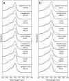
UV-visible absorption spectra of wild-type and selected mutants of RpBphP3–521 (a) and RpBphP2–505 (b). Spectra are in order of appearance in the text from the top down. Spectra were measured in solution in the dark-adapted state (solid lines) and light-illuminated state (dashed lines). The corresponding mutants in RpBphP3 and RpBphP2 are shown side by side. Spectra on the bottom of each panel are from RpBphP3-CBD (1–337) and RpBphP2-CBD (1–321). Numbers in parentheses represent the estimated half-time of dark reversion for each sample.
Fig. 4.
Fig. 4.
VDW surface of the RpBphP3-CBD structure. Residues Tyr-272, Asp-216, and Leu-207-Phe-210-Phe-212 in the 15Ea pocket (colored in maroon) form a continuous surface patch and shield ring D of the chromophore (in cyan) in the GAF domain.

References

    1. Rockwell NC, Su YS, Lagarias JC. Annu Rev Plant Biol. 2006;57:837–858. - PMC - PubMed
    1. Heyne K, Herbst J, Stehlik D, Esteban B, Lamparter T, Hughes J, Diller R. Biophys J. 2002;82:1004–1016. - PMC - PubMed
    1. Chen E, Lapko VN, Lewis JW, Song PS, Kliger DS. Biochemistry. 1996;35:843–850. - PubMed
    1. Borucki B, von Stetten D, Seibeck S, Lamparter T, Michael N, Mroginski MA, Otto H, Murgida DH, Heyn MP, Hildebrandt P. J Biol Chem. 2005;280:34358–34364. - PubMed
    1. Giraud E, Zappa S, Vuillet L, Adriano JM, Hannibal L, Fardoux J, Berthomieu C, Bouyer P, Pignol D, Vermeglio A. J Biol Chem. 2005;280:32389–32397. - PubMed

Publication types

Associated data