. 2007 Sep;171(3):872-81.
doi: 10.2353/ajpath.2007.060972. Epub 2007 Jul 19.

Oncostatin M gene therapy attenuates liver damage induced by dimethylnitrosamine in rats

Affiliations

Oncostatin M gene therapy attenuates liver damage induced by dimethylnitrosamine in rats

Tetsuhiro Hamada et al. Am J Pathol. 2007 Sep.

Abstract

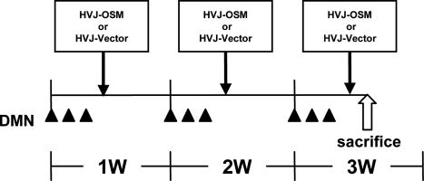
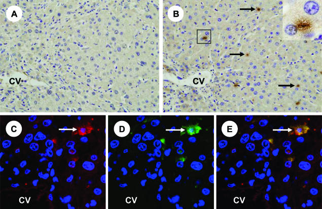
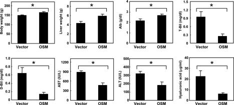
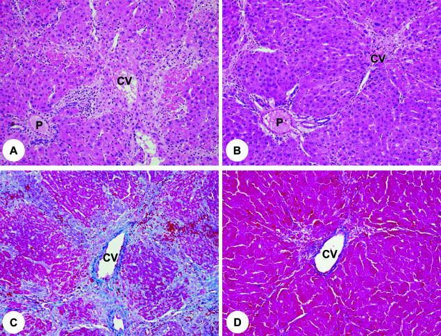
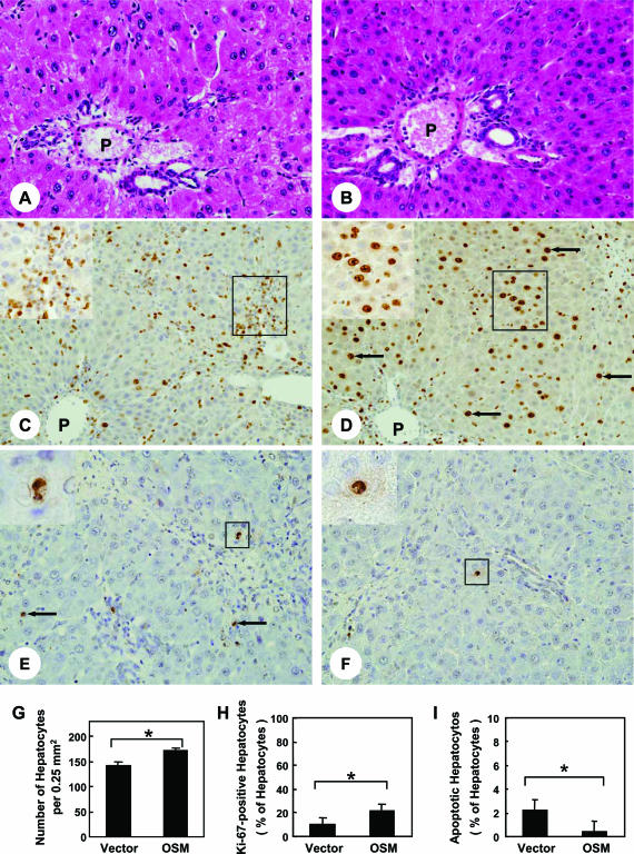
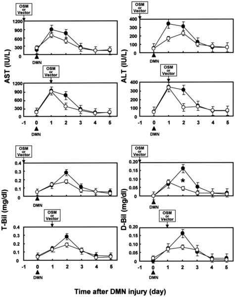
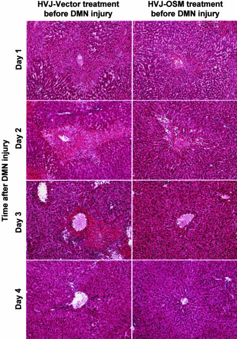
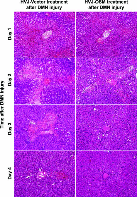
To assess the usefulness of oncostatin M (osm) gene therapy in liver regeneration, we examined whether the introduction of OSM cDNA enhances the regeneration of livers damaged by dimethylnitrosamine (DMN) in rats. Repeated injection of OSM cDNA enclosed in hemagglutinating virus of Japan envelope into the spleen resulted in the exclusive expression of OSM protein in Kupffer cells of the liver, which was accompanied by increases in body weight, liver weight, and serum albumin levels and the reduction of serum liver injury parameters (bilirubin, aspartate aminotransferase, and alanine aminotransferase) and a serum fibrosis parameter (hyaluronic acid). Histological examination showed that osm gene therapy reduced centrilobular necrosis and inflammatory cell infiltration and augmented hepatocyte proliferation. The apoptosis of hepatocytes and fibrosis were suppressed by osm gene therapy. Time-course studies on osm gene therapy before or after DMN treatment showed that this therapy was effective not only in enhancing regeneration of hepatocytes damaged by DMN but in preventing hepatic cytotoxicity caused by subsequent treatment with DMN. These results indicate that OSM is a key mediator for proliferation and anti-apoptosis of hepatocytes and suggest that osm gene therapy is useful, as preventive and curative means, for the treatment of patients with liver damage.


Figures

Figure 1
Figure 1
Schedule of DMN administration and HVJ-OSM or HVJ-Vector injection. Rats were given DMN intraperitoneally at 15 mg/kg body weight for 3 consecutive days per week for 3 weeks. On day 4 of each week, HVJ-OSM or HVJ-Vector was injected into the spleen. On day 5 of the last week, rats were sacrificed for analysis. w, week.
Figure 2
Figure 2
Expression of OSM in the liver of a rat injected with HVJ-OSM. A: Immunostaining of OSM in the liver of a rat injected with HVJ-Vector. B: Immunostaining of OSM in the liver of a rat injected with HVJ-OSM. Arrows indicate representative OSM-expressing cells. Inset in B is a higher magnification of the squared region, and shows an OSM-expressing cell. C: Fluorescein for ED2, which is a marker of Kupffer cells (red). D: Fluorescein for OSM (green). E: Merged confocal image of C and D. Coexpression of ED2 and OSM is shown as yellow. White arrows indicate representative coexpression of ED2 and OSM. C to E are photographs of the same section, taken by confocal laser-scanning microscope. CV, central vein. Original magnifications: ×200 (A and B); ×600 (inset).
Figure 3
Figure 3
Effects of OSM on body weight, liver weight, serum albumin levels, serum liver injury parameters, and serum, a fibrosis parameter in rats with DMN-induced liver damage. Vector: Rats injected with HVJ-Vector. OSM: Rats injected with HVJ-OSM. Alb, albumin. Data represent the mean of six rats. Bars are standard errors. *P < 0.05, significant difference by t-tests.
Figure 4
Figure 4
Histology of livers of rats injected with HVJ-Vector or HVJ-OSM. A and C: Liver sections of a rat injected with HVJ-Vector. B and D: Liver sections of a rat injected with HVJ-OSM. Sections of A and B were stained with H&E, and those of C and D with Azan. P, portal vein; CV, central vein. Original magnifications: ×90 (A and B); ×100 (C and D).
Figure 5
Figure 5
Effects of OSM on proliferation and apoptosis of hepatocytes in rats with DMN-induced liver damage. A, C, and E: Liver sections of a rat injected with HVJ-Vector. B, D, and F: Liver sections of a rat injected with HVJ-OSM. Sections of A and B were stained with H&E, those of C and D were immunostained with Ki-67 antibody, and those of E and F were subjected to TUNEL staining. Arrows in D and E indicate representative Ki-67-positive and apoptotic hepatocytes, respectively. The inset in C is a high magnification of the squared region and shows inflammatory cells expressing Ki-67 antigen. The inset in D is a high magnification of the squared region and shows hepatocytes expressing Ki-67 antigen. Each inset in E and F is a high magnification of the squared region and shows apoptotic hepatocytes. P, portal vein. G and H show the number of total hepatocytes per portal field and the proportion of Ki-67-positive hepatocytes to total hepatocytes, respectively. I shows the proportion of TUNEL-positive hepatocytes to total hepatocytes. Data represent the mean of six rats. Bars are standard errors. *P < 0.05, significant difference by t-tests. Vector: Rats injected with HVJ-Vector. OSM: Rats injected with HVJ-OSM. Original magnifications: ×150 (A and B); ×100 (C and D); ×170 (E and F); ×160 (C and D, insets); ×520 (E and F, insets).
Figure 6
Figure 6
Time-course studies of the effect of OSM treatment before or after DMN injury on serum liver injury parameters in rats with DMN-induced liver damage. (•): Rats injected with HVJ-Vector. (○): Rats injected with HVJ-OSM. Data represent the mean of three rats. Bars are standard errors. *P < 0.05, significant difference by t-tests.
Figure 7
Figure 7
Time-course studies on the liver histology of rats injected with HVJ-Vector or HVJ-OSM before DMN injury. Sections were stained with H&E. Original magnifications, ×40.
Figure 8
Figure 8
Time-course studies on the liver histology of rats injected with HVJ-Vector or HVJ-OSM after DMN injury. Sections were stained with H&E. Original magnifications, ×40.

References

    1. Fausto N, Laird AD, Webber EM. Liver regeneration. 2. Role of growth factors and cytokines in hepatic regeneration. FASEB J. 1995;9:1527–1536. - PubMed
    1. Michalopoulos GK, DeFrances MC. Liver regeneration. Science. 1997;276:60–66. - PubMed
    1. Lindroos PM, Zarnegar R, Michalopoulos GK. Hepatocyte growth factor (hepatopoietin A) rapidly increases in plasma before DNA synthesis and liver regeneration stimulated by partial hepatectomy and carbon tetrachloride administration. Hepatology. 1991;13:743–750. - PubMed
    1. Rubin RA, O’Keefe EJ, Earp HS. Alteration of epidermal growth factor-dependent phosphorylation during rat liver regeneration. Proc Natl Acad Sci USA. 1982;79:776–780. - PMC - PubMed
    1. Webber EM, FitzGerald MJ, Brown PI, Bartlett MH, Fausto N. Transforming growth factor-alpha expression during liver regeneration after partial hepatectomy and toxic injury, and potential interactions between transforming growth factor-alpha and hepatocyte growth factor. Hepatology. 1993;18:1422–1431. - PubMed

Publication types