. 2007 Sep;171(3):906-16.
doi: 10.2353/ajpath.2007.070028. Epub 2007 Jul 19.

Pemphigus IgG causes skin splitting in the presence of both desmoglein 1 and desmoglein 3

Affiliations

Pemphigus IgG causes skin splitting in the presence of both desmoglein 1 and desmoglein 3

Volker Spindler et al. Am J Pathol. 2007 Sep.

Abstract

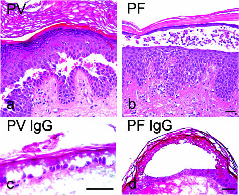
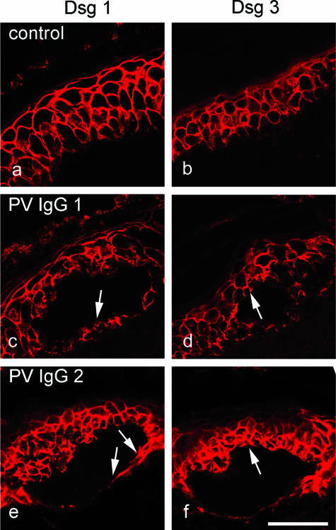
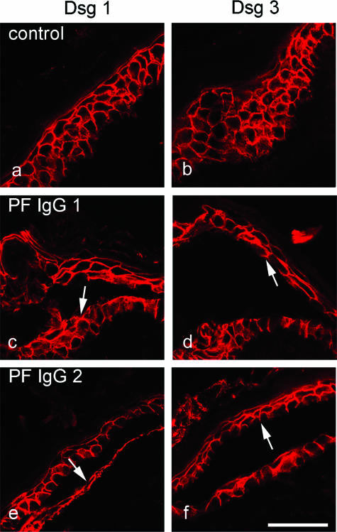
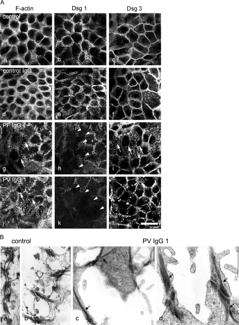
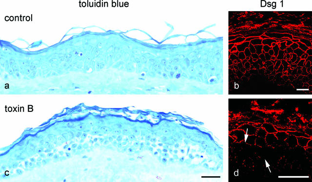
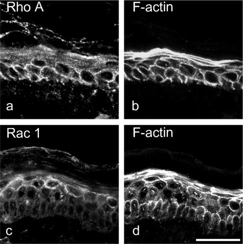
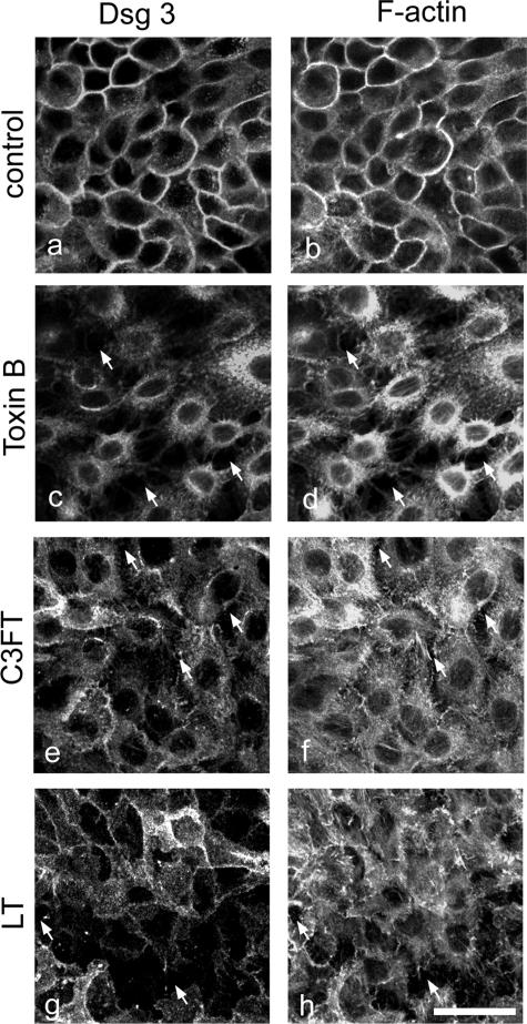
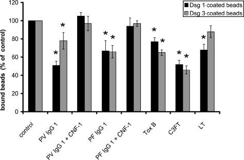
According to the desmoglein (Dsg) compensation concept, different epidermal cleavage planes observed in pemphigus vulgaris and pemphigus foliaceus have been proposed to be caused by different autoantibody profiles against the desmosomal proteins Dsg 1 and Dsg 3. According to this model, Dsg 1 autoantibodies would only lead to epidermal splitting in those epidermal layers in which no Dsg 3 is present to compensate for the functional loss of Dsg 1. We provide evidence that both pemphigus foliaceus-IgG containing Dsg 1- but not Dsg 3-specific antibodies and pemphigus vulgaris-IgG with antibodies to Dsg 1 and Dsg 3 were equally effective in causing epidermal splitting in human skin and keratinocyte dissociation in vitro. These effects were present where keratinocytes expressed both Dsg 1 and Dsg 3, demonstrating that Dsg 3 does not compensate for Dsg 1 inactivation. Rather, the cleavage plane in intact human skin caused by pemphigus autoantibodies was similar to the plane of keratinocyte dissociation in response to toxin B-mediated inactivation of Rho GTPases. Because we recently demonstrated that pemphigus-IgG causes epidermal splitting by inhibition of Rho A, we propose that Rho GTPase inactivation contributes to the mechanisms accounting for the cleavage plane in pemphigus skin splitting.


Figures

Figure 1
Figure 1
Histology from pemphigus patients whose sera were used for this study revealed typical blister localization (a and b). The H&E-stained paraffin sections showed suprabasal epidermal cleavage in the PV histology and superficial blistering in the PF lesion. In human skin in vitro (c and d), PV-IgG-induced splitting was suprabasally located, whereas PF-IgG-induced cleavage was found within the spinous layer. Bar = 50 μm in all panels.
Figure 2
Figure 2
In cultured intact human epidermis, PV-IgG caused epidermal splitting where both Dsg1 and Dsg3 were present. Skin was immunostained for Dsg 1 (a, c, and e) and Dsg 3 (b, d, and f). In untreated skin, Dsg 1 and Dsg 3 displayed broad overlapping distribution patterns (a and b). Dsg 1 was expressed throughout the epidermis with a decreasing gradient from superficial to basal layers. Despite its basal prominence, Dsg 3 was present in all layers except the upper granular layer. Incubation with PV-IgG containing antibodies against both Dsg 1 and Dsg 3 lead to suprabasal splitting (c–f). Note that Dsg1 was present in the basal layer underneath the split (arrows in c and e) and that Dsg 3 was found above the cleavage plane (arrows in d and f). Bar = 50 μm.
Figure 3
Figure 3
Similar to PV-IgG, PF-IgG also caused epidermal splitting where both Dsg1 and Dsg3 were present. Skin was immunostained for Dsg 1 (a, c, and e) and Dsg 3 (b, d, and f). Compared with untreated skin (a and b), incubation with PF-IgG containing only Dsg 1-specific antibodies resulted in splitting where expression of Dsg 1 as well as Dsg 3 was found (c–f). Dsg 1 was present underneath the cleavage plane (arrows in c and e), and Dsg 3 immunoreactivity was detected above the split (arrows in d and f). Bar = 50 μm.
Figure 4
Figure 4
Pemphigus IgG induced cell dissociation in cultured human keratinocytes (HaCaT cells) expressing both Dsg 1 and Dsg 3. A: Cells were stained for F-actin using ALEXA-phalloidin to detect sensitively cell dissociation and to visualize effects on the actin cytoskeleton (a, d, g, and j) and for Dsg 1 (b, e, h, and k) or for Dsg 3 (c, f, i, and l) to label desmosomes. In untreated monolayers (a–c), after incubation with control IgG from a healthy volunteer (d–f), staining of F-actin and Dsg 1 and Dsg 3 was continuous along the cell borders. Incubation with PF-IgG 1 or PV-IgG 1 resulted in keratinocyte dissociation (arrows in g and j) and profound alteration of the actin cytoskeleton and loss of Dsg 1 immunoreactivity in areas of cell dissociation (arrowheads in h and k). After incubation with PF-IgG 1, staining for Dsg 3 was lost at gap margins (arrows in i), whereas PV-IgG 1 resulted in fragmentation of Dsg 3 immunoreactivity, indicating a general loss of desmosomes. Bar = 20 μm. B: The effect of PV-IgG 1 on desmosomes was characterized by transmission electron microscopy (n = 3). In controls (a and b), keratinocyte cell borders were aligned, and numerous desmosomes were visible. After treatment with PV-IgG 1 (c and d), large gaps were present, and desmosomes were reduced in number and restricted to filopodial processes between neighboring cells. These desmosomes were linked to thick keratin filament bundles. Bar = 600 nm.
Figure 5
Figure 5
Inhibition of Rho GTPases by toxin B caused keratinocyte dissociation in the deep epidermis. Staining of semithin sections of human skin with toluidine blue (a) revealed profound cell dissociation in deep epidermal layers in response to inhibition of Rho A, Rac 1, and Cdc42 by toxin B (c). Compared with untreated skin (b), immunostaining for Dsg 1 appeared fragmented and reduced in the affected areas (arrows in d). Bar: 100 μm (a and c); 20 μm (b and d).
Figure 6
Figure 6
Distribution of Rho A and Rac 1 is different in human epidermis. Skin was stained for Rho A (a), Rac 1 (c), or for F-actin (b and d) using ALEXA-phalloidin to visualize the entire epidermis. Rho A displayed membrane localization primarily in the deep epidermis (a), whereas Rac 1 was expressed predominantly in superficial epidermal layers (c). Bar = 50 μm.
Figure 7
Figure 7
Inhibition of Rho GTPases resulted in cell dissociation and loss of desmosomes in keratinocyte monolayers. HaCaT cells were stained for Dsg 3 (a, c, e, and g) to label desmosomes and for F-actin using ALEXA-phalloidin (b, d, f, and h) to detect sensitively cell dissociation and to visualize effects on the actin cytoskeleton. Compared with untreated cells (a and b), inhibition of all three GTPases Rho A, Rac 1, and Cdc42 by toxin B caused cell dissociation (arrows in c and d), loss of Dsg 3 at cell membranes (c), and complete disruption of actin organization (d). Specific inhibition of Rho A by C3FT and Rac 1 inhibition by LT caused similar effects like PF-IgG (compare with Figure 4, g and i) but less pronounced than toxin B (e–h). Bar = 20 μm.
Figure 8
Figure 8
Pemphigus IgG and inhibition of Rho GTPases reduced desmoglein-mediated adhesion. Adhesion of beads coated with Dsg 1 or Dsg 3 was assayed using laser tweezers. Beads were allowed to settle on the cell surface for 30 minutes before binding was probed (control). After incubation with pemphigus IgG for 120 minutes in the absence or presence of CNF-1 to activate Rho GTPases or after incubation with bacterial toxins for 180 minutes to inhibit Rho GTPases, binding was tested again. Compared with controls, PV-IgG 1 and PF-IgG 1 significantly reduced adhesion of Dsg 1- and Dsg 3-coated beads to the cell surface, which was completely blocked by simultaneous activation of Rho GTPases. Similarly, inhibition of Rho A, Rac 1, and Cdc42 by toxin B as well as specific inhibition of Rho A by C3FT resulted in significant loss of bead binding. Inhibition of Rac 1 by LT caused significant reduction of Dsg 1-mediated binding only. Significance compared with controls is indicated by asterisks (P < 0.05).

Similar articles

Cited by

References

    1. Amagai M. Desmoglein as a target in autoimmunity and infection. J Am Acad Dermatol. 2003;48:244–252. - PubMed
    1. Amagai M, Ahmed AR, Kitajima Y, Bystryn JC, Milner Y, Gniadecki R, Hertl M, Pincelli C, Fridkis-Hareli M, Aoyama Y, Frusic-Zlotkin M, Muller E, David M, Mimouni D, Vind-Kezunovic D, Michel B, Mahoney M, Grando S. Are desmoglein autoantibodies essential for the immunopathogenesis of pemphigus vulgaris, or just “witnesses of disease”? Exp Dermatol. 2006;15:815. - PubMed
    1. Payne AS, Hanakawa Y, Amagai M, Stanley JR. Desmosomes and disease: pemphigus and bullous impetigo. Curr Opin Cell Biol. 2004;16:536–543. - PubMed
    1. Stanley JR, Amagai M. Pemphigus, bullous impetigo, and the staphylococcal scalded-skin syndrome. N Engl J Med. 2006;355:1800–1810. - PubMed
    1. Grando SA. Cholinergic control of epidermal cohesion. Exp Dermatol. 2006;15:265–282. - PubMed

Publication types

MeSH terms