. 2007 Sep;171(3):777-89.
doi: 10.2353/ajpath.2007.070056. Epub 2007 Jul 19.

A thrombospondin-1 antagonist of transforming growth factor-beta activation blocks cardiomyopathy in rats with diabetes and elevated angiotensin II

Affiliations

A thrombospondin-1 antagonist of transforming growth factor-beta activation blocks cardiomyopathy in rats with diabetes and elevated angiotensin II

Souad Belmadani et al. Am J Pathol. 2007 Sep.

Abstract

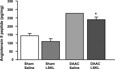
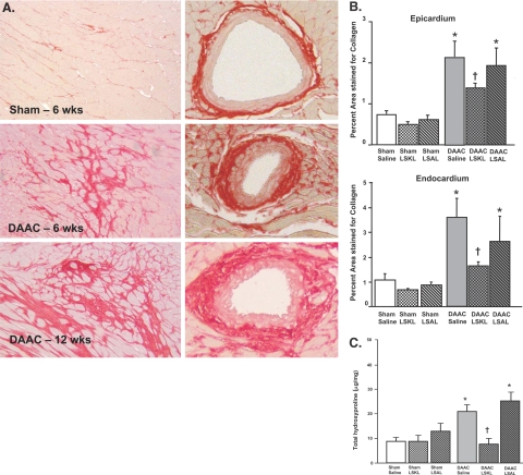
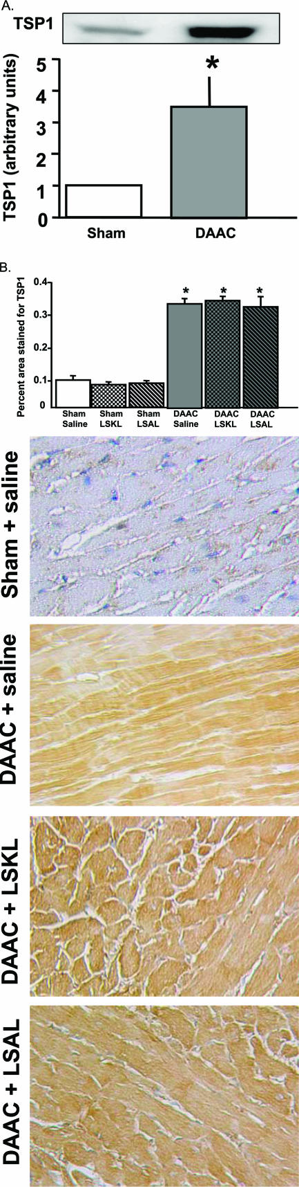
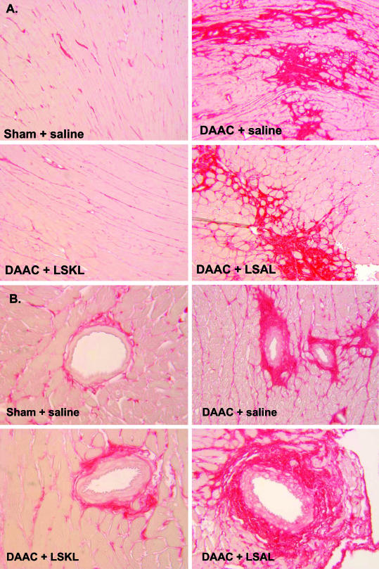
In diabetes and hypertension, the induction of increased transforming growth factor-beta (TGF-beta) activity due to glucose and angiotensin II is a significant factor in the development of fibrosis and organ failure. We showed previously that glucose and angiotensin II induce the latent TGF-beta activator thrombospondin-1 (TSP1). Because activation of latent TGF-beta is a major means of regulating TGF-beta, we addressed the role of TSP1-mediated TGF-beta activation in the development of diabetic cardiomyopathy exacerbated by abdominal aortic coarctation in a rat model of type 1 diabetes using a peptide antagonist of TSP1-dependent TGF-beta activation. This surgical manipulation elevates initial blood pressure and angiotensin II. The hearts of these rats had increased TSP1, collagen, and TGF-beta activity, and cardiac function was diminished. A peptide antagonist of TSP1-dependent TGF-beta activation prevented progression of cardiac fibrosis and improved cardiac function by reducing TGF-beta activity. These data suggest that TSP1 is a significant mediator of fibrotic complications of diabetes associated with stimulation of the renin-angiotensin system, and further studies to assess the blockade of TSP1-dependent TGF-beta activation as a potential antifibrotic therapeutic strategy are warranted.


Figures

Figure 1
Figure 1
Angiotensin II peptide is increased in the hearts of DAAC rats. Levels of angiotensin II (ANG II) peptide were measured in tissues from the left ventricles of sham and DAAC rats treated with either saline or LSKL peptide. Sham + saline (n = 6), Sham + LSKL (n = 6), DAAC + saline (n = 2), and DAAC + LSKL (n = 4). Data are expressed as the mean ± SEM. *P < 0.001 DAAC + LSKL versus sham + saline. The difference between Sham + saline and Sham + LSKL is not statistically significant (P < 0.09) by Mann-Whitney analysis.
Figure 2
Figure 2
Collagen deposition is increased in the hearts of rats with DAAC. A: Picric acid-Sirius red staining of the myocardium. Sections of the left ventricle (left) and cardiac vasculature (right) from sham and DAAC rats at 6 weeks and of DAAC rats at 12 weeks were analyzed for interstitial collagen content by picric acid-Sirius red staining. Original magnification, ×10. B: Quantification of increased collagen deposition in the epi- and endocardium of DAAC rats at 12 weeks. Interstitial collagen deposition in both epicardium and endocardium was measured by analysis of picric acid-Sirius red staining as described in Materials and Methods. Sham + saline (n = 8), Sham + LSKL (n = 8), Sham + LSAL (n = 8), DAAC + saline (n = 5), DAAC + LSKL (n = 8), and DAAC + LSAL (n = 5). Data are expressed as the mean percent area staining for collagen ± SEM. *P < 0.001 versus sham. P < 0.001 versus DAAC. C: Left ventricular hydroxyproline content is increased at 12 weeks in DAAC rats. The hydroxyproline content was measured in the left ventricles of Sham + saline (n = 8), Sham + LSKL (n = 8), Sham + LSAL (n = 8), DAAC + saline (n = 5), DAAC + LSKL (n = 8), and DAAC + LSAL (n = 5) rats. Data are expressed as the mean ± SEM. *P < 0.001 versus sham group. P < 0.001 versus DAAC.
Figure 3
Figure 3
TSP1 is increased in the hearts of DAAC rats. A: Immunoblot of TSP1 in extracts of left ventricle at 12 weeks. Equal amounts of protein extracted from the left ventricles of sham and experimental animals were separated by sodium dodecyl sulfate-polyacrylamide gel electrophoresis (6% gel), transferred to polyvinylidene difluoride membranes, and immunoblotted with antibody to TSP1 and developed by enhanced chemiluminescence. The bands were quantified by densitometry (n = 4). Data are expressed as the mean ± SEM. *P < 0.001 versus sham. B: Immunohistochemical staining for TSP1 in left ventricle. Sections were reacted with antibody specific for TSP1, color developed with diaminobenzidine, and counterstained with hematoxylin. The TSP1 immunostaining was quantified by morphometric analysis of 15 to 20 fields in each of three different regions of the left ventricle as described in Materials and Methods. Original magnification, ×20. Sham + saline (n = 8), Sham + LSKL (n = 8), Sham + LSAL (n = 8), DAAC + saline (n = 5), DAAC + LSKL (n = 8), and DAAC + LSAL (n = 5). Data are expressed as the mean percent area staining for TSP1 ± SEM. *P < 0.001 versus sham.
Figure 4
Figure 4
TGF-β signaling is increased in hearts of DAAC rats. A: Immunohistochemical staining for active TGF-β in the left ventricle 12 weeks after disease induction. Three different regions of the left ventricle were analyzed, and 10 to 15 fields were examined in each region. Sections were reacted with antibody specific for active TGF-β, color-developed with diaminobenzidine, and counterstained with hematoxylin. The percent area stained for active TGF-β was quantified as described in Materials and Methods. Original magnification, ×20. Sham + saline (n = 8), Sham + LSKL (n = 8), Sham + LSAL (n = 8), DAAC + saline (n = 5), DAAC + LSKL (n = 8), and DAAC + LSAL (n = 5). B: Immunohistochemical staining for nuclear phosphorylated Smad 2 in the left ventricle at 12 weeks. Tissue sections of the left ventricles were immunostained for phosphorylated Smad2, and the percentage of total nuclei stained positive (brown staining) was determined as described in Materials and Methods. Ten to 15 fields were examined in each region, and 80 to 128 nuclei were counted per field. Original magnification, ×20. Sham + saline (n = 8), Sham + LSKL (n = 8), Sham + LSAL (n = 8), DAAC + saline (n = 5), DAAC + LSKL (n = 8), and DAAC + LSAL (n = 5). Data are expressed as mean ± SEM. *P < 0.001 versus sham. P < 0.001 versus DAAC. C: Immunoblot for total and phosphorylated Smad 2 in extracts of the left ventricle at 12 weeks. Equal amounts of protein extracted from the left ventricles of sham and experimental animals were separated by sodium dodecyl sulfate-polyacrylamide gel electrophoresis (10% gels), transferred to polyvinylidene difluoride membranes, and immunoblotted separately with antibodies to phosphorylated Smad 2 (phospho-Smad2) or to Smad 2 (Smad2) and developed by enhanced chemiluminescence. The bands were quantified by densitometry (n = 4).
Figure 5
Figure 5
LSKL peptide treatment reduces fibrosis in hearts of DAAC rats. Sections of left ventricular myocardium (A) or vasculature (B) from DAAC rats after 12 weeks of disease induction and 6 weeks of treatment with saline, LSKL, or LSAL peptide were analyzed for interstitial collagen content by picric acid-Sirius red staining. Original magnification, ×10.

Similar articles

Cited by

References

    1. Fonarow GC, Srikanthan P. Diabetic cardiomyopathy. Endocrinol Metab Clin North Am. 2006;35:575–599, ix. - PubMed
    1. Asbun J, Villarreal FJ. The pathogenesis of myocardial fibrosis in the setting of diabetic cardiomyopathy. J Am Coll Cardiol. 2006;47:693–700. - PubMed
    1. Factor SM, Bhan R, Minase T, Wolinsky H, Sonnenblick EH. Hypertensive-diabetic cardiomyopathy in the rat: an experimental model of human disease. Am J Pathol. 1981;102:219–228. - PMC - PubMed
    1. Poornima IG, Parikh P, Shannon RP. Diabetic cardiomyopathy: the search for a unifying hypothesis. Circ Res. 2006;98:596–605. - PubMed
    1. Factor SM, Minase T, Bhan R, Wolinsky H, Sonnenblick EH. Hypertensive diabetic cardiomyopathy in the rat: ultrastructural features. Virchows Arch A Pathol Anat Histopathol. 1983;398:305–317. - PubMed

Publication types

MeSH terms