. 2007 Sep;171(3):947-61.
doi: 10.2353/ajpath.2007.070049. Epub 2007 Jul 19.

RAGE expression in rhabdomyosarcoma cells results in myogenic differentiation and reduced proliferation, migration, invasiveness, and tumor growth

Affiliations

RAGE expression in rhabdomyosarcoma cells results in myogenic differentiation and reduced proliferation, migration, invasiveness, and tumor growth

Francesca Riuzzi et al. Am J Pathol. 2007 Sep.

Abstract

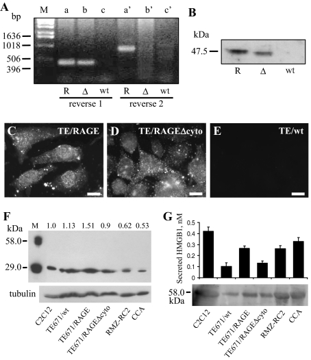
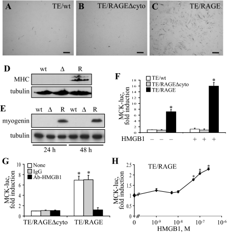
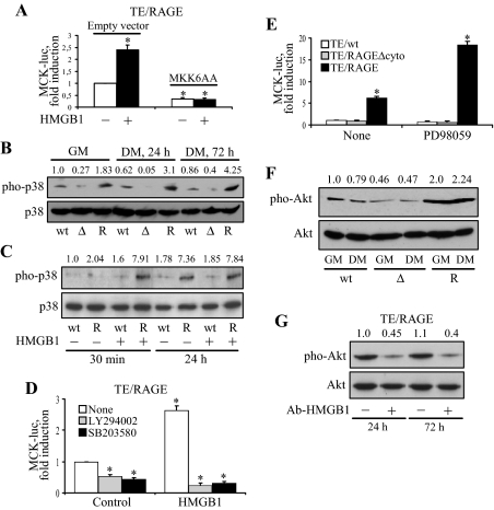
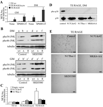
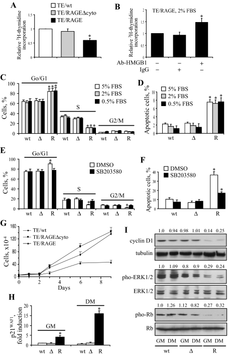
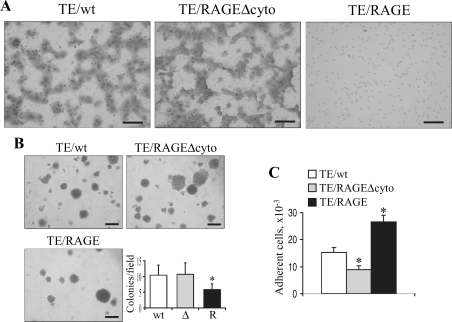
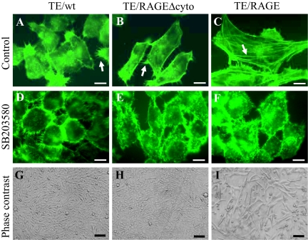
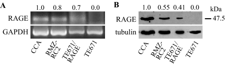
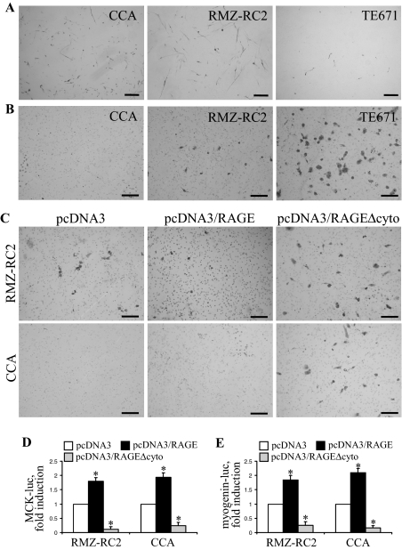
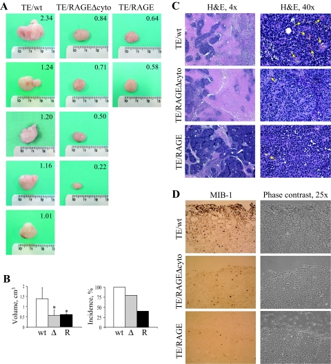
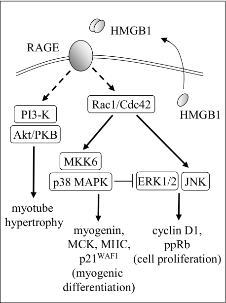
Activation of receptor for advanced glycation end products (RAGE) by its ligand, HMGB1, stimulates myogenesis via a Cdc42-Rac1-MKK6-p38 mitogen-activated protein kinase pathway. In addition, functional inactivation of RAGE in myoblasts results in reduced myogenesis, increased proliferation, and tumor formation in vivo. We show here that TE671 rhabdomyosarcoma cells, which do not express RAGE, can be induced to differentiate on transfection with RAGE (TE671/RAGE cells) but not a signaling-deficient RAGE mutant (RAGEDeltacyto) (TE671/RAGEDeltacyto cells) via activation of a Cdc42-Rac1-MKK6-p38 pathway and that TE671/RAGE cell differentiation depends on RAGE engagement by HMGB1. TE671/RAGE cells also show p38-dependent inactivation of extracellular signal-regulated kinases 1 and 2 and c-Jun NH(2) terminal protein kinase and reduced proliferation, migration, and invasiveness and increased apoptosis, volume, and adhesiveness in vitro; they also grow smaller tumors and show a lower tumor incidence in vivo compared with wild-type cells. Two other rhabdomyosarcoma cell lines that express RAGE, CCA and RMZ-RC2, show an inverse relationship between the level of RAGE expression and invasiveness in vitro and exhibit reduced myogenic potential and enhanced invasive properties in vitro when transfected with RAGEDeltacyto. The rhabdomyosarcoma cell lines used here and C2C12 myoblasts express and release HMGB1, which activates RAGE in an autocrine manner. These data suggest that deregulation of RAGE expression in myoblasts might concur in rhabdomyosarcomagenesis and that increasing RAGE expression in rhabdomyosarcoma cells might reduce their tumor potential.


Figures

Figure 1
Figure 1
Characterization of TE671/wt (embryonal type), TE671/RAGEΔcyto, and TE671/RAGE cells. A: RT-PCR products for human RAGE and RAGEΔcyto in TE671/RAGE (R), TE671/RAGEΔcyto (Δ), and TE671/wt (wt) cells. Shown are PCR products obtained with human RAGE reverse primer 1 (lanes a–c) and reverse primer 2 (lanes a′–c′). No PCR products can be seen with reverse primer 2 in the case of TE671/RAGEΔcyto cells because of specificity of this primer for a region present in full-length RAGE and absent from RAGEΔcyto. No PCR products can be seen with either primer in the case of TE671/wt cells. B: TE671/wt, TE671/RAGEΔcyto, and TE671/RAGE cells in GM were solubilized and analyzed for expression of RAGE and RAGEΔcyto by Western blotting using an anti-RAGE extracellular domain antibody (Chemicon). Note the faster migrating immunoreactive band in the TE671/RAGEΔcyto lane. C–E: TE671/wt, TE671/RAGEΔcyto, and TE671/RAGE cells were cultivated in GM and processed for immunofluorescence with the anti-RAGE extracellular domain antibody above. F and G: Lysates from C2C12 myoblasts, TE671/wt, TE671/RAGE, TE671/RAGEΔcyto, RMZ-RC2, and CCA cells (F) and culture media from these same cell lines (G) were analyzed for HMGB1 by Western blotting. Notice that purified HMGB1 (M, in F) shows two bands, one corresponding to monomeric (29-kd) and the other one to dimeric (58-kd) HMGB1, that intracellular HMGB1 migrates as a monomer exclusively (F), and that extracellular (released) HMGB1 migrates as a dimer exclusively (G). Semiquantitative analysis of HMGB1 was performed using the culture media of the cell lines as indicated (G). One representative experiment of three is shown (A–F). The numbers on top of lanes in F refer to the HMGB1-to-tubulin ratio. Averages of three independent experiments ± SD (G). Bars = 20 μm (C–E).
Figure 2
Figure 2
Enforced expression of RAGE in TE671 cells activates the myogenic program on stimulation with HMGB1. A–C: TE671/wt, TE671/RAGEΔcyto, and TE671/RAGE cells were cultivated in DM for 4 days, fixed, and subjected to immunocytochemistry for detection of MHC. D: Same as in A–C except that the cells were solubilized and subjected to Western blotting for detection of MHC. R, wt, and Δ stand for TE671/RAGE, TE671/wt, and TE671/RAGEΔcyto cells, respectively. E: Same as in A–C except that the cells were cultivated in DM for 24 and 48 hours before Western blotting for detection of myogenin. F: TE671/wt, TE671/RAGEΔcyto, and TE671/RAGE cells were transiently transfected with MCK-luc reporter gene, switched to DM, cultivated for 24 hours in the absence or presence of added HMGB1 to 100 nmol/L, and harvested to measure luciferase activity. G: Same as in F except that TE671/RAGEΔcyto and TE671/RAGE cells were cultivated in the presence of either 2.5 μg/ml nonimmune IgG or 2.5 μg/ml anti-HMGB1 antibody for 48 hours. H: Same as in F except that TE671/RAGE cells were cultivated in the presence of added HMGB1 to the concentrations indicated. One representative experiment of three is shown (A–E). Averages of three independent experiments ± SD (F–H). *Significantly different from control (first column from left in F and G; TE671/RAGE cells in the absence of additions in H) (P < 0.01). Bars = 200 μm (A–C).
Figure 3
Figure 3
Enforced expression of RAGE in TE671 cells activates p38 MAPK and Akt on stimulation with HMGB1. A: TE671/RAGE cells were transiently transfected with MCK-luc reporter gene and MKK6AA, switched to DM, cultivated for 24 hours under these conditions in the absence or presence of added HMGB1 to 100 nmol/L, and harvested to measure luciferase activity. B: TE671/wt (wt), TE671/RAGEΔcyto (Δ), and TE671/RAGE (R) cells were cultivated in GM for 24 hours or in DM for 24 or 72 hours and processed for detection of phosphorylated and total p38 MAPK by Western blotting. C: TE671/wt (wt), TE671/RAGEΔcyto (Δ), and TE671/RAGE (R) cells were cultivated in DM for 30 minutes or 24 hours in the absence or presence of added HMGB1 to 100 nmol/L and processed for detection of phosphorylated and total p38 MAPK. D: TE671/RAGE cells were transiently transfected with MCK-luc reporter gene, switched to DM, cultivated for 24 hours in the absence or presence of added HMGB1 to 100 nmol/L plus or minus the PI3-K inhibitor LY294002 or the p38 MAPK inhibitor SB203580 and harvested to measure luciferase activity. E: TE671/wt, TE671/RAGEΔcyto, and TE671/RAGE cells were transiently transfected with MCK-luc reporter gene, switched to DM, cultivated for 24 hours in the absence or presence of the mitogen-activated protein kinase kinase inhibitor PD98059, and harvested to measure luciferase activity. F: TE671/wt, TE671/RAGEΔcyto, and TE671/RAGE cells were cultivated in GM or DM for 24 hours and processed for detection of phosphorylated and total Akt by Western blotting. G: Same as in F except that TE671/RAGE cells were cultivated in DM for 24 hours or 72 hours in the presence of either 2.5 μg/ml nonimmune IgG or 2.5 μg/ml anti-HMGB1 antibody. One representative experiment of three is shown (B, C, F, and G). The numbers on top of lanes in B, C, F, and G refer to the phosphorylated-to-total ratio of the pertinent kinase relative to the respective control (first lane from left on each blot). Averages of three independent experiments ± SD (A, D, and E). *Significantly different from control (first column from left in A, D, and E) (P < 0.01).
Figure 4
Figure 4
Enforced expression of RAGE in TE671 cells results in relief of the inhibitory effect of JNK on myogenic differentiation. A: TE671/wt, TE671/RAGEΔcyto, and TE671/RAGE cells were transiently transfected with MCK-luc reporter gene and cultivated in GM for 24 hours or DM for 24 hours in the absence or presence of the JNK inhibitor, SP600125. B: TE671/wt (wt), TE671/RAGE (R), and TE671/RAGEΔcyto (Δ) cells in DM (a–f) and GM (a′–f′) were analyzed for JNK phosphorylation and in the absence (a–c, a′c′) or presence (df, d′f′) of 2 μmol/L SB203580 (p38 MAPK inhibitor) by Western blotting. C: TE671/wt, TE671/RAGEΔcyto, and TE671/RAGE cells were transiently transfected with MCK-luc reporter gene and either N17Cdc42 or N17Rac1, switched to DM, cultivated for 24 hours, and harvested to measure luciferase activity. D: TE671/RAGE cells were transiently transfected with N17Cdc42, N17Rac1, or MKK6AA, cultivated in DM for 24 hours, and processed for detection of phosphorylated and total p38 MAPK by Western blotting. E: Same as in D except that TE671/RAGE cells were cultivated in DM for 4 days and that one additional sample was treated with the p38 MAPK inhibitor, SB203580. The cells were then fixed and subjected to immunocytochemistry for detection of MHC. One representative experiment of three is shown (B, D, and E). Averages of three independent experiments ± SD (A and C). *Significantly different from control (first column from left in A and C) (P < 0.01). Bars = 200 μm (E).
Figure 5
Figure 5
Enforced expression of RAGE in TE671 cells reduces proliferation and increases apoptosis. A: TE671/wt, TE671/RAGEΔcyto, and TE671/RAGE cells were subjected to a [3H]thymidine assay. B: Same as in A except that TE671/RAGE cells were cultivated in the presence of either 2.5 μg/ml nonimmune IgG or 2.5 μg/ml anti-HMGB1 antibody for 48 hours before [3H]thymidine assay. C and D: TE671/wt (wt), TE671/RAGEΔcyto (Δ), and TE671/RAGE (R) cells were cultivated for 72 hours in the presence of the FBS concentration indicated and subjected to FACS analysis to measure the fraction of cells in the various phases of the cell cycle (C) and apoptosis (D). E and F: Same as in C and D, respectively, except that the cells were cultivated in 2% FBS in the presence of the p38 MAPK inhibitor, SB203580, or vehicle [dimethylsulfoxide (DMSO)]. G: TE671/wt, TE671/RAGEΔcyto, and TE671/RAGE cells were cultivated in GM for the days indicated and processed by the 3-(4,5-dimethylthiazol-2-yl)-2,5-diphenyltetrazolium assay. H: TE671/wt (wt), TE671/RAGEΔcyto (Δ), and TE671/RAGE (R) cells were transiently transfected with p21WAF1-luc reporter gene, switched to DM, cultivated for 48 hours and harvested to measure luciferase activity. I: TE671/wt (wt), TE671/RAGEΔcyto (Δ), and TE671/RAGE (R) cells were cultivated for 24 hours in either GM or DM and processed for detection of cyclin D1, phosphorylated and total ERK1/2, and phosphorylated and total Rb by Western blotting. A Western blot of tubulin is included below the cyclin D1 blot to show total protein loading in individual lanes. One representative experiment of three is shown (I). The numbers on top of lanes in I refer to the relative density of cyclin D1, phosphorylated ERK1/2 and phosphorylated Rb to tubulin, total ERK1/2, and total Rb, respectively. Averages of three independent experiments ± SD (A–H). *Significantly different from control (first column from left in A, B, D, F, and H) or from internal control (first column from left in each group in C and E) (P < 0.01).
Figure 6
Figure 6
Enforced expression of RAGE in TE671 cells reduces migration, colony formation, and invasiveness and increases adhesiveness. A: TE671/wt, TE671/RAGEΔcyto, and TE671/RAGE cells were subjected to a migration assay in Boyden chambers. B: In a colony formation assay, a ∼50% decrease in the number of colonies was observed in the case of TE671/RAGE cells (R). C: In a cell adhesion assay, nearly twice as many TE671/RAGE cells adhered to the support after 3 hours as TE671/wt and TE671/RAGEΔcyto cells. One representative experiment of three is shown (A and B). *Significantly different from control (first column from left in B and C, n = 3) (P < 0.01). Bars: 100 μm (A); 500 μm (B).
Figure 7
Figure 7
Enforced expression of RAGE in TE671 cells increases cell volume and restores stress fiber formation in a p38 MAPK-dependent manner. A–F: TE671/wt, TE671/RAGEΔcyto, and TE671/RAGE cells were cultivated in GM for 48 hours in the absence or presence of the p38 MAPK inhibitor SB203580 (2 μmol/L for 24 hours), fixed, and incubated with fluorescein isothiocyanate-phalloidin to detect F-actin. Well-structured stress fibers (arrow in C) can be seen in TE671/RAGE cells that also exhibit a smaller number of filopodia than do TE671/wt and TE671/RAGEΔcyto cells (arrows in A and B). Note the absence of stress fibers in TE671/wt and TE671/RAGEΔcyto cells and the larger size of TE671/RAGE cells compared with TE671/wt and TE671/RAGEΔcyto cells. Treatment with SB203580 results in collapse of F-actin onto plasma membranes in the three TE671clones, acquisition of a round shape, decrease in cell volume, and appearance of numerous filopodia. G–I: TE671/wt, TE671/RAGEΔcyto, and TE671/RAGE cells were cultivated in DM for 3 days and analyzed by phase-contrast microscopy. One representative experiment of three with similar results is shown (A–I). Bars: 20 μm (A–F); 250 μm (G–I).
Figure 8
Figure 8
CCA (embryonal type) and RMZ-RC2 (alveolar type) RMS cells express RAGE. A and B: Expression of RAGE in CCA, RMZ-RC2, TE671, and TE671/RAGE cells was investigated by RT-PCR (A) and Western blotting (B). The numbers on top of lanes in A and B refer to the densities of RAGE mRNA relative to GADPH mRNA (A) and of RAGE protein relative to tubulin (B). One representative experiment of three with similar results is shown.
Figure 9
Figure 9
CCA and RMZ-RC2 RMS cells show reduced invasiveness compared with TE671 cells, express MHC in DM, and exhibit increased invasiveness and reduced MCK and myogenin induction when transfected with RAGEΔcyto. A: CCA, RMZ-RC2, and TE671 cells were cultivated in GM for 24 hours, switched to DM for 4 days, and subjected to immunocytochemistry to detect MHC. B: CCA, RMZ-RC2, and TE671 cells were subjected to an invasiveness assay. C: CCA and RMZ-RC2 cells were transiently transfected with RAGE or RAGEΔcyto and subjected to an invasiveness assay. D and E: CCA and RMZ-RC2 cells transiently transfected with either RAGE or RAGEΔcyto were transfected with MCK-luc or myogenin-luc reporter gene. One representative experiment of three is shown (A–C). *Significantly different from control (first column from left in each group in D and E) (P < 0.01). Bars: 250 μm (A); 100 μm (B and C).
Figure 10
Figure 10
Inoculation of TE671/RAGE cells into mice results in reduced tumor volume and incidence. Immunocompromised mice were injected with TE671/wt, TE671/RAGEΔcyto, or TE671/RAGE cells, and tumor mass formation was followed for 4 to 6 weeks. Tumor masses were analyzed for volume (A and B), histopathology (C), and proliferating cells (D). Ample zones of necrosis are detected in TE671/wt and TE671/RAGEΔcyto tumors compared with TE671/RAGE tumors (C, left panels), and TE671/RAGEΔcyto tumors and TE671/RAGE tumors exhibit a remarkably reduced vasculature compared with TE671/wt tumors (arrows in C, right panels). In addition, a much larger number of proliferating cells is detected in TE671/wt tumors compared with TE671/RAGEΔcyto tumors that, in turn, show a larger number of proliferating cells than TE671/RAGE tumors, as investigated by Ki-67 (MIB1) immunohistochemistry (D). *Significantly different from control (first column from left in the average volume panel in B) (n = 5, P < 0.05).
Figure 11
Figure 11
Schematic representation of the proposed effects of enforced expression of RAGE in TE671 RMS cells. RAGE engaged by HMGB1 signals to Rac1/Cdc42 and to PI3-K via unknown intermediates, thereby stimulating myogenic differentiation through activation of the MKK6/p38 MAPK module and myotube hypertrophy through the PI3-K/Akt module. HMGB1/RAGE-dependent activation of p38 MAPK also causes inactivation of ERK1/2 and JNK with consequent inhibition of proliferation and decrease in cell survival. Shown are also monomeric HMGB1 within myoblasts and the RMS cell lines used in this study and released HMGB1, which forms disulfide cross-linked dimers in the extracellular medium.

Similar articles

Cited by

References

    1. Andrés V, Walsh K. Myogenin expression, cell cycle withdrawal, and phenotypic differentiation are temporally separable events that precede cell fusion upon myogenesis. J Cell Biol. 1996;132:657–666. - PMC - PubMed
    1. Arnold HH, Winter B. Muscle differentiation: more complexity to the network of myogenic regulators. Curr Opin Genet Dev. 1998;8:539–544. - PubMed
    1. Chargé SB, Rudnicki MA. Cellular and molecular regulation of muscle regeneration. Physiol Rev. 2004;84:209–238. - PubMed
    1. Buckingham M. Myogenic progenitor cells and skeletal myogenesis in vertebrates. Curr Opin Genet Dev. 2006;16:525–532. - PubMed
    1. Lassar AB, Skapek SX, Novitch B. Regulatory mechanisms that coordinate skeletal muscle differentiation and cell cycle withdrawal. Curr Opin Cell Biol. 1994;6:788–794. - PubMed

Publication types

MeSH terms

Substances