. 2007 Dec;37(6):651-9.
doi: 10.1165/rcmb.2006-0428OC. Epub 2007 Jul 19.

Integrin beta6 mediates phospholipid and collectin homeostasis by activation of latent TGF-beta1

Affiliations

Integrin beta6 mediates phospholipid and collectin homeostasis by activation of latent TGF-beta1

Laura L Koth et al. Am J Respir Cell Mol Biol. 2007 Dec.

Abstract

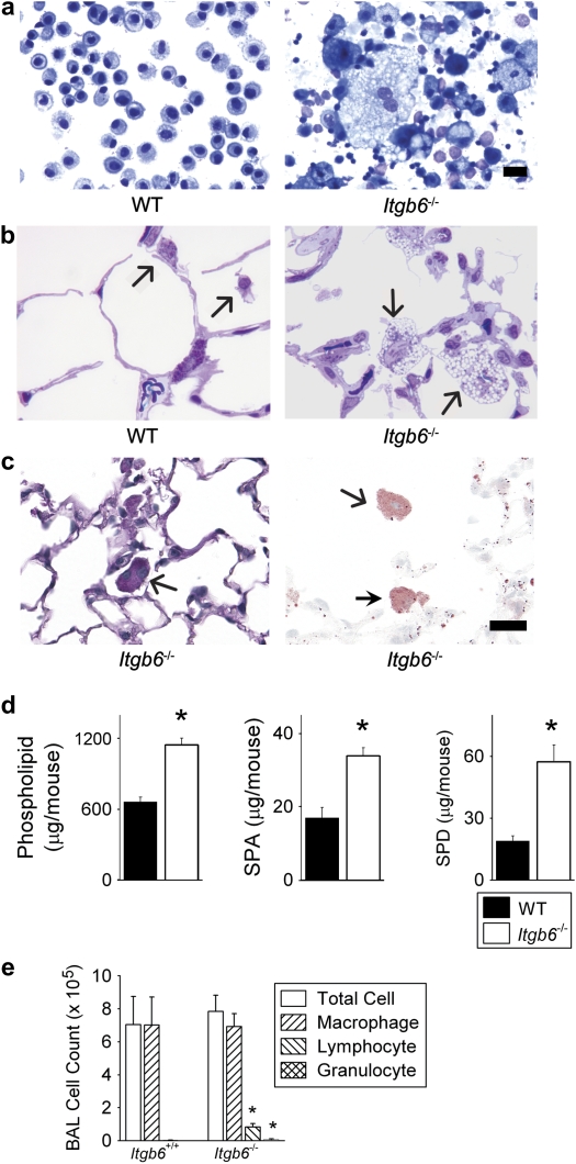
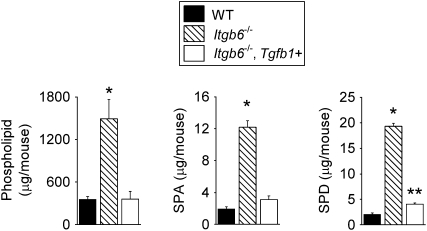
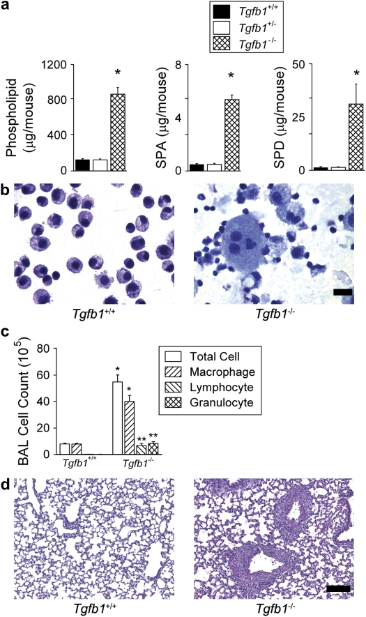
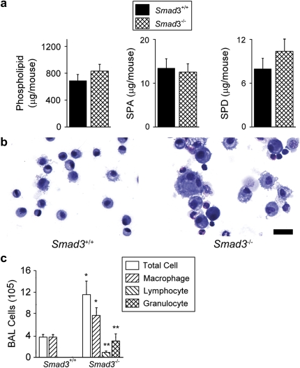
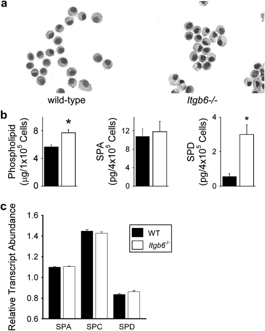
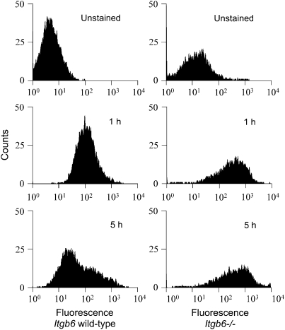
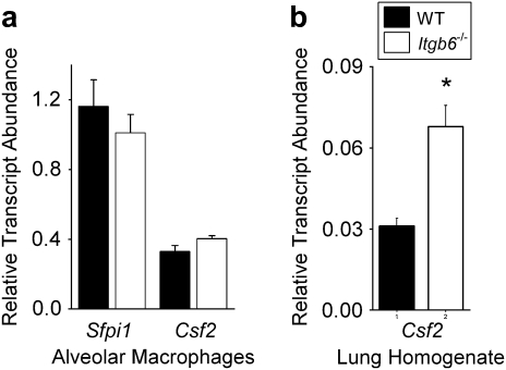
Surfactant lines the alveolar surface and prevents alveolar collapse. Derangements of surfactant cause respiratory failure and interstitial lung diseases. The collectins, surfactant proteins A and D, are also important in innate host defense. However, surfactant regulation in the postnatal lung is poorly understood. We found that the epithelial integrin, alphavbeta6, regulates surfactant homeostasis in vivo by activating latent transforming growth factor (TGF)-beta. Adult mice lacking the beta-subunit of alphavbeta6 (Itgb6-/-) developed increased bronchoalveolar lavage phospholipids and surfactant proteins A and D, and demonstrated abnormal-appearing alveolar macrophages, reminiscent of the human disease pulmonary alveolar proteinosis. Using lung-specific expression of constitutively active TGF-beta1 in Itgb6-/- mice, we found that TGF-beta1 was sufficient to normalize these abnormalities. Tgfbeta1-deficient mice also demonstrated increased phospholipids and surfactant proteins A and D, but mice lacking the key TGF-beta signaling molecule, SMAD3, did not. Therefore, integrin-mediated activation of latent TGF-beta1 regulates surfactant constituents independent of intracellular SMAD3. In vivo increases in surfactant protein A and D were not associated with increases in mRNA for these proteins in alveolar tissue from Itgb6-/- mice. On the other hand, isolated alveolar macrophages from Itgb6-/- mice were defective in processing phospholipids in vitro, suggesting that reduced surfactant clearance contributes to altered surfactant homeostasis in these mice in vivo. These findings show that alphavbeta6 and TGF-beta1 regulate homeostasis of phospholipids and collectins in adult mouse lungs and may have implications for anti-fibrotic therapeutics that inhibit active TGF-beta in the lung.


Figures

<b>Figure 1.</b>
Figure 1.
Integrin β6 subunit-deleted (Itgb6−/−) mice spontaneously develop elevated bronchoalveolar lavage (BAL) phospholipid and collectin levels associated with large foamy alveolar macrophages and inflammation. (a) Representative BAL fluid cytospins from Itgb6−/− mice revealed enlarged alveolar macrophages with cytoplasmic vacuolization and extracellular debris. (b) Representative high-power microscopic images of lung tissue showed enlarged size and numerous vacuoles in macrophages from Itgb6−/− mice; this was not observed in any of the sections from littermate controls. Arrows indicate alveolar macrophages. (c) PAS (left) and Oil red O (right) staining of inflated lung sections from Itgb6−/− mice demonstrate cytoplasmic staining (PAS stains glycoprotein pink; Oil red O stains lipid red). Arrows indicate alveolar macrophages. (d) Total phospholipids, SP-A, and SP-D in BAL fluid from wild-type (WT) littermates and Itgb6−/− mice expressed as mean ± SEM (n = 6 per group). Results are representative of three separate experiments; *P < 0.005. (e) BAL fluid cell counts and differentials in wild-type littermates and Itgb6−/− mice; * P < 0.008. Mice studied in b were 6 months old, while all other data were collected from 2-month-old mice. Scale bars indicate 20 μm.
<b>Figure 2.</b>
Figure 2.
Increases in surfactant constituents in Itgb6−/− mice are normalized by doxycycline-induced lung expression of active TGF-β1. Total phospholipid, SP-A, and SP-D in BAL fluid from 2-month-old wild-type (WT), Itgb6−/−, and Itgb6−/− mice carrying a transgene for active TGF-β1 (Itgb6−/−, Tgfb1+). Data are expressed as mean ± SEM (n = 4–6 per group). Doxycycline treatment was started in all groups at 3 weeks of age and continued until the mice were killed at 2 months of age. Results are representative of three separate experiments. *P < 0.008 compared with all other groups; **P = 0.02 compared with WT.
<b>Figure 3.</b>
Figure 3.
Tgfb1-deficient mice have elevated levels of surfactant constituents, enlarged, foamy alveolar macrophages and lung inflammation. (a) Total phospholipids, SP-A, and SP-D in BAL fluid from 12-day-old wild-type (Tgfb1+/+), heterozygote (Tgfb1+/−), and Tgfb1-deleted (Tgfb1−/−) littermate mice expressed as mean ± SEM (n = 6 per group). *P ⩽ 0.005 compared with all other groups. (b) BAL fluid cytospins from 12-day-old wild-type littermates and Tgfb1-deleted mice. Alveolar macrophages from heterozygote mice appeared similar to wild type (not shown) (scale bar, 20 μm). (c) BAL fluid cell counts and differentials in 12-day-old wild-type (Tgfb1+/+) and Tgfb1-deleted (Tgfb1−/−) littermate mice. (d) Representative hematoxylin and eosin–stained lung sections from wild-type littermates and Tgfb1-deleted mice, showing normal-appearing lung parenchyma with areas of peribronchovascular inflammation (scale bar, 100 μm).
<b>Figure 4.</b>
Figure 4.
Smad3-deficient mice have normal levels of surfactant constituents in the setting of lung inflammation. (a) Total phospholipids, SP-A, and SP-D in BAL fluid from 4-month-old wild-type (Smad3+/+) and Smad3-deficient (Smad3−/−) littermate mice expressed as mean ± SEM (n = 10–11 per group). Differences in means were not statistically significant. (b) BAL cytospins from 4-month-old wild-type littermates and Smad3-deficient mice (Scale bar, 20 μm). (c) Two-month-old Smad3-deficient mice show increased numbers of BAL fluid macrophages, lymphocytes, and granulocytes expressed as mean ± SEM (n = 5–9 per group). *P ⩽ 0.05; **P ⩽ 0.007.
<b>Figure 5.</b>
Figure 5.
Alveolar epithelial cells from Itgb6−/− mice contained increased phospholipids and SP-D with no detectable change in surfactant protein transcript levels. (a) Representative cytospins of alveolar epithelial cells from 4-month-old Itgb6−/− mice and littermate controls. (b) Total phospholipids and SP-A and SP-D in alveolar epithelial cells isolated from Itgb6−/− mice and littermate controls. Data are expressed as average μg or pg per designated cell number with n = 5 per group. (c) RNA was isolated from laser captured alveolar tissue from Itgb6−/− mice and littermate controls. Messenger RNA levels of SP-A, -C, and -D were measured using real-time PCR (n = 4 per group). Values for wild-type littermates are represented by solid bars and for Itgb6−/− mice by open bars. Data are expressed as means ± SEM.
<b>Figure 6.</b>
Figure 6.
Itgb6−/− mice demonstrated abnormal handling of lipid by alveolar macrophages. FACS histogram plot of alveolar macrophages from wild-type (left column) and Itgb6−/− mice (right column) after incubation with a mixture of natural surfactant and rhodamine-DPPE as described in Materials and Methods. Macrophages were exposed to 50 μg/ml rhodamine-DPPE for 1 hour, then washed, and a fraction of the cells were then allowed to incubate for an additional 4 hours. Wild-type macrophages demonstrate a decay in fluorescence intensity indicating a loss of rhodamine-DPPE at 5 total hours of incubation that was not observed with Itgb6−/− macrophages. Similar results were obtained using different concentrations of rhodamine-DPPE. Minimal nonspecific binding of rhodamine-DPPE was seen in macrophages incubated at 4°C (data not shown). Ten thousand cell counts were collected for each sample and counts shown are gated to exclude lymphocytes.
<b>Figure 7.</b>
Figure 7.
Alveolar macrophages from Itgb6−/− mice and control littermates had similar mRNA levels of GM-CSF and PU.1 while Itgb6−/− lung homogenate showed increased expression of GM-CSF compared with controls. (a) Total RNA was isolated from purified BAL macrophages, and expression of Sfpi1 (PU.1) and Csf2 (GM-CSF) were measured using real-time PCR (n = 5 per group). (b) Lung homogenate total RNA was analyzed for Csf2 (GM-CSF). Data are expressed as relative transcript abundance normalized to GAPDH ± SEM. Values for wild-type littermates are represented by solid bars and for Itgb6−/− mice by open bars.

Comment in

References

    1. Rooney SA, Young SL, Mendelson CR. Molecular and cellular processing of lung surfactant. FASEB J 1994;8:957–967. - PubMed
    1. Trapnell BC, Whitsett JA. GM-CSF regulates pulmonary surfactant homeostasis and alveolar macrophage-mediated innate host defense. Annu Rev Physiol 2002;64:775–802. - PubMed
    1. Rider ED, Ikegami M, Jobe AH. Localization of alveolar surfactant clearance in rabbit lung cells. Am J Physiol 1992;263:L201–L209. - PubMed
    1. Wright JR. Clearance and recycling of pulmonary surfactant. Am J Physiol 1990;259:L1–L12. - PubMed
    1. Nogee LM. Genetics of pediatric interstitial lung disease. Curr Opin Pediatr 2006;18:287–292. - PubMed

Publication types

MeSH terms